
Abstract
This study describes an extremely sensitive and simple assay to measure small volumes of solutions, <1 nL. The assay takes advantage of the Sandell-Kolthoff reaction in which yellow cerium(IV) is reduced to colorless cerium(III) in the presence of arsenic(III) and catalytic quantities of iodide ion. The reaction is linear with respect to the rate of Ce(IV) reduction and the quantity of I– present. Typical assays can measure 10-100 picomoles of iodide in a sample. When I– is substituted for chloride ion in standard biological buffers, such as Tris-buffered saline, the assay can be used to determine the volume of solution printed in a microarray.
Introduction
Microfabrication techniques are playing an increasingly important role in the production of the latest diagnostic devices and sensors. Many of these devices require the functionalization of part of the sensor surface with a biologically active compound, such as an antibody or enzyme. To achieve this, techniques such as microcontact printing, quill-pen spotting, and inkjet printing have been developed to impart a small quantity of biological material to surfaces.1–6
In many instances it is important to measure the quantity of biological material printed by these techniques. The quantity of active material is readily measured using an activity assay for the biological material, for example, an enzyme assay if an enzyme is printed, or an immunoassay if an antibody is printed. However, these methods do not necessarily measure the quantity of biological material actually delivered to the surface, since denaturing can occur at various points in the printing process. Denaturing could occur during printing, during adsorption on the surface, or during the storage time between printings and assay. Even if an excess of material is delivered to a surface, the resulting surface concentration of active material could be low if significant damage occurs during printing.
We recently measured damage to the enzyme horseradish peroxidase (HRP) caused by ink-jet printing. 7 An internal standard, fluorescein sodium salt, was added to solutions containing HRP, and these solutions were printed using an ink-jet. The solutions were printed directly into microplate wells containing a buffer solution. The fluorescence of the internal standard measures the total enzyme printed, and a standard assay measures the quantity of active enzyme. Comparing these measurements allowed us to determine the fraction of peroxidase that denatured due to printing. We found that denaturing of the peroxidase varied linearly with the rate of compression of the piezoceramic during printing.
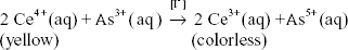
In this report we describe an internal standard, sodium iodide, which is less intrusive, more sensitive, and easily assayed. The increased sensitivity of this assay when compared to the commonly used fluorescent standards, allows for the measurement of less than one nanoliter in total volume. Thus, when employing microprinting techniques that deliver very small volumes, such as electrostatic printing, which can deliver less than one picoliter of volume per spot8–10 (Nishioka, Holloway and Sokolik, unpublished results), the use of iodide as an internal standard is required. Sodium iodide is substituted for the sodium chloride present in commonly used biological buffers, such as Tris-buffered saline (TBS). After microprinting, the small quantities of iodide are measured by exploiting the reaction first described by Sandell and Kolthoff. 11 In this reaction iodide catalyzes the reduction of cerium(IV) to cerium(III):
Taking advantage of this reaction and mechanistic studies performed by other investigators12–16 we have developed a rapid, reliable, and convenient assay for detecting picomole quantities of iodide present as an internal standard in solution. The iodide assay is conveniently performed in polystyrene UV-transparent 96-well microplates at room temperature.
Experimental
Assay Procedure
The assay is performed by the sequential addition of 100 μl each of solutions containing arsenic(III), the sample containing the iodide standard/sample, and cerium(IV). The order of addition is extremely important as is mixing, accomplished by pipetting rapidly up and down three times, after each component is added. Absorbance at 350 nm is measured following addition of cerium(IV), and is measured again after 2.0 minutes. The rate of cerium(IV) reduction is then calculated over this time period; this rate varies linearly with the quantity of iodide present in the sample (see Fig. 1). The Molar absorptivity of cerium(IV) is 4282.5 M-1 cm-1, the path length for 300 μL of solution in a typical 96-well UV-transparent microplate is 0.88 cm. The rate is constant over the first 3 minutes of the reaction; therefore absorbance change can be measured over any time within this period.
Calibration curve for determining picomoles of iodide by measuring the rate of Ce(IV) reduction. The line is the linear best fit to the average of five separate experiments, y = 0.929x + 26.6. Error bars are the 95% confidence intervals. Data were obtained at room temperature (20 °C-21 °C). The y-intercept value represents the uncatalyzed rate of Ce(IV) reduction (the rate in the absence of iodide ion).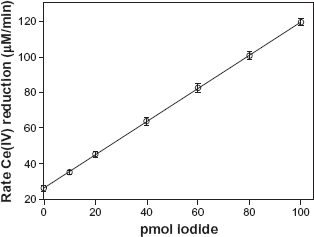
Assay Solutions
The 15.0 mM arsenic(III) solution is prepared by dissolving 0.297 g of As2O3 in 100 mL of 0.3 M NaOH with heat. After dissolution, 2.0 mL of concentrated sulfuric acid and 6.0 g NaCl are added, and the solution brought to a final volume of 200 mL with water. The 100 mM iodide stock solution is prepared by dissolving 1.5 g of NaI in 100 mL water. A working standard solution of iodide ion of 1 μm (1 pmol/μl) is prepared by serial dilution of the stock solution with the appropriate buffer. The 2.4 mM cerium(IV) solution is prepared by dissolving 0.194 g of Ce(SO4)2 • 4H2O in 200 mL of 0.9 M sulfuric acid.
Buffers
Buffers used were as follows: PBS: 10 mM phosphate buffered saline pH 7.4, TBS: 50 mM Tris buffered saline pH 7.6 (2-Amino-2-(hydroxymethyl)-1,3 propanediol), HBS: 50 mM HEPES buffered saline pH 7.4 (2-[4-(2-Hydroxyethyl)-1 piperazinyl]ethanesulfonic acid).
Chip Preparation
<P100> silicon chips were heat cleaned at 500 °C for 11 hours. For self assembled monolayered-C18 (SAM-C18) coating, heat-cleaned chips were immersed in a 1% (v/v) solution of octadecyltrichlorosilane in dicyclohexyl for 5 minutes, then washed 5 times with toluene and ethanol, and air dried in a clean hood. The advancing contact angle of water on these surfaces was 75°-78°. For polyethylene glycol (PEG) coating, heat-cleaned chips were immersed in a 1% (v/v) solution of 2-(methoxy(polyethyleneoxy) propyltrichlorosilane in dyclyclohexyl for 5 minutes, then washed 5 times with toluene and ethanol, and air dried in a clean hood. Water spreads on these surfaces. The PEG contains between 6 and 9 ethylene oxide residues.
Spotting Solution and Enzymec Assay
Solutions were prepared consisting of 1.0 mg/mL HRP and 0.3 M sodium iodide in phosphate buffer, Tris buffer, or HEPES buffer (the sodium iodide substitutes for saline). Other solutions contained in addition 10% w/v trehalose. These solutions were diluted 1:5,000 with the appropriate saline buffer (PBS, TBS, or HBS). Two microliters of each solution were then spotted on chips, delivering 0.4 ng HRP and 120 picomoles iodide. To produce HRP standard activity curves, each spotting solution was diluted 3:1, 1:1, and 1:3 in the appropriate buffer. These solutions and a negative control were assayed using 3, 3', 5, 5'-tetramethylbenzidine (TMB) substrate in a microplate reader. The rise in absorbance was linear over the first 15 minutes. Linear calibration plots of absorbance versus concentration were plotted for each spotting solution. Samples were assayed in a similar fashion, and the calibration plots were used to determine active HRP concentration. Further details are given in reference (1).
Results and Discussion
Figure 1 plots the effect of iodide concentration on the rate of cerium(IV) reduction. The data in Figure 1 are used to determine the quantity of iodide in samples and are readily reproduced. Because of room temperature variations we routinely measured the calibration curve for each set of samples but only minor deviations from the curve in Figure 1 were observed.
The application of this assay in microprinting is demonstrated as follows. Microprinting of proteins (using electrospray) typically involves the deposition of low volumes (~1 nL) of a buffered saline solution. If iodide is substituted for chloride in buffered saline, then 1 nL of a 0.14 M iodide solution is deposited, equaling 140 picomoles of iodide. In order to quantitatively deliver a representative amount of iodide we used a standard micropipette to deposit a relatively large volume (2 μL) of a dilute iodide solution (100 μm NaI), thus delivering 200 picomoles of iodide to the sample surface. We deposited solutions consisting of 100 μm sodium iodide dissolved in one of three biological buffers: phosphate, Tris, or HEPES.
These solutions were deposited on silicon chips diced from a silicon wafer. Three surfaces were tested: the bare surface, which consists of a thin silica layer, a surface coated with a self-assembled monolayer of octadecylsilane (SAM-C18), and a surface coated with polyethylene glycol (PEG). These surfaces represent a high-energy oxide surface, a low energy alkyl surface, and a bio-compatible low-adsorbing surface respectively.
Three samples of each surface were spotted with 200 picomoles of iodide and subsequently dried overnight in a desiccator. Drying was performed in order to determine whether the dehydration would adversely affect iodide recovery. The dried spots were redissolved in 210 μl of the appropriate buffer (without iodide). Each recovered solution was assayed twice using 50 μl of the recovered solution + 50 μl of the appropriate buffer (PBS, TBS, or HBS). Measured rates were ~70 μm/min, corresponding to ~50 pmoles iodide (~200 pmol iodide in the entire sample).
Percentage iodide recovered from silicon chips.
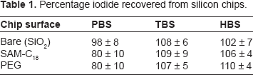
From our data we can conclude that small quantities of iodide can be delivered and completely recovered from a variety of surfaces using TBS and HEPES buffers. Iodide can thus serve as an internal standard in these buffers. Recovery of iodide from surfaces using PBS buffer may present problems and should be used with caution. The recovery of the iodide may be affected by the charge of the main buffering species. Using the zwitterionic HEPES or positively charged Tris may provide for better recovery when compared to the negatively charged phosphate, but addition studies will need to be performed to assess the buffer effect of iodide recovery.
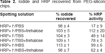
We investigated the use of the iodide standard in the presence of the enzyme horseradish peroxidase (HRP, type 12). We spotted silicon chips coated with PEG using solutions containing iodide and HRP in different buffers. Some solutions also contained trehalose to preserve enzyme activity.
We spotted 2 μl of each solution onto PEG-silicon chips, delivering 0.4 ng HRP and 120 picomoles of iodide to the surface. Samples were spotted in triplicate. Samples were dried 4 hours in a desiccator, then regenerated in 210 μl of the appropriate buffer. Aliquots from each solution were assayed for HRP activity and iodide.
Iodide and HRP chips. recovered from PEG-silicon chips.
When adding non-native chemicals to biological samples, care should be taken to ensure that there is no adverse affect on the biomolecules. The addition of iodide ion to biological samples seems to be relatively benign. We have analyzed the activity of HRP, alkaline phosphatase and β-galactosidase in the presence of iodide ion with no measured loss of activity (unpublished results). Although the reaction of iodine with sulfhydral groups and aromatic rings of tyrosine and histidine is well described, 17 it is unlikely under the conditions of the assay that the iodide ion would be oxidized to iodine which could then react with proteins in the sample.
There are also some compounds that are known to interfere with the iodide-catalyzed reaction. Ru(IV) and Os(VIII) can also catalyze the reduction of Ce(IV), while Ag(I) and Hg(II) ions significantly inhibit the reaction at low concentrations.11,16 However, these ions can be controlled for in the buffered solutions typically used in microprinting applications and therefore are of little concern. Other more common ions, such as PO43-, SO42-, ClO–, Cl–, Br–, Fe3+, Al3+, Mg2+, Cu2+, and Zn2+ have no affect on the assay although any species that absorbs in the region of 350 nm will interfere.11,16
The results summarized in Tables 1 and 2 indicate that iodide can be used as an internal standard to provide a measure of the volume and quantity of material printed. We recommend its use in microprinting applications that deposit sub-nanoliter volumes of solution as follows. (1) Substitute sodium iodide for the saline salt used in the biological buffer in the print solution. (2) Assay the biological activity of the print solution and determine the ratio of activity to iodide concentration. (3) Print the solution. (4) Remove the printed iodide from the substrate and assay. (5) Assay the biological activity of the printed sample. (6) Compare the biological activity/iodide ratio after printing to the ratio of the original solution.
In summary, the use of this iodide assay could provide valuable insights on the microprinting process, specifically the loss of activity of a printed biological material. A properly designed assay could distinguish loss due to the actual printing process, denaturing from drying, or denaturing due to adsorption.
Footnotes
Acknowledgements
This work was supported by a Denison University Robert C. Good Faculty Fellowship, the Denison University Research Foundation, Denison University Anderson Research Assistantship, and the National Institutes of Health, AREA grant 1-R15-GM62781-01 and SBIR grant 1-R43-GM080803-01.
Author(s) have provided signed confirmations to the publisher of their compliance with all applicable legal and ethical obligations in respect to declaration of conflicts of interest, funding, authorship and contributorship, and compliance with ethical requirements in respect to treatment of human and animal test subjects. If this article contains identifiable human subject(s) author(s) were required to supply signed patient consent prior to publication. Author(s) have confirmed that the published article is unique and not under consideration nor published by any other publication and that they have consent to reproduce any copyrighted material. The peer reviewers declared no conflicts of interest.