
Abstract
Direct detection and analysis of biomolecules and cells in physiological microenvironment is urgently needed for fast evaluation of biology and pharmacy. The past several years have witnessed remarkable development opportunities in vitro organs and tissues models with multiple functions based on microfluidic devices, termed as “organ-on-a-chip”. Briefly speaking, it is a promising technology in rebuilding physiological functions of tissues and organs, featuring mammalian cell co-culture and artificial microenvironment created by microchannel networks. In this review, we summarized the advances in studies of heart-, vessel-, liver-, neuron-, kidney- and Multi-organs-on-a-chip, and discussed some noteworthy potential on-chip detection schemes.
Introduction
The capability of traditional platforms, such as well plate, does not match researchers’ demands for precise and creative experiments any more. With various advantages, such as low cost, versatility, miniaturization, preciseness, and flexibility in handling biological samples, microfluidics has drawn tremendous attention in scientific and engineering academia and has become a great platform to replace the conventional one. Specially designed microfluidic devices, which are called organ-on-a-chip(s), 1 provide a controllable culture microenvironment for living cells in micrometer-sized chambers for modeling physiological functions of tissues and organs. Currently, a series of “suborgans” were created that imitated the construction of minimal functional units and kept partial function of organs or tissues in vivo. These emulative organ or tissue models provide a powerful tool for biological and pharmaceutical researches. In this mini review, we introduce some recent advances in the novel organ-on-a-chip researches that can be extended to pharmaceutical analysis and new drug development. Also, this review covers related techniques of organs and tissues that are of great interest to drug developers, such as heart, vessel, liver, neuron, and kidney.
Heart-on-a-Chip
Cardiovascular diseases (CVDs) are the first cause of death globally. It is estimated that 17.5 million people died from CVDs in 2012, representing 31% of global death. 2 In a mammalian body, in the circulatory system, heart is responsible for pumping blood, and has a most important feature of autorhythmicity, modulated by the endocrine and nervous systems. 3 Autorhythmic heartbeat leads to cyclical fluctuant mechanical stress at the endothelial cell (EC) surface, which is important for vascular phenomena such as vessel remodeling, endothelial permeability, vasoregulation, blood formation, and vascular pathology, including platelet thrombus formation and atheroma. With the help of microfluidic chip technology, replicating functional cardiovascular organ model in vitro is becoming a reality.
In Xiao's report, 4 human embryonic stem cell (hESC) and primary neonatal rat cardiomyocytes were used to generate a microfabricated cardiac biowire bioreactor that was attractive for pharmaceutical testing. The spontaneous beating of the cardiac biowires could be retarded by the treatment of nitric oxide, which was carried by the medium into the chamber. Furthermore, the integrated carbon rod electrodes offered electrical stimulation for further improving the phenotype of cardiomyocytes (Fig. 1). Nguyen and colleagues reported a heart-on-chip methodology with which an accurate controllable physiologic mechanical stimulation was managed to rebuild a simulative physiological condition that holds significant promise for immature cardiomyocytes for generating functional cardiac patches in vitro for replacement of injured cardiac tissues. 5 In another example, 6 a microfluidic device that generates cardiac-like flow in a continuous closed culture system was reported with advantages of miniaturization, low circulatory volume (2-3 µL).
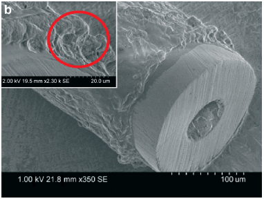
SEM image for the thick layer of cardiac tissue attached to the tubing surface after remodeling. Primary and hESC-derived cardiomyocytes were used to generate cardiac biowires and beat spontaneously. Reproduced from Xiao Y, Zhang B, Liu H, et al. Microfabricated perfusable cardiac biowire: a platform that mimics native cardiac bundle. Lab Chip. 2014;14(5):869-882, with permission from the Royal Society of Chemistry.
Vessel-on-a-Chip
The network of blood vessels distributes throughout the whole body of vertebrates and connects all the organs and tissues. Even the structure and dimension of different vessels are distinct, and the foundation of the blood circulation system is still the vascular ECs that build selective permeability barrier among tissue and blood. ECs involve in multiple physiological functions (such as barrier, clotting, inflammation, angiogenesis, vasoconstriction–vasodilation, and tumor metastasis) and become a hotspot in organ-on-a-chip research.
A functional and perfusable 3D microvascular network on a chip was reported recently 7 (Fig. 2). In that report, ECs were spatially controlled cocultured with stromal fibroblasts, pericytes, or cancer cells to generate normal microvessel development and tumor vasculature angiogenesis. Further research in the regulating angiogenesis mechanism was achieved. It was realized with the powerful microfluidic system that incorporated the whole milieu of soluble factors, produced by cells in situ and allowed potential drugs to target angiogenesis in disease 8 (Fig. 3).
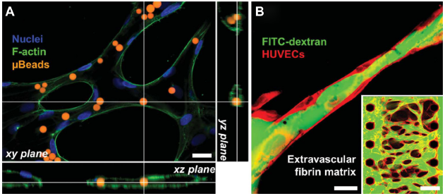
Introduction of solutions containing red fluorescent microbeads (A) or fluorescein isothiocyanate (FITC)-dextran (70 kDa, green) (B) into the perfusable microvessels. Human umbilical vein endothelial cells (HUVECs) were cocultured with human normal lung fibroblasts (LFs) to form interconnected network within ECM environment. Reproduced from Kim S, Lee H, Chung M, Jeon NL. Engineering of functional, perfusable 3D microvascular networks on a chip. Lab Chip. 2013;13(8):1489-1500, with permission from the Royal Society of Chemistry.
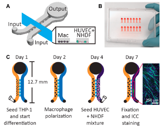
Microfluidic device design and workflow to analyze biomolecular signaling in angiogenesis. (A) Macrophages and the HUVEC/Primary neonatal normal human dermal fibroblast (NHDF) mixture were seeded in left and right channels, respectively. (B) An array of 14 microculture devices. (C) Workflow showing cell seeding, polarization, and ICC. Reprinted with permission from Theberge AB, Yu J, Young EW, Ricke WA, Bushman W, Beebe DJ. Microfluidic multiculture assay to analyze biomolecular signaling in angiogenesis. Anal Chem. 2015;87(6):3239-3246. Copyright 2015 American Chemical Society.
Vascular smooth muscle cell (VSMC) is another functional cell composing the majority of the wall of blood vessels, responsible for systolic–diastolic to regulate the blood volume and pressure. A vascular muscular thin film was generated, based on genipin and extracellular matrix (ECM) proteins modified PDMS, sufficiently mimicking the circumferential alignment of VSMCs in arteries and successfully maintaining tissue contractility and integrity throughout two weeks. 9 Three cell types forming different layers of the vessel wall were patterned on a membrane that was stress-induced, rolling into a tube, precisely mimicking blood vessels, and many other tubular structures. 10 This study provided a new strategy to rebuild tissues in vitro.
Liver-on-a-Chip
In human body, liver is a major metabolism organ, and its functions include regulation of glycogen storage, decomposition of red blood cells, plasma protein synthesis, hormone production, and detoxification. 11 Ample vascular and bile network provides supplement of nutrient–oxygen and elimination of metabolites and waste products. The hepatocyte makes up to 70%-85% of the liver's mass and is the main functional parenchymal cell of the liver. Therefore, hepatocytes are seen as the representative model for liver in artificial liver in vitro. Unfortunately, even though it is well known that the liver keeps the capability to regenerate in vivo,12,13 the isolated hepatocyte degeneration is unavoidable during the culture in vitro in weeks.
With the development of lab-on-a-chip technique, it has been demonstrated that a continuous medium flow allows a feasible exchange of oxygen and wastes, respectively, with the outside and creates a more physiological environment in stimulating the expression of detoxifying genes. 14 Moreover, with a liver-on-a-chip method, appropriate interaction between cell–cell, cell–ECM, and cell-soluble factors contribute to stabilize primary hepatocyte functions.15,16 Also, micro-bioreactor based on hepatocytes is considered to be a promising in vitro model for artificial liver, in which hepatocytes were 3D cultured in different approaches, such as spheroids, sandwich gels, porous scaffolds, or encapsulation in natural or synthetic hydrogels.17–26 In another report, rat hepatocytes were isolated by Li and coworkers, and these cells were then encapsulated in polyethylene glycol diacrylate, which maintained liver-specific function for over 50 days while responding to prototypic small molecules and drug–drug interactions. Even more, this microtissue could be manufactured using a microfluidic droplet generator 27 (Fig. 4).

Microfluidic encapsulation of hepatocyte pucks. (A) A microtissue of hepatocyte pucks and fibroblast were generated using a (B) microfluidic droplet generating device. (C) Phase image and viability staining of an individual microtissue. (D) Albumin secretion of different kinds of microtissue in 16 days. (E) Coculture array of microtissues seeded together onto micropatterned islands. Reproduced with permission from Li CY, Stevens KR, Schwartz RE, Alejandro BS, Huang JH, Bhatia SN. Micropatterned cell-cell interactions enable functional encapsulation of primary hepatocytes in hydrogel microtissues. Tissue Eng Part A. 2014;20(15-16):2200-2212. The publisher for this copyrighted material is Mary Ann Liebert, Inc. publishers.
Neuron-on-a-Chip
Central nervous system (CNS) and peripheral nervous system (PNS) form the nervous system with the basic unit of neuron. Neuron, also called nerve cell, is electrically excitable. These highly differentiated cells do not undergo cell division, and therefore makes traumatic injuries and degenerative diseases in the CNS insufficiently or completely untreatable. 28 Neurons are generally generated by neural stem/progenitor cells (NSPCs),29–31 and that makes researchers place great importance on NSPCs differentiation into the desired neural cell types when repairing the injury.32–36
On a microfluidic device fabricated by Shamloo and coworkers, 37 rat NSPCs were isolated and cultured within varying densities of collagen matrices, and then, the effect of varying concentrations of nerve growth factor on migration and differentiation was detected. 37 Moreover, a method for observing migration out of the embryonic medial ganglionic eminence (MGE) in real-time was described 38 (Fig. 5). Live neurons were costained with different intracellular probes to monitor the distribution of cellular organelles, including Golgi and nuclear. Then, an efficient method to transfect cells in MGE explants with adeno-associated virus expressing reporter proteins was established. In another work, NSPCs were cultured on an indium tin oxide surface, and cell differentiation, process development, and functionality of differentiated neuron were successfully controlled by the electrical stimulation. 39

Experimental models to study MGE neurons migration from the explants of mouse embryos. (A) Brains of the E15 embryos isolated from female mice and sliced to dissect MGE explants (B). MGE explants were placed in a Matrigel coated glass dish (C) and neurons migrated out after 48 hours (D). MGE explants and neurons were costained with neuronal marker βIII-tubulin (red), centromere marker pericentrin (green), and the nuclear marker ToPro3(blue) (E). MGE explant and cortical explant were placed in one of the wells of a microfluidic device, respectively (F). Neurons migrated away from MGE explant toward the cortical explant within microchannels filled with Matrigel (G–I). Reproduced from Nery FC, da Hora CC, Yaqub U, et al. New methods for investigation of neuronal migration in embryonic brain explants. J Neurosci Methods. 2015;239:80-84, with permission from Elsevier.
Kidney-on-a-Chip
Kidney is the most important organ for drug metabolism and elimination; therefore, drug-induced nephrotoxicity is one of the most frequently addressed problems in drug safety evaluation.
There were some early works exploring on nephrotoxicity screening platform-based microfluidics. With the similar PDMS champing porous membrane structure, Jang and Suh used rats, collecting duct epithelial cells to build kidney tubules on chip and observed their transportation ability on the chip 40 (Fig. 6). In an independent report, Ferrell used opossum kidney epithelial cells to build the chip and analyzed the mechanism of albumin transportation. 41 Also, Jang KJ et al applied human primary renal tubules epithelial cells on their microfluidic device and measured a number of important parameters, such as albumin transport, glucose reabsorption, brush border alkaline phosphatase activity, cisplatin toxicity, and P-gp efflux transporter activity 42 on chip. All of these works used only one kind of cells in forming partial physiological environment, and then characterized their systems with a series of biomarkers.

Fabrication and operation of a multilayer microfluidic device (MMD) for efficient culture and analysis of renal tubular cells. (A) Sandwich structure of MMD. Photograph (B) and schematic of the device (C) on a culture dish containing outside tubular fluid. (D) Microscope image of IMCD cells grown confluently after seeding three days within the MMD. Reproduced from Jang KJ, Suh KY. A multi-layer microfluidic device for efficient culture and analysis of renal tubular cells. Lab Chip. 2010;10(1):36-42, with permission from the Royal Society of Chemistry.
Multi-Organs-on-a-Chip
Not only efficacy but also pharmacokinetics, toxicity, and security characteristics decide the fates of candidate drugs. Unfortunately, the high throughput, traditional drug screening platforms, such as 96-well plate, are excessively simple, which is greatly different from the complex conditions in vivo and incapable of evaluating drugs accurately and comprehensively. Numerous candidates are eliminated during animal and clinical test, which is one of the reasons why drug development process is so costly, time-consuming, and inefficient.
Multi-organ-on-a-chip is probably a novel strategy to increase efficiency and precision of drug screening before animal test. Being different from single organ chips, these elaborate microfluidic devices integrate several cells from different organs and tissues according to the way in vivo, attempting to simulate human body. For example, 43 in Abacia and Shuler's report, a multi-organ-on-a-chip that was designed according to actual blood velocity and organ proportions had been proved to be a platform suitable for studying physiologically based pharmacokinetics/pharmacodynamics. In another example, 44 intestine, liver, skin, and kidney tissues were cocultured and sustainable over at least 28 days for absorption, distribution, metabolism, and excretion profiling. In this way, multi-organs-on-a-chip devices are capable of providing comprehensive information of candidate drugs and may bring revolutionary changes in the pharmaceutical industry.
Conclusion and Perspectives
At present, more and more research groups regard microfluidic device as an important platform for their potential in-life sciences. In cell- and tissue-level researches in vitro, the primary cells, immortalization cell lines, and stem cells are the major experiment models. Among them, immortalization cell lines are most easily cultured but anamorphic frequently happens on well plate and resulted in deviation from reality. Primary cells are more effective models but hard to maintain in traditional culture method and also induction of stem cells is under scrutiny. The microfluidic device has shown its ability on controlling microenvironment, which is urgently needed for primary and stem cells research. On the other hand, considered to be the substitute of experiment animal in physiological and pharmaceutical research, “human-on-chip” is the ultimate goal with the combination of different functional organ chips. Multiple elements can be integrated in one chip, such as different cells, ECM, fluid, droplet, valve, even sensors, and more useful features. Benefited by “design as you wish,” prospect of microfluidic device is limitless.
Organ-on-a-chip holds many advantages over traditional in vitro cytology experiment tools (such as 96-well plate and transwell chamber) and general cell culture systems based on microchip. For example, the cell microenvironment is highly maneuverable and closer to it is in vivo; organ-on-a-chip keeps capacity to reproduce the function and structure of the organ; multi-organs-on-a-chip are able to simultaneously evaluate efficacy, toxicity, and pharmacokinetics, which are otherwise impossible to evaluate with traditional drug screening tools. Although powerful, at the current stage, organ-on-a-chip cannot completely replace the traditional drug screening tools. One important reason is lack of on-chip detection schemes. Off-chip HPLC and mass spectrometer, which are of low efficiency, were widely used for determination of biomolecules on the organ-on-a-chip. Separation of molecules of interest cannot be conducted on the organ-on-a-chip yet. Direct observation of cells on a chip with an optical microscope is somehow difficult because of low transparency of the membrane and thickness of organ-on-a-chip beyond the working distance of the objective. And it is analysts’ responsibility to solve these problems on the way, by cooperating with the biologists. And here we would expect some solutions, for example, electrophoresis of different modes can be integrated with an organ-on-a-chip to address separation issue; the design of an organ-on-a-chip can be updated to fit the short working distance of high magnification objective; coupling of organ-on-a-chip with mass spectrometer can be carried out; electrode can be integrated in the organ-on-a-chip for in situ monitoring of biological process, etc. In summary, development of an on-chip detection approach will accelerate the commercialization of organ-on-a-chip, and it will eventually speed up the progress in pharmacological studies and new drug development.
Author Contributions
Wrote the first draft of the manuscript: FA. Contributed to the writing of the manuscript: YQ, XL, and YL. Made critical revisions and approved the final version: RZ. All the authors reviewed and approved the final manuscript.