
Abstract
A gas chromatography tandem mass spectrometry method for quantification of buprenorphine (BUP) and norbuprenorphine (NBUP) in brain and plasma samples from mice was developed and validated. Analytes were extracted from the brain or plasma by solid phase extraction and quantified within 20 minutes. Calibration was achieved by linear regression with a 1/x weighting factor and d4-buprenorphine internal standard. All products were linear from 1 to 2000 ng/mL with a correlation of determination >0.99. Assay accuracy and precision of back-calculated standards were within ±10%. The lower limit of quantification for both BUP and NBUP from the brain and plasma was 1 ng/mL. This sensitive and specific method can be used for the investigation of BUP mechanism of action and clinical profile.
Introduction
Buprenorphine (BUP), a semi-synthetic opioid, has been approved for pain management and as effective maintenance therapy for heroin addiction. However, following its marketing, forensic studies reported several cases of fatalities because of asphyxia, attributed to BUP misuse or concomitant intake of psychotropic drugs such as benzodiazepines or ethanol.1–4 Consistently, acute poisonings with severe respiratory depression and typical opioid features requiring admission to the intensive care unit have been attributed to BUP.5,6 In humans, BUP metabolism by cytochrome P450 widely produces an active metabolite, norbuprenorphine (NBUP) with potent respiratory depressant effects.7,8 Recently, the inhibition of P-glycoprotein-mediated efflux of NBUP at the blood–brain barrier was shown to significantly enhance BUP-related respiratory effects. 9 However, to date, no data exist regarding differences in BUP and NBUP-related toxicity attributable to gender and strain in mice. The purpose of the present study was to develop a specific measure of the plasma and brain levels of BUP and NBUP in samples from mice. One LC/MS method was developed to determine the two compounds in rat brain tissue and plasma. As there is no reported GC/MS method validation that measures BUP and NBUP concentrations in animals, we proposed in this study to validate a GC/MS method for the simultaneous quantification of both compounds in mice brain and plasma samples with a new extraction procedure.
Materials and Methods
Chemicals and materials
BUP (B-902), NBUP (N-912), and d4-buprenorphine (d4-BUP, B-901) freebase (Fig. 1) in methanol (100 μg/mL) were purchased from Cerilliant Co (Round Rock, TX, USA). Sterile water Versylene® was obtained from Fresenius Kabi (Sevres, France). Sodium acetate trihydrate, GC capillary grade methanol Lichrosolv®, and GC capillary grade ethyl acetate Lichrosolv® were obtained from Merck (Darmstadt, Germany). Ammonia solution (25%) was purchased from VWR (Fontenay-sous-Bois, France). GC capillary grade dichloromethane Chromasolv® was obtained from Riedel-de-Haën (Steelze, Germany). Isopropyl alcohol Chromasolv® was obtained from Carlo Erba (Val de Reuil, France). Clean Screen solid phase extraction columns (CSDAU133, 130 mg/3 mL) were purchased from UCT (Bristol, PA, USA). The derivatizing agent utilized was bis(trimethylsilyl) trifluoroacetamide (BSTFA) + 1% trimethylchlorosilane (TMCS) (Sylon BFT, Sigma-Aldrich, Saint-Quentin Fallavier, France).
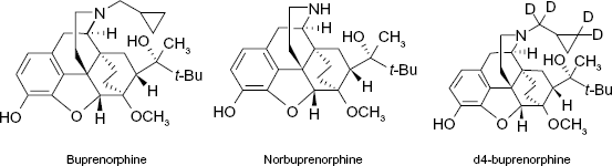
Chemical structures of BUP, NBUP, and d4-BUP.
Animals
Male and female FVB (20-25 g) and male Swiss mice (20-25 g) were purchased from Janvier (Genest, France). Animals were housed in well-ventilated cages at 20-22 °C with 45-65% relative humidity and maintained under a 12-hour dark/light cycle (light from 8:00 a.m. to 8:00 p.m.) for at least one week before the experiments. Food and water were provided ad libitum. Following each experiment, mice were euthanized using a carbon dioxide chamber. All animals were treated in accordance with the ethical guidelines established by the National Institutes of Health and the French Ministry of Agriculture. Protocols followed the animal facility experimental procedures of the Paris-Descartes University were approved by the institutional ethics committee.
Gas Chromatography– Mass Spectrometry equipment
The Thermo Focus DSQ II gas chromatograph/mass spectrometric system was used for GC separation and detection. The system was equipped with an Uptibond® UB5 premium column (30 m × 0.25 mm × 0.25 μm). The instrument was programed at 200-220 °C at 30 °C/minute and held for three minutes before being programed to 390 °C at 15 °C/minute and held for 17 minutes, for a total analysis time of 20 minutes. The transfer line temperature was maintained at 280 °C. 1 μl of the derivatized extract was injected. The injection port temperature was held at 250 °C and operated in the pulsed splitless mode. The instrument utilized electron impact ionization and was operated in the selected ion monitoring (SIM) mode. Ions with m/z 468 (NBUP-TMS), m/z 450 (BUP-TMS), and m/z 454 (d4-BUP-TMS) were monitored.
Sample collection
Whole-brain was removed from each animal, quick-frozen, kept frozen at −20 °C, and shipped overnight on dry ice. The brains were stored at −70 °C until analysis. Blood samples were collected in heparinized tubes, plasma separated, and handled as described for the brain.
Sample preparation
The frozen sample was thawed at room temperature for 15 minutes, and then 1 mL of sample was transferred to a 5 mL polypropylene tube. 2 mL 0.1 N acetate buffer (pH 5) and 0.1 mL of internal standard stock solution (1 μLg/mL) of d4-BUP was added. The mixture was vortexed and then loaded onto a Clean Screen solid phase extraction column that was preconditioned with 3 mL methanol and 3 mL sterile water, and then equilibrated with 2 mL 0.1 N acetate buffer. The mixture was added in the column. The column was washed with 2 mL sterile water, 3 mL 0.1 N acetate buffer, and 3 mL methanol. The column was then dried in vacuum for 10 minutes. The analytes were collected in a 5 mL glass tube by elution with a fresh mixture of 3 mL dichloromethane, isopropyl alcohol, and ammonia solution (25%) (78/20/2). The solvent was then evaporated under a gentle stream of nitrogen. The residue was reconstituted with 20 μL ethyl acetate and 20 μL BSTFA with 1% TMS, vortexed briefly and transferred to an autosampler vial insert for GC/MS analysis.
Preparation of Calibration standards
Working solutions of BUP and NBUP (1.0 and 10 μg/mL) were made in methanol. Internal standard stock solution of d4-BUP (0.1 μg/mL) was made in methanol. The stock solutions were stored in 2 mL glass vials at −80 °C. Matrix based (brain and plasma) calibration standards and quality controls samples were prepared by spiking analyte free brain homogenate and plasma with known concentrations of BUP and NBUP, followed by sample preparation and GC/MS analysis.
BUP and NBUP calibrators were prepared at 1, 2, 5, 10, 20, 50, 100, 500, 1000, and 2000 ng/mL in each matrix. The 1-100 ng/mL calibrators were prepared from the 1.0 μg/mL BUP and NBUP standards. The 500-2000 ng/mL calibrators were prepared from 10 μLg/mL BUP and NBUP standards. Plasma and brain calibrators were freshly prepared on each day.
Validation procedure
The linearity of the method was determined by utilizing 13 calibrators of different concentrations in each matrix. Linearity was determined by linear regression of calibrator concentration versus peak-area ratio of either BUP or NBUP peak area divided by the peak area of d4-BUP. The limit of detection (LOD) for each analyte was determined as the lowest concentration yielding signal-to-noise ratios of at least 3:1 with correct relative ions intensities and a retention time within ±0.2 minutes of the average calibrator retention time. The specificity of the method was evaluated by comparing calibration standards to the blank and zero samples (three independent batches of plasma and brain were used). Accuracy, the degree of closeness of measurements of a quantity to that quantity's true value, and precision, the degree to which repeated measurements under unchanged conditions show the same results, were assessed by adding analytes to a series of three replicates in the plasma or brain of three concentrations (10, 500, and 1000 ng/mL) of each analyte and determining their concentrations from linear regression of matrix matched calibration curves. The interday accuracy and interday precision were measured on three different days. The deviation (%) of the mean concentration from nominal concentration served as the measure of accuracy. The related standard deviation (RSD%), the absolute value of the coefficient of variation, served as the measure of precision. Recovery was determined by adding analytes to a series of six replicates in the plasma or brain with low (10 ng/mL) and high concentrations (1000 ng/mL) of each analyte that were extracted and compared to low and high concentrations of each analyte that were not extracted. Recovery was expressed as a percentage of the mean peak area of the extracted replicates divided by the mean peak area of non-extracted replicates. The stability of samples in the autosampler tray was tested using blank plasma and brain samples spiked with 10 ng BUP, 10 ng NBUP, and 100 ng d4-BUP. The spiked samples were by the procedures described above followed by evaporation, reconstitution with 20 μL ethyl acetate and 20 μL BSTFA with 1% TMS, transfer to vial insert, and placement in the autosampler tray. Every hour 1 μl of the solution was injected from the same sample. The response ratios of BUP and d4-BUP of successive injections were compared to that of the first injection. Identical calculations were performed for NBUP.
Data analysis
Data were collected and analyzed utilizing Thermo Electron GC/MS Solution software (Xcalibur™, version 1.4.2; Thermo Electron Corporation, San Jose, CA, USA).
Results
The representative chromatogram and spectrum of BUP and NBUP in plasma are shown in Figure 2. The same chromatogram was performed in brain matrix.
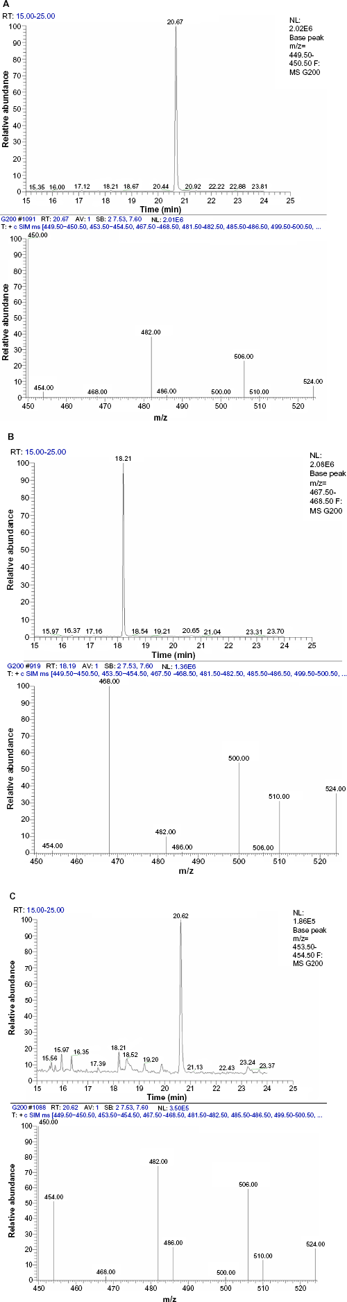
Typical chromatogram and spectrum of BUP (200 ng/ml, A), NBUP (200 ng/ml, B), and d4-BUP (C) spiked in plasma.
Selectivity
The extracted chromatograms of blank brain and plasma samples (sample processed without standards), and zero brain and plasma samples (blank sample processed with internal standard) were compared. The absence of analyte peaks in the chromatograms of blank and zero samples, and the absence of internal standard peak in the chromatograms of blank samples indicated the selectivity of the method.
Recovery
The recoveries of BUP and NBUP from plasma extractions were determined to be 76 and 85%, respectively. The recoveries of BUP and NBUP from brain extractions were determined to be 72 and 66%, respectively.
Linearity
BUP and NBUP were linear over the range 1-2000 ng/mL with a R2 of 0.999 and 0.997, respectively. The lower limits of detection of both BUP and NBUP were 1 ng/mL for the brain and plasma.
Accuracy and precision
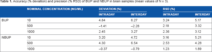
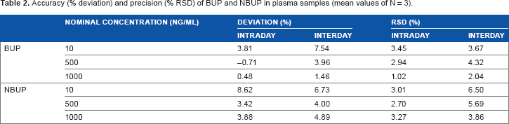
Results of precision (represented by % RSD) and accuracy (represented by % deviation) of the method are given in Tables 1 and 2. Accuracy for BUP in brain samples ranged from −1.41 to 6.27% and the precision ranged from 2.18-5.17%; for NBUP, the accuracy ranged from −0.79 to 6.54% and the precision ranged from 1.23 to 5.21%. The accuracy for BUP in plasma samples ranged from −0.71 to 7.54% and the precision ranged from 1.02 to 4.32%; for NBUP, the accuracy ranged from 3.42 to 8.62% and the precision ranged from 2.70 to 6.50%.
Accuracy (% deviation) and precision (% RSD) of BUP and NBUP in brain samples (mean values of N = 3).
Accuracy (% deviation) and precision (% RSD) of BUP and NBUP in plasma samples (mean values of N = 3).
Scope of the Method
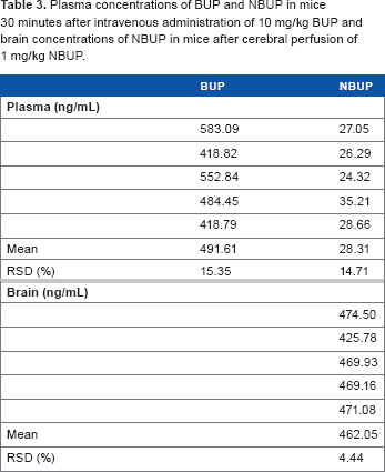
The developed method was applied in our laboratory to quantify plasma concentrations of BUP and NBUP in mice administered 10 mg/kg BUP by intraperitoneal administration, and to measure brain concentrations of NBUP in mice administered 1 mg/kg NBUP by in situ brain perfusion. The main objectives were to asses P-glycoprotein involvement in NBUP transport in vivo and study its role in the modulation of BUP-related respiratory effects in mice. The complete results of this study have been already published. 3 The results of samples collected 30 minutes after administration of BUP or NBUP are given in Table 3 and the representative chromatograms are shown in Figure 3.
Plasma concentrations of BUP and NBUP in mice 30 minutes after intravenous administration of 10 mg/kg BUP and brain concentrations of NBUP in mice after cerebral perfusion of 1 mg/kg NBUP.
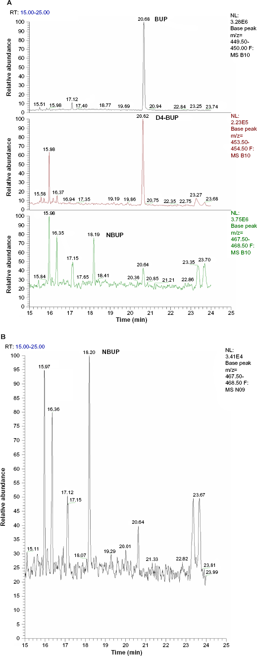
Representative chromatograms of plasma sample from a mice 30 minutes after intraperitoneal administration of 10 mg/kg BUP (A) and brain sample from a mice 30 minutes after cerebral perfusion of 1 mg/kg NBUP (B).
Discussion
The method developed in this study is the first GC/MS method to determine simultaneously BUP and NBUP in brain and plasma samples of mice providing a SPE extraction procedure and showing such a low LOQ. It shows improved higher sensitivity, accuracy, and precision than the previously published method. 10 The wide linear range (1-2000 ng/mL) for both BUP and NBUP in brain and plasma samples allows the analysis in most research studies. The proposed GC/MS method satisfies sensitivity requirements using elementary equipment, available at common laboratories that perform everyday routine analysis, at a significantly low cost. The developed method can be used in the determination of BUP and NBUP for pharmacokinetic studies, for therapeutic drug level monitoring, or for the investigation of forensic studies.
Author Contributions
Conceived and designed the experiments: FC, JS. Analyzed the data: FC, JS. Wrote the first draft of the manuscript: FC, JS. Contributed to the writing of the manuscript: FC, JS. Agree with manuscript results and conclusions: FC, JS. Jointly developed the structure and arguments for the paper: FC, JS. Made critical revisions and approved final version: FC, JS. All authors reviewed and approved of the final manuscript.
Disclosures and Ethics
As a requirement of publication the authors have provided signed confirmation of their compliance with ethical and legal obligations including but not limited to compliance with ICMJE authorship and competing interests guidelines, that the article is neither under consideration for publication nor published elsewhere, of their compliance with legal and ethical guidelines concerning human and animal research participants (if applicable), and that permission has been obtained for reproduction of any copyrighted material. This article was subject to blind, independent, expert peer review. The reviewers reported no competing interests.