
Abstract
Background
Leptospirosis is a health problem that causes death in Indonesia. In 2017, Boyolali District was reported that the number of leptospirosis cases reached 40.62 per 100,000 population with a CFR of 23.52%. The determination of risk factors and Leptospira bacteria's presence in the body of water plays an essential role in the transmission of leptospirosis.
Design and methods
This study aims to determine the risk factors and Leptospira bacteria's presence in water bodies in Boyolali District. This research is descriptive research with a survey method using a cross-sectional design and an analytical study using an observational method with a case-control approach. The sample was 100 water samples from wells, rivers, and paddy fields in endemic and non-endemic areas of leptospirosis. This study's population was 34 people with leptospirosis in Boyolali Regency in January 2017 - August 2018.
Results
There was a significant relationship between leptospirosis incidence in the Boyolali Regency with garbage, the pet presence, a history of injuries, and field activity. Leptospira bacteria are found in rivers (18.18%) and rice fields (6.67%), while in sub-districts with cases occur almost every year. Leptospira are found in wells (18.18%) and rice fields (6.67%).
Conclusions
People should pay more attention to home sanitation and the surrounding environment to avoid leptospirosis.
Keywords
Introduction
Leptospirosis is a worldwide zoonotic disease caused by pathogenic Leptospira species.1,2 The infectious agent is capable of infecting both humans and animals. The primary reservoirs of the organisms are cattle, horses, canines, and rodents. 2 The genus Leptospira belongs to the Sprirochaete phylum, which includes organism that causes significant human disease.3-7
Leptospirosis disease is an infection in animals. Infection in humans occurs accidentally, inadvertently in contact with materials contaminated by leptospirosis bacteria. Humans are contaminated through direct contact with urine, blood, or tissue of infected animals and indirect contact through contaminated environments. Although leptospirosis transmission from rats to humans can be through direct contact with the tissue of mice infected with Leptospira sp. bacteria, frequent transmission occurs through contact with water or moist soil contaminated with infected mouse urine. Leptospira bacteria in the body of water play an essential role in the transmission of leptospirosis.6,8-10
Leptospirosis is included as a neglected disease. Outbreaks of leptospirosis due to El Niño floods in India, Southeast Asia, Central and South America, as well as the United States, cause leptospirosis to receive attention, particularly in the diagnosis, treatment, and prevention of leptospirosis transmission, both from the World Health Organization (WHO) and Ministry of Health in developing countries. 7 Leptospirosis is widespread in the world, both in cold, subtropical, and tropical climates. Data from the International Leptospirosis Society (ILS) states that Indonesia is declared a world-class leptospirosis incident in the world for mortality. The number of leptospirosis cases increases from 1.43 per 100,000 population in 2015 to 3.21 per 100,000 population in 2016 in Indonesia. 11
Boyolali district is an endemic area of leptospirosis in Indonesia, where leptospirosis cases first occurred in 2012 and during the last five years reported cases with an increase in the number of cases each year. In 2016 the number of leptospirosis cases in the Boyolali district decreased, 19.7 per 100,000 population in 2015 to 6.25 per 100,000 population in 2016. However, CFR for cases of Leptospirosis in Boyolali District is high. In 2014 CFR is reported to be 35%, and by 2015 CFR is reported to be 55%. In 2017, based on Boyolali District Health Office data, it has been reported that the number of leptospirosis cases reached 40.62 per 100,000 population with a CFR of 23.52%. Most leptospirosis cases in the Boyolali district clustered in the eastern part of the Ngemplak subdistrict, Nogosari district, Sambi district, and Simo district.12,13
The biotic and abiotic environmental factors influence leptospirosis incidents. Land use affects rat populations, such as densely populated and rundown housing, where rat nests are detected. Homes close to the rice fields are also a risk for where the mice run away for food. High rainfall makes water puddles around the house that increase the risk of water puddles become Leptospira contaminated. The altitude of the place affects flooding resulting in many puddles around the house. The vegetation around becomes the determinant of the rat haven, the denser the vegetation, the better it is for a rat sanctuary. The house's location near the river affects leptospirosis, which is related to the behavior of people who bathe and wash in a waterway and can cause puddles around the house if the river water overflows. Homes that are close to landfills are at risk for moon hiding places during the day. Occupational risk factors such as being farmers, veterinarians, and rodent controls put individuals at risk of this disease.8,14,15
Significance for public health
Leptospirosis is one of the zoonoses that is still causing death in Indonesia. In its development, leptospirosis does occur in developing countries and developed countries, with the contributing factor, is human trafficking. Analysis of various risk factors and the presence of Leptospira bacteria are essential in the transmission of leptospirosis. Research shows that activity rice fields activity, history of wounds, garbage, and pets are associated with leptospirosis incidence. Researchers found the presence of Leptospira bacteria in rivers, rice fields, and wells.
This research object is to determine the risk factors for the transmission of leptospirosis in humans. The second purpose was to know the existence of Leptospira sp. bacteria in the water body, human, and reservoir using PCR method of sensitivity 90-100%.
Design and Methods
This research is an analytical study using an observational method with a case-control approach. This study's population was 34 people with leptospirosis in Boyolali Regency in January 2017 - August 2018. The comparison between the case group sample and the control group sample was 1:1, 34 controls, and 34 cases. Data analysis was calculated by univariate and bivariate using chisquare.
Analysis of Leptospira bacteria's presence was carried out by taking water samples from water bodies in the environment. Water sampling was done by using the purposive sampling technique. One hundred water samples taken from wells are commonly used by irrigation channels used by respondents to clean themselves and the fields where respondents work. Each water sample was taken with a volume of 150 ml placed in a dark glass bottle. Water samples are stored at 4°C and processed within 24 hours. Water samples were taken measured temperature, pH, and dissolved oxygen. Each water sample was filtered with one nitrocellulose membrane (Millipore) whose pore size was 0.22 μm. Next, the membrane is cut into small pieces and inserted into a microcentrifuge tube. DNA isolation of the nitrocellulose membrane is used to filter the water sample. The DNA isolation was calculated using the Genomic DNA Mini Kit isolation kit (Geneaid). The Polymerase Chain Reaction (PCR) process performed on DNA samples was obtained using primers: RpoB-R- CGCATCCTCRAAGTTGTAWCCTT using Go Taq Green Master Mix (Promega) and rpoB-F-CCTCATGGGTTCACAATATCA. Eleven PCR Stages were as follows: predenaturation 94°C for 2 minutes, followed by 40 amplification cycles consisting of denaturation at 94°C for 30 s, annealing at 55° C for 1 min, extension at 72°C for 1 min followed by a final extension for 20 min at 72°C. Electrophoresis performed the analysis of PCR results using agarose 1.5% at 100 volts for 15 min. UV transilluminators Positive samples presented specific DNA specifications when electrophoresis results showed that the DNA bands were in the 600 bp position.
Results and Discussion
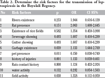
Table 1 shows the respondent characteristics in this study, where most of the respondents were male. The majority were in the 46-65 age group with the lowest age was 18 years, the highest age group was 91 years, and an average age of 48.82 years. The majority of respondents are not in school. The majority of respondents’ occupations are farmers. There was a significant relationship between leptospirosis incidence in the Boyolali Regency with garbage, the pet presence, a history of injuries, and field activity. The incidence of leptospirosis in Boyolali Regency has no relationship with the rat presence, the existence of rivers, the existence of rice fields, the showing of sewerage, the showing of gutters, a history of contact with rats, and water activity (Table 2).
Distribution of the respondent characteristics.

Determine the risk factors for the transmission of leptospirosis in the Boyolali Regency.
One hundred water samples consist of well water, irrigation channels, and paddy fields coming from sub-districts where leptospirosis cases occur every year, sub-districts almost every year occur leptospirosis cases and sub-district without leptospirosis in Boyolali District. Six positive water samples of the rpoB gene were obtained from two wells, two rivers, and two rice fields. The result indicates that the Leptospira sp. from infected animals contaminates the water bodies. The prevalence of Leptospira sp. in water samples is tabulated in Table 3.
Results of water sampling and Leptospira sp. detection based on Leptospira rpoB gene detection (n=100).
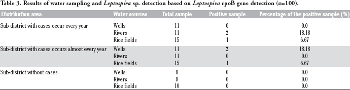
The study revealed that Leptospira bacteria exist in sub-district with cases that occur almost and every year. In sub-districts with the incident that occur every year, Leptospira bacteria live in rivers (18.18%) and rice fields (6.67%). While in sub-districts with cases that occur almost every year, Leptospira survive in wells (18.18%) and rice fields (6.67%). The bacteria survive in river irrigation channels. There are rats’ holes on one of the positive rivers. The water in the river was relatively stagnant. The presence of rats around the river can contaminate the water in the river. Leptopsires survive weeks or months in moist and warm soil, stagnant water at neutral or slight pH.16-20
Based on observation, the floor material of two positive wells of Leptospira is from the soil. A soil well is easy to contaminate through non-water-resistant floors. 21 Leptospira bacteria contamination in the rice fields is a cause. A rice field is a place for the availability of food for rats. Moreover, the soil in the rice fields is wet. It is suitable for Leptospira sp. to survive for a whole period. The rice field's water puddles presence can allow Leptospira sp. to spread the soil's contaminated urine. A previous study revealed that Leptospira serovar, Hardjo, has a higher survival rate in moist soil at pH 6.9-7.4.8,22 The temperature was an important factor for the survival of Letopsires in nature. 21 Leptospires are obligate aerobes with an optimum growth temperature of 28 to 30°C. 23 In this study, Leptospira sp. was found at a temperature of 24-31°, pH 6.8-7.4, and DO 4-12 ppm (Table 4). These results show the ability of Leptospira sp. to survive in a variety of environmental conditions. Our results are consistent with the theory that the ability to survive Leptospira sp. in the environment is affected by variations in soil and water conditions in contaminated areas. 4 Water and rice fields in the leptospirosis case areas have more optimal temperatures than leptospirosis-free areas. Temperature is influenced by external factors such as weather, wind, and currents. Water temperature variations are caused by natural processes such as biochemistry and microbiological processes (geothermal sources).24,25
Results of water sampling and Leptospira sp. detection based on Leptospira rpoB gene detection (n=100).
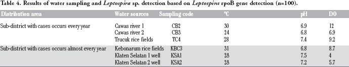
Optimal temperature makes Leptospira sp. survive last longer. The previous study found that The leptospires can survive for ten months in adverse conditions (4°C) and up to 20 months when stored at 30°C. 26 The length of life of Leptospira bacteria in water can be affected by water pH. Optimum growth of Leptospira bacteria occurred in the range of pH 7.2-7.6. 27 The examination found Leptospira bacteria's existence at a relatively acidic pH that is pH 6.8-6.9.
This research is in line with research conducted by Benacer in Malaysia, that Leptospira bacteria found in water with acidic pH that is 5.77-6.63. State of environmental conditions that are not suitable for breeding bacteria Leptospira can cause bacteria are inhibited growth even become deadly. 28 Based on research found Leptospira bacteria in DO 4-12 ppm. The oxygen concentration is influenced by many factors, including water temperature, photosynthesis rate, turbidity and water depth, degree of water turbulence or wave action, and the amount of oxygen used by respiration and decay of organic matter. The oxygen concentration is the limiting factor of growth. Based on the rat survey field results, the trap success results were obtained by 8%, where the standard density of rats can be said to be a high-density level if it passes the 7% for locations inside the house and 2% outside the home. The density of mice at the study site was high because it exceeded 7%. The number of trappings installed also influences this. Trap Success can be influenced by various factors, including traps, human activities, bait types, and laying traps. The ideal installation of mouse traps in each area of 10 m 2 is given one trap. For example, type 45 homes require a minimum of 3 traps or more mousetrap.5,10,29 Based on the bait type, baked coconut bait and meatballs are the most eaten bait by mice because rats are more attracted to food that is oily and has a strong odor, thus attracting mice's attention and then being caught in the rat trap.
Then for the sex of the catch, the mice were obtained more males than females. There was no significant difference between the number of catches of female and male mice. Sex observation is only used to measure the mobility of mice in the area. The results of Ramadhan stated that male rats were trapped more than females. 30 However, this is different from Priyambodo's statement that female rats are more comfortable to catch than male rats because, in their group, female rats are individuals food seekers for their children. 21 According to Nasir and Mahmud females will be caught more during the breeding and breastfeeding seasons because the females need large amounts of food. 31 So it can be concluded that the number of males being caught compared to females at the study site may be influenced by the seizure of territorial territory (power), home range, and feed availability. 23 Many trapped male rats indicate that male rats carry high mobility.
In implementing the density survey of mice in the study site, there were more chisses (Suncus murinus) than rat catches, where the number of chisses (Suncus murinus) was ten and the rat species were seven tails. The number of chisels in the research location is probably due to a residential area close to gardens and rice fields. Many insects around the housing enter the homes of residents and provoke the arrival of Cecurut (Suncus murinus) into the house because we know that Cecurut (Suncus murinus) is an animal insectivorous.
Identification results of mice caught trapping
1. Rattus norvegicus
The results of the survey of mice, which were then identified, were one female Rattus norvegicus 1. The Rattus norvegicus type has the following identification key characteristics: T = 190 mm, H + B = 250mm, HF = 42 mm, E = 17.5 mm, W = 260 gr. Mammary / nipple formula 3 + 3.
According to Zumarotus Solichah, Rattus norvegicus has a rough and slightly long hair texture, blunt nose shape, large body, cylindrical body shape slightly enlarged on the back, upper body color gray-black-brown, lower body color gray-brown (pale), the color of the upper tail is dark. 32 The dark bottom is slightly pale, W = 150-600 grams, H + B 150-250 mm, T = 160-210 mm (shorter than the head length + body), TL 310-460 mm, E = 18-24 mm, HF = 40-47 mm and nipples 3 + 3 pairs. 24
The classification of Rattus norvegicus is as follows:
Kingdom: Animalia
Phylum: Chordata
Class: Mammals
Order: Rodentia
Family: Muridae
Genus: Rattus
Species: Rattus norvegicus 25
2. Rattus tanezumi
The survey results of mice, which were then identified, were found in 1 male Rattus tanezumi. These mice have W = 150 grams, H + B = 155 mm, T = 150 mm, HF = 32 mm and E = 20 mm. Do not have mammary glands. This mouse is a type of house mouse.
Following the identification key, Rattus tanezumi mice have the following morphological characteristics: TL = 220-370 mm, T = 101-180 mm, HF = 20-39 mm, E = 13-23 mm, and have the formula mamae 2 + 3 = 10 The color of the body over the dark brown body and lower gray dark brown belly hair. When viewed from its proximity to humans, Rattus tanezumi rats are domestic mice, namely, activities carried out in human homes or commensal or synanthropic mice.
The classification of Rattus tanezumi is as follows:
Kingdom: Animalia
Phylum: Chordata
Class: Mammals
Order: Rodentia
Family: Muridae
Genus: Rattus
Species: Rattus tanezumi 21
3 Mus musculus
Mus musculus rats are home mice that have a smaller size than other types of mice. The identification lab results obtained 1 female Mus musculus species with morphological characteristics: HB = 100 mm, T = 110 mm, HF = 30 mm, and E = 12. Having the formula of mamae 2 + 3 = 10.
As stated in the identification key book, said that these mice have TL = less than 175 mm, T = 81-108 mm, HF = 12-18 mm, E = 8-12 mm and with the formula mamae 2 + 3 = 10. Upper and lower gray body hair color.
Mus musculus classification is as follows:
Kingdom: Animalia
Phylum: Chordata
Class: Mammals
Order: Rodentia
Family: Muridae
Genus: Mus
Species: Mus musculus 21
4 Rattus exulans
The survey results of mice, which were then identified, were found in male Rattus exulans. These mice have W = 85 grams, H + B = 140 mm, T = 145 mm, HF = 27 mm and E = 189mm. Do not have mammary glands. This mouse is a type of field mouse.
By the identification key, Rattus tanezumi mice have the following morphological characteristics: TL = 220-370 mm, T = 101-180 mm, HF = 20-39 mm, E = 13-23 mm, and have the formula mamae 2 + 3 = 10 The color of the body over the dark brown body and lower gray dark brown belly hair. When viewed from its proximity to humans, Rattus tanezumi rats are domestic mice; namely, activities carried out in human homes or commensal or synanthropic mice.
The classification of Rattus tanezumi is as follows:
Kingdom: Animalia
Phylum: Chordata
Class: Mammals
Order: Rodentia
Family: Muridae
Genus: Rattus
Species: Rattus exulans 21
5. Suncus murinus
In the implementation of the lab, 10 Suncus murinus were obtained. Suncus murinus itself is not a group of rodent orders because Suncus murinus is an insectivorous or Insectivorous animal. Suncus murinus existence around the housing indicates the number of insects around the area of housing.
Morphologically, Suncus murinus generally has a longer and more pointed snout than the group of mice. The front legs have five long, clawed fingers, in contrast to mice with short fingers on the front legs and are flat toed. All cecal teeth will round out or form sharp-pointed cones, while the mouse's front incisors will rot and curve (have a cutting edge like a chisel) and separate from the molars relatively larger by a wide gap.
The classification of Suncus murinus is as follows:
Kingdom: Animalia
Phylum: Chordata
Class: Mammals
Order: Soricomorpha
Family: Soricidae
Genus: Suncus
Species: Suncus murinus 21
Based on the results of examining the presence of Leptospira bacteria in mice, they are all negative, likewise the results of the examination in humans.
Conclusions
The density of rats in housing is still not too high because rats’ density is high if it exceeds 7% trap success while trap success is 8%. The types of mice that are scattered around the location obtained Rattus norvegicus, Rattus tanezumi, Rattus exulans, and Mus musculus. Suncus murinus is also found, but it is not an order of Rodentia, so it is not a rat. The presence of Leptospira bacteria in mice they are all negative. Likewise, the results of the examination in humans. Leptospira bacteria are found in sub-district cases with cases that occur every year, and sub-district cases occur almost every year. In sub-districts with cases that occur every year, Leptospira bacteria were found in rivers (18.18%) and rice fields (6.67%). While in sub-districts with cases occur almost every year. Leptospira were found in wells (18.18%) and rice fields (6.67%). The bacteria were found in river irrigation channels. One of the positive rivers found rats holes, water in the river was relatively stagnant.
Footnotes
The author reports no conflicts of interest in this work.
Acknowledgments
We thank Diponegoro University for its research funding. We acknowledge all the study participants, Health Office of Boyolali, Health Research and Development Unit, Banjarnegara, to make this study possible.