
Abstract
Background
Porcine reproductive and respiratory syndrome virus (PRRSV) is an economically important pathogen and causes significant economic losses to the swine industry worldwide each year. Current vaccination strategies do not effectively prevent and control the virus. Consequently, it is necessary to develop novel antiviral strategies. Carrageenan, extracted from marine red algae, exhibits anti-coagulant, anti-tumour, anti-virus and immunomodulatory activities.
Methods
We investigate the inhibitory effect of iota-carrageenan (CG) on PRRSV strain CH-1a via antiviral assay and viral binding, entry and release assays.
Results
We found that CG effectively inhibited CH-1a replication at mRNA and protein levels in both Marc-145 cells and porcine alveolar macrophages (PAMs). The antiviral activity of CG occurred during viral attachment and entry in virus life cycle. In addition, CG suppressed viral release in Marc-145 cells, as well as blocked CH-1a-induced apoptosis during the late period of infection. Furthermore, CG inhibited CH-1a-induced NF-κB activation, thus interfering with cytokine production in Marc-145 cells and PAMs, which contributes to its anti-PRRSV activity.
Conclusions
Taken together, our data imply that CG might be an ideal candidate that is worthwhile developing into a new anti-PRRSV prophylactic and therapeutic drug.
Introduction
Porcine reproductive and respiratory syndrome (PRRS) is a highly contagious swine illness that causes severe losses in the swine industry worldwide [1]. It emerged in North America and Canada in late 1980s [2,3]. The disease is characterized by respiratory disease, weight loss and poor growth performance in pigs of all ages, and late-term abortions in sows [3]. The causative agent is PRRS virus (PRRSV), which belongs to the order Nidovirales, family Arteriviridae, together with equine arteritis virus (EAV), lactate dehydrogenase-elevating virus (LDV) and simian haemorrhagic fever virus (SHFV) [4,5]. It is an enveloped, single-stranded, positive-sense RNA virus, which has approximately 15 kb genome encoding at least 11 open reading frames (ORFs). ORF1a and ORF1b accounting for about 75% of the total length of the viral genome encode the non-structural proteins (NSP), which mainly act as RNA replicase and RNA polymerase [6,7]. Based on genetic and antigenic differences, PRRSV can be classified into two genotypes, type 1 PRRSV (European genotype) and type 2 PRRSV (North American genotype) [8]. Previous studies have shown that PRRSV-infected pigs have prolonged viraemia and persistent infection, they may shed virus for several months [9,10]. In addition, the modified live attenuated vaccine can revert to virulence and cause more severe infection instead of prevention [11,12]. Consequently, the prevalence of PRRSV infection is still high in swine industry worldwide, which highlights the need for novel and effective strategies against PRRSV infection [13].
Carrageenan, extracted from marine red algae, is abundant water-soluble sulfated galactan [14]. It is generally recognized as safe by the Food and Drug Administration, and has been extensively used in the food, cosmetic and pharmaceutical industries as an emulsifier, stabilizer or thickener [15]. Previous studies have shown that carrageenan exhibits anti-coagulant, anti-tumour and immunomodulatory activities [15,16]. More importantly, it shows potent inhibitory effect on different viruses such as influenza A virus, dengue virus-2, human rhinovirus, herpes simplex virus-1 and so on [16–18]. However, whether carrageenan is capable of inhibiting PRRSV replication has not yet been investigated. In this study, we demonstrated that iota-carrageenan (CG) exhibited potent inhibitory effect against PRRSV infection both in Marc-145 cells and porcine alveolar macrophage (PAMs) and found that CG is a promising inhibitor of PRRSV infection.
Methods
Cells and viruses
PAMs were isolated from the lungs of 4 to 6-week-old PRRSV-negative piglets (Guangxi State Farms, Guangxi, China) by lung lavage [4] and cultured in RPMI-1640 supplemented with 100 U/ml penicillin and 100 ng/ml streptomycin sulfate. Animals were euthanized and carcasses were treated innocuously. All animal experiments were approved by the Institutional Animal Care and Use Committee of Sun Yat-sen University. Marc-145 cells (China Center for Type Culture Collection, Wuhan, China), a PRRSV-permissive cell line, were cultured in Dulbecco's modified Eagle's medium (DMEM; Life Technologies Corporation, Carlsbad, CA, USA) supplemented with 10% of heat-inactivated fetal bovine serum (FBS; PAA, Pasching, Austria). Classical North American type PRRSV (N-PRRSV) strain CH-1a (GenBank accession number AY032626.1) was propagated and titrated in PAMs or Marc-145 cells and used throughout the study.
Cell viability analysis
The viability of Marc-145 cells and PAMs treated with CG (Sigma, St. Louis, MO, USA) was detected by the alamarBlue assay (Invitrogen, Carlsbad, CA, USA). Cells were seeded in a 96-well plate (1x10 4 cells/well), then CG were added at different concentrations. Mock-treated cells were set up simultaneously. After 48 h incubation, alamarBlue agent was added (10 μl/well) and plates were incubated for 3 h. The absorbance was measured with an ELISA microplate reader (BioTek, Winooski, VT, USA) at 490 nm wavelength and normalized to the control for each sample.
Quantitative real-time reverse-transcription polymerase chain reaction (qRT-PCR)
Total RNA was extracted from cells using TRIzol Reagent (Magen, Guangzhou, China) according to the manufacturer's instructions. cDNA was synthesized using a RT-PCR kit (Promega, Madison, WI, USA). Relative analysis of gene expression was calculated by 2−ΔΔCT method [19,20]. GAPDH mRNA served as an internal reference. Serial 10-fold dilutions of CH-1a ORF7 fragment were used to construct a standard curve. Data are presented as fold changes in gene expression. Specific primers for the quantitative analysis of mRNAs were designed and are listed in Table 1.
List of primers for qRT-PCR
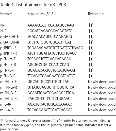
F, forward primer; R, reverse primer. The ‘m’ prior to a primer name indicates it is for a monkey gene, and the ‘p’ prior to a primer name indicates it is for a porcine gene.
Western blot analysis
Cells were collected and lysed in RIPA lysis buffer (Beyotime Biotechnology, Shanghai, China) for 30 min, subjected to SDS-PAGE, then transferred to nitrocellulose (Millipore, Boston, MA, USA). Bradford assays were used to quantify the protein contents. After blocking of membranes (blocking solution: 5% skim milk powder in TBS with 0.5% Tween-20 [TBST]) for 2 h at 37°C, antibodies: anti-PRRSV N protein monoclonal antibody (1:1,000; Jeno Biotech, Inc., Chuncheon, South Korea), p-IκBα mAb (1:200; Cell Signal Technology, Danvers, MA, USA) and anti-GAPDH (1:1,000; Cell Signal Technology) were applied overnight at 4°C. Horseradish-peroxidase-conjugated anti-mouse IgG (1:2,000; Cell Signal Technology) and anti-rabbit IgG (1:2,000; Cell Signal Technology) were used to detect primary antibodies. The signals were detected using ECL Plus chemiluminescence reagent (Pierce, Rockford, IL, USA).
Immunofluorescence assay (IFA)
Cells were fixed with 4% paraformaldehyde at room temperature (RT) for 15 min. After three washes with PBS, cells were permeabilized with Triton X-100 for 15 min and blocked with PBS containing 1% BSA for 30 min, then incubated with anti-PRRSV N protein monoclonal antibody (1:200; Jeno Biotech, Inc.) or anti-cleaved caspase-3 (1:200; Cell Signal Technology) overnight at 4°C. After three washes with PBS, FITC-conjugated secondary antibody (1:1,000; Cell Signal Technology) was added and incubated for 3 h at RT. The nuclei were stained with Hoechst dye 33342 (Life Technologies, Carlsbad, CA, USA) and examined by fluorescence microscopy (Nikon, Tokyo, Japan).
Antiviral assay
The inhibitory effect of treatment with CG was analysed with two different approaches. Co-treatment: cells with 60–70% confluence were infected with CH-1a (multiplicity of infection [MOI]=0.1) in the presence or absence of CG for 24 h; and post-treatment: cells with 60–70% confluence were infected with CH-1a (MOI=0.1) for 4 h at 37°C, and then the viral inoculum was removed and fresh medium containing various concentrations of CG was added. The cells were then further incubated for 24 h. The inhibitory effect was detected by virus yield titration, IFA, qRT-PCR and western blot. The viral titres were assessed as the 50% tissue culture infectious dose (TCID50) [19].
Virus release assay
Cells were infected with CH-1a at an MOI of 0.1 at 37°C for 24 h. After three washes with PBS, cells were then treated with CG at 37°C for another 4 h. The supernatants were harvested to titrate the viral yields and to quantify viral particles by TCID50 and qRT-PCR.
Virus attachment assay
Cells were cooled at 4°C for 45 min, and then inoculated with CH-1a at an MOI of 0.1 in the presence or absence of CG at 4°C for 4 h. After rinsed with PBS three times to remove unbound viral particles, the cells were shifted to 37°C for 24 h. Cell lysates were collected for western blot and qRT-PCR analysis. The supernatants were harvested for TCID50.
Virus entry assay
Marc-145 cells were initially infected with CH-1a at an MOI of 0.1 for 4 h at 4°C. After the cells were washed three times with PBS, they were treated with or without CG at 37°C for 6 h. After three washes with PBS, cells were cultured for 24 h at 37°C. Cell lysates were collected for qRT-PCR and western blot analysis. The supernatants were collected for TCID50.
Apoptosis assay
An annexin V-FITC/Propidium Iodide (PI) assay (Beyotime Biotechnology, Shanghai, China) was used to detect apoptosis. Cells were inoculated with CH-1a at an MOI of 0.1 in the presence or absence of CG (30 mg/ml) for 48 h. After three washes with cold PBS, cells were incubated with FITC-conjugated annexin V and PI for 20 min in the dark. Finally, cells were analysed by fluorescence microscope (Carl Zeiss, Jena, Germany) and flow cytometry (BD FACSCalibur, Franklin Lake, NJ, USA).
Statistical analysis
All experiments were performed at least three times with reproducible results. Significant differences between experimental groups were determined by Student's t-test and analysis of variance (ANOVA). P-values of less than 0.05 were considered statistically significant.
Results
CG Suppresses CH-1a in Marc-145 Cells
To determine the antiviral activity of CG against CH-1a replication, cytopathic effect (CPE) observation and IFA assays were performed. CPE were initially observed daily during the incubation period. CG significantly abrogated the CH-1a-induced CPE in a dose-dependent manner in Marc-145 cells challenged with CH-1a (MOI=0.1) at 72 h (Figure 1A). The specificity of CPE was further confirmed by IFA at 24 h post infection (hpi). Virus-infected cells were markedly decreased by CG treatment in Marc-145 cells in a dose-dependent manner (Figure 1B), suggesting that infection and spread of CH-1a to the neighbouring cells are suppressed by CG.

CG inhibits CH-1a infection and replication in Marc-145 cells
To further examine its antiviral activity on CH-1a replication, we tried to assess the inhibitory effect of CG on virus production with two different approaches as described in Methods. As shown in Figure 1C and 1D, in co-treatment approach, the transcription and translation levels of the viral N protein were significantly reduced in cells treated with CG in comparison to those without CG treatment. Consistently, the production of viral progeny was significantly decreased at 24 hpi by CG in a dose-dependent manner (Figure 1G). Previous studies have shown that the expression of viral N protein is located in cytoplasm and nucleus in cells infected with PRRSV [21], we tried to investigate the effect of CG on viral N protein in infected cells. As shown in Figure 1E and 1F, the expression of N protein in both nucleus and cytoplasm fractions in virus-infected cells was significantly reduced by CG at 24 hpi when administered with co-treatment method. Consistent with these findings, CG markedly decreased the mRNA and protein levels of viral N gene when administered with the post-treatment method (Figure 1H and 1I). To determine whether the concentration-dependent cytotoxicity of CG could affect CH-1a replication, the cytotoxicity of different concentrations of CG was determined using alamarBlue (Thermo Scientific, Waltham, MA, USA). As shown in Figure 1J, cells incubated in medium containing CG at the concentration up to 50 mg/ml retained approximately relative viability of 100% after treated for 48 h. Taken together, these data demonstrate that CG strongly inhibits CH-1a replication in Marc-145 cells.
CG Inhibits Apoptosis Induced by PRRSV during the Late Stage of Infection
Research demonstrates that PRRSV activates anti-apoptotic pathways during the early phase of infection, and that virus-infected cells die from apoptosis during the late period of infection [22]. To examine the effect of CG on apoptosis during the late stage of infection, an Annexin V fluorescein isothiocyanate (FITC)/propidium iodide (PI) assay and IFA with anti-cleaved caspase-3 mAb were performed. As shown in Figure 2A, in the presence of CG (30 mg/ml), the apoptosis of virus-infected cells was remarkably reduced at 48 hpi. Consistently, cleaved caspase-3-specific staining was notably decreased in CG-treated Marc-145 cells compared with the control without CG treatment at 48 hpi (Figure 2B). These findings demonstrate that CG can block virus-induced apoptosis during the late stage of infection.

The effect of CG on CH-1a-induced apoptosis during the late period of infection
CG Blocks viral Particle Release
To investigate the effect of CG on viral particle release in Marc-145 cells, an assay described previously [23] was performed to quantify the viral particles in the supernatant. As shown in Figure 3A, the copies of released extracellular viral RNA were significantly reduced in the presence of CG (30 mg/ml) in Marc-145 cells at 6 hpi. Consistent with these findings, the production of viral progeny in the supernatant was significantly decreased by CG (Figure 3B). Collectively, these results suggest that CG can inhibit the release of CH-1a viral particle.

CG inhibits the release of viral particles
CG Blocks Viral Attachment and Entry
We have identified that CG can inhibit the replication of CH-1a, but the underlying molecular mechanism that the antiviral activity of CG against PRRSV remains unknown. The primary process of virus reproduction includes attachment and entry during virus life cycle. To characterize which stage of viral life cycle is interrupted by CG, an viral attachment assay was initially performed to examine whether CG can block CH-1a binding to Marc-145 cell surface. As shown in Figure 4A and 4B, the mRNA and protein levels of CH-1a N gene were markedly diminished by CG (30 mg/ml) during the period of viral attachment. Furthermore, upon CG treatment (30 mg/ml), the titre of CH-1a was significantly decreased compared with the control without CG treatment (Figure 4C), suggesting that CG is able to notably decrease the quantity of infectious viral particles attached to cell surface. Consistent with the above findings, CG (30 mg/ml) inhibited viral N gene at the transcription and translation levels as well as virus yield during the period of viral entry (Figure 4D–4F). Taken together, CG is capable of blocking viral attachment and entry during virus life cycle.

CG blocks viral attachment and internalization during virus life cycle
Inhibition of NF-κB Activation by CG Contributes to its Anti-PRRSV Activity
Previous studies have shown that PRRSV can activate NF-κB pathway, which is necessary for viral gene transcription and replication [24]. To determine the effect of CG on virus-induced activation of NF-κB, confocal microscopy assay was performed in Marc-145 cells. As expected, in CH-1a-infected cells, nuclear accumulation of p65 was observed (arrowheads, Figure 5A). Nevertheless, upon CG (30 mg/ml) treatment, endogenous p65 mainly existed in cytoplasm in virus-infected cells, which is similar to the findings of uninfected mock-treated cells (top panel). To further confirm that CG is able to inhibit PRRSV-induced NF-κB activation, the level of IκBα phosphorylation was also tested using p-IκBα mAb. As shown in Figure 5B, the level of IκBα phosphorylation induced by CH-1a was significantly inhibited by CG (30 mg/ml) in virus-infected Marc-145 cells. PRRSV can activate NF-κB pathway and subsequent inflammatory cytokine production [24]. To investigate the effect of CG on PRRSV-induced cytokine upregulation, qRT-PCR was conducted to assess the expression of interferon (IFN)-α, IFN-β and interleukin (IL)-6 in Marc-145 cells and PAMs. As shown in Figure 5C–5E, CG (30 mg/ml) notably suppressed the expression of IFN-α, IFN-β and IL-6 upregulated by CH-1a in Marc-145 cells. Consistent with above data, the upregulation of IFN-α and IFN-β induced by CH-1a was decreased in the presence of CG (30 mg/ml) in PAMs (Figure 5F and 5G). Taken together, these data demonstrate that CG inhibits PRRSV-induced NF-κB activation, thus interfering with cytokine production, which contributes to its anti-PRRSV activity.

CG inhibition of NF-κB activation contributes to its anti-PRRSV activity
CG Inhibits CH-1a Replication in PAMs
Since CG exerted potent inhibitory effect against PRRSV infection in Marc-145 cells, we examined whether CG can inhibit CH-1a replication in PAMs, the main target cells for PRRSV in vivo. Cell cytotoxicity assay was firstly performed, CG was noncytotoxic up to 50 mg/ml when PAMs were treated with CG for 48 h (Figure 6A). Next the effect of CG on CH-1a infection in PAMs was assessed. Consistent with the results in Marc-145 cells, CG (30 mg/ml) caused significant decrease in the transcription and translation levels of the viral N protein at 24 hpi (Figure 6B and 6C). Collectively, CG markedly inhibits CH-1a replication in PAMs as well.

Anti-PRRSV activity of CG in PAMs
Discussion
PRRS is one of the most economically important diseases to the pig industry worldwide. It has quickly spread to many countries and has become a major problem in preventing swine diseases in China [25–28]. Its high variation and persistent infection make it difficult to control [29,30]. Current PRRS vaccines such as modified live vaccines (MLVs) fail to effectively prevent and control this disease. Therefore, there is an urgent need for safer and more effective strategies to control PRRSV. Recently, marine oligosaccharides and their derivatives have been attracting increasing interest in developing potential antiviral drugs [31,32]. Carrageenan is a high-molecular-weight linear polysaccharide and widely exists in red algae. Previous reports have shown that carrageenan possesses antiviral activities effective against different viruses [32,33]. However, to our knowledge, this is the first study to report the inhibitory effect of CG against PRRSV infection and replication in vitro. We have demonstrated that CG exerted potent antiviral activity against CH-1a infection by multiple pathways such as inhibition of virus RNA and protein synthesis, virus progeny production, viral particle release and PRRSV-induced apoptosis in the late period of infection in Marc-145 cells (Figures 1, 2 and 3). Since Marc-145 cells are of monkey origin (not of porcine origin) [34], we also investigated whether CG can block CH-1a replication in PAMs, which are the main target cells for PRRSV in vivo. Consistent with the findings obtained with Marc-145 cells, CG remarkably suppressed CH-1a infection in PAMs (Figure 6).
Previous studies have shown that CG can inhibit the replication of many viruses such as HPV, dengue virus and influenza A H1N1 virus by preventing virus adsorption to host cells [18]. CG inhibits attachment mainly by binding with viral glycoproteins or envelope proteins, which might be attributed to its chemical structure that is similar to haparan sulfate, a cell surface receptor for many viruses [32]. In this study, the molecular mechanisms of CG-mediated inhibition against CH-1a were characterized during the attachment and the entry stage of virus life cycle. We found that CG blocked viral adsorption and entry of virus life cycle (Figure 4), indicating that the interactions between viral particles and receptors on the cell membrane might be blocked by CG. However, the precise molecular mechanisms regarding how CG affects virus binding to the cell surface is still unclear and accordingly would be our next issue to be addressed in future. In addition, we found that CG inhibited PRRSV-induced NF-κB activation, thus interfering with cytokine production, which contributes to its anti-PRRSV activity (Figure 5). Further research should be undertaken to clarify the molecular mechanisms of inhibition of NF-κB activation by CG.
In conclusion, our results suggest that CG is a safe and effective means of inhibiting PRRSV infection in vitro by affecting viral attachment and internalization. CG may be suitable serving as a promising antiviral candidate for the prevention and treatment of PRRSV infection. Future studies are needed to determine the antiviral activity of CG against PRRSV in vivo in order to facilitate further development of CG as a virostatic agent.
Footnotes
Acknowledgements
This work was supported by National Natural Science Foundation of China (31601917 and 31872329), Natural Science Foundation of Guangdong Province (2014A030312011), and Science and Technology Planning Project of Guangzhou (201804020039).
The authors declare that they have no competing interests. None of the authors has any financial or personal relationships that could inappropriately influence or bias the content of the paper.