
Abstract
Background
In previous research, we have demonstrated that sodium tanshinone IIA sulfonate (STS) has anti-porcine reproductive and respiratory syndrome virus (PRRSV) activity, but whether autophagy is involved in this process is still unknown. In this study, the autophagy effect of STS against PRRSV infection was investigated in vitro.
Methods
Quantitative real-time PCR (qRT-PCR) and western blot was used to evaluate the inhibition ability of STS on the mRNA expression levels on cell autophagy genes, that is Beclin1, ATG5 and ATG7. Simultaneously, the effect of STS on N protein/gene expression was assessed by indirect immuno-fluorescence assay (IFA), qRT-PCR and western blot.
Results
The results indicated that STS inhibits autophagy induced by PRRSV. In addition, STS effectively suppresses PRRSV's N protein replication and N gene expression in Marc-145 cells infected with PRRSV in a time-dependent manner.
Conclusions
Our results suggest that STS exhibits anti-PRRSV activity in vitro by suppressing autophagy-related genes, which may provide a theoretical basis for further pharmacological agent development regarding PRRSV infection.
Introduction
Porcine reproductive and respiratory syndrome virus (PRRSV), a member of Nidovirales within the Arteriviridae, is a single-stranded RNA virus causing severe reproductive failure, respiratory distress, high morbidity and mortality in pigs [1,2]. Although vaccination is a common immune method that plays an important role in controlling the disease process, still many problems exist, such as improper immune effect, virus variation and strengthened virulence [3–6]. Furthermore, live-attenuated vaccine has the potential risk of virulence reversion, and may induce a new generation virus from genetic mutations [4].
In eukaryocytes, autophagy is a widely existing conservative mechanism. Autophagy is recognized both as a unique cell death pathway and as an adaptation to various stresses that supports cell growth and survival [7]. An increasing number of studies demonstrated that autophagy is involved in the infection processes of a variety of pathogens. It can be hijacked by various viruses to facilitate their replication, including PRRS virus (PRRSV), influenza A virus, foot-and-mouth disease virus, pseudorabies virus, etc [8–12]. In addition, several studies have reported that PRRSV has evolved strategies to exploit autophagy for its own replication [12–15].
It is well known that many traditional medicinal plants and herbs have potent antiviral activity, such as Scutellaria baicalensis, Matrine etc [16–18]. Tanshinone IIA is a natural compound derived from salvia miltiorrhiza, well known for treating cardiovascular disease [19]. We have proved that the water-soluble derivative of tanshinone IIA, sodium tanshinone IIA sulfonate (STS), can inhibit Marek's disease virus (MDV) infection effectively in vitro and in vivo [20,21]. Moreover, our previous study also demonstrated that STS can inhibit PRRSV infection in vitro through directly inactivating and/or disturbing the replication of PRRSV [22,23]. In this study, we further investigated the anti-PRRSV mechanisms of STS through exploring N gene/protein expression of PRRSV and PRRSV-induced cell autophagy.
Methods
Cells, viruses and STS
Marc-145 cells were purchased from the China Institute of Veterinary Drug Control (Beijing, China). Cells were grown in DMEM supplemented with 10% FCS and 1% penicillin-streptomycin at 37°C with 5% CO. PRRSV vaccine (JXAI-R, No. 1012001; Guangdong Dahuanong Animal Health Products Co., Ltd, Guangdong, China) was propagated in Marc-145 cells. Virus titres were 1x107.5 50% tissue culture infective dose (TCID50)/ml, determined by the Reed-Muench formula. STS and ribavirin were obtained from the National Institute for Food and Drug Control (Beijing, China). According to previous results, the maximum STS concentration used was 62.5 μg/ml, and when the concentration was lower than 15.625 μg/ml, there was almost no anti-PRRSV activity [22]. In this study, 62.5 μg/ml and 31.25 μg/ml STS were tested.
qRT-PCR
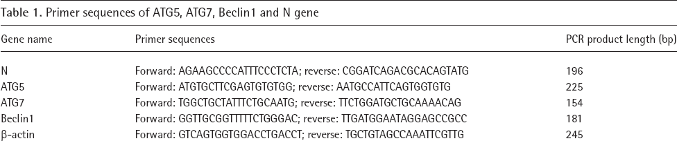
Marc-145 cells were planted into six-well plates with a density of 2x10 5 /ml. When the cells grew to a confluent monolayer, the culture medium was discarded and 2 ml 100 TCID50 PRRSV were added into the cells for 2 h to complete the infection process. Marc-145 cells were treated with 62.5 μg/ml and 31.25 μg/ml STS, and cell samples were collected at 12, 24, 36 h after PRRSV infection. Ribavirin (125 μg/ml) was used as a positive medicine treatment control group. qRT-PCR was used to quantify the expression of cell autophagy related genes (ATG5, ATG7, Beclin1 [ATG6]) and PRRSV N gene. The gene primer sequences and PCR fragment length were shown in Table 1. Total RNAs were extracted using Trizol reagent (Invitrogen, Carlsbad, CA, USA), followed by DNase I treatment and cDNA was synthesized using PrimeScript® RT Master Mix kit (TAKARA, Dalian, China). qRT-PCR was performed using Bio-Rad IQ5 QPCR System with SYBR® Premix Ex Taq™ kit (TAKARA). The relative quantities of ATG5, ATG7 and Beclin1 mRNA were calculated based on the 2−ΔΔCt method and normalized to β-actin. In addition, the PRRSV N gene copies were determined by absolute fluorescence quantification.
Primer sequences of ATG5, ATG7, Beclin1 and N gene
Western blot
Cell samples were prepared as described in the qRT-PCR section. Cells were rinsed with cold PBS and collected, total protein was extracted using a total protein extraction kit (Applygen, Beijing, China). Protein samples were subjected to 12% SDS-PAGE and transferred into PVDF transfer membrane (0.22 μm; Millipore, Bedford, MA, USA). Membrane was blocked with 5% non-fat milk in TBST for 1 h at room temperature, and then incubated with a polyclonal antibody against the Beclin1 (1:400; Abcam, Cambridge, UK), ATG5 (1:400; Abcam), ATG7 (1:400; Abcam), N (1:1,000; gifted by Z Wang, Shanxi Agriculture University, Taigu, China) and β-actin (1:1,000; Cowin Biotech, Beijing, China) at 4°C overnight. After being washed 3x with TBST, membrane strips were incubated with secondary antibodies corresponding to their respective primary antibodies at 1:5,000 dilutions at 37°C for 1 h with gentle shaking. Following another three washes with TBST, the protein bands were visualized using enhanced chemiluminescence (Cowin Biotech) and exposed to autoradiography film.
Immunofluorescence assay (IFA)
Marc-145 cells were planted in 384-well plates infected with 100 TCID50 PRRSV, and then treated with ribavirin and STS. After 36 hpi, cells were fixed by pre-cooled acetone and methanol (1:1) for 30 min at −20°C. After being rinsed 3x by PBS, the fixed cells were incubated with a monoclonal antibody against the N protein (ORF7) of the PRRSV for 2 h at 37°C. Unbounded antibodies were washed away by PBS, and sample was incubated with FITC-labelled goat anti-mouse antibody (Bioss, Beijing, China) for 1 h at 37°C. DAPI was used for nuclei stained. Fluorescence was detected using a fluorescence microscopy (Olympus, Shinjuku-Ku, Japan).
Statistical analysis
Data were analysed by one-way ANOVA with GraphPad Prism 5 software (GraphPad Software Inc., San Diego, CA, USA). All data were expressed as mean +sd and P<0.05 was considered significant difference.
Results
Effect of STS on PRRSV N gene and protein expression
qRT-PCR and western blot were used to analyse the effect of STS on the expression of PRRSV N gene and protein. qRT-PCR results showed that when compared with PRRSV group, STS reduced N gene copies and N mRNA level significantly when treated with 62.5 μg/ml STS (Figure 1A; P<0.05). Using 62.5 μg/ml STS, there was almost no detectable band (N protein; Figure 1B). When the concentration of STS was decreased, the inhibition on N protein expression declined, but with the action time of STS extending, the inhibitory effects were more obvious. These results were further demonstrated by IFA. At 36 h, only light green fluorescence could be detected in 62.5 μg/ml STS treatment group (Figure 1C). The staining results further confirmed that STS could effectively reduce PRRSV replication in a dose-dependent and time-dependent manner the STS possessed a higher inhibitory ability than ribavirin.

Effect of STS on PRRSV N gene and protein expression
STS Inhibited the Expression of ATG5, ATG7 and Beclin1 Gene
To determine whether STS could effectively inhibit ATG expression, ATG5, ATG7 and Beclin1 mRNA expression levels in all groups were measured by qRT-PCR. As shown in Figure 2A, comparing with cell control, the expression of ATG5 was significantly enhanced in PRRSV control at all time points (P<0.05), while a continuous ATG5 suppression in STS 62.5 μg/ml treatment group has been found at 12, 24 and 36 h (P<0.05), 31.25 μg/ml STS could inhibit the ATG5 gene expression at 12 h and 36 h (P<0.05). As shown in Figure 2B, the expression of ATG7 mRNA was significantly higher in the PRRSV group at all time points, especially at 12 and 36 h (P<0.05). The ATG7 mRNA level was reduced when the infected cells were treated with 31.25 μg/ml and 62.5 μg/ml STS for 12 h, 24 h and 36 h. Compared with the ribavirin-positive control group, STS possessed a higher inhibitory ability at 36 h. As shown in Figure 2C, PRRSV enhanced the expression of Beclin1 at 24 and 36 h (P<0.05), and STS could potently inhibit the Beclin1 gene expression at 12, 24 and 36 h.

STS inhibited the expression of ATG5, ATG7 and Beclin1 gene
STS Inhibited the Expression of ATG5 and Beclin1 Protein
The corresponding expression of ATG5, ATG7 and Beclin1 protein levels were analysed by western blot. As shown in Figure 3, comparing with cell control, the expression of ATG5 and Beclin1 protein in PRRSV control were significantly increased (P<0.05), which is consistent with the results of qRT-PCR. The ATG7 protein expression in PRRSV control declined while the ATG7 gene expression increased. Moreover, the western blot results indicated that the ATG5 and Beclin1 protein levels were reduced in STS treatment groups compared with PRRSV control group at 12, 24 and 36 h. The inhibitory effect of 62.5 μg/ml STS on ATG5 and Beclin1 protein expression showed better inhibitory effects.

STS inhibited the expression of ATG5 and Beclin1 protein
Discussion
Our previous reports suggested that STS possessed anti-PRRSV activity in Marc-145 cells model [22,23]; however, the latent mechanism was still unknown. Autophagy is an evolutionarily conserved lysosomedependent degradation pathway that acts in the maintenance of cellular homeostasis and plays a crucial role in viral replication and pathogenesis [8–15]. Autophagy-related genes (ATGs) play an important role in induction, maturation, degradation and recirculation of autophagy. Therefore, ATGs were often used to evaluate antiviral activity of medicines [24].
In the process of nidovirus proliferation, the formation of autophagic double-membrane structure is induced by the expression of non-structural protein, then the replicase complex positions to the autophagy membrane and promotes the synthesis of virus protein, such as Equine arteritis virus (EAV) and PRRSV [25,26]. Liu et al. [13] indicate that autophagy induced by PRRSV infection plays a role in sustaining the replication of PRRSV in host cells. Inhibiting the autophagy decreases the virus titres. Some studies have shown that knockdown of Beclin1 [7,15], ATG5 [12] or ATG7 [15] gene could reduce the replication of PRRSV. Li et al. [11] also suggested that HBV infection induces the host's autophagy machinery to enhance HBV replication. The inhibition of autophagosome formation by the small interfering RNA duplexes targeting the genes critical for autophagosome formation (Beclin1 and ATG5 genes) markedly suppressed HBV production. Taken together, ATG5, ATG7 and Beclin1 play a key role in the formation process of autophagosome double-membrane structure. In this study, we demonstrated that STS at a higher concentration (62.5 μg/ml) inhibits the expression of cell autophagy-related protein ATG5, ATG7 and Beclin1 in PRRSV-infected cells. It suggested that STS inhibits autophagosome formation by supressing expression of ATG5, ATG7 and Beclin1 gene, which may affect the localization of viral replicase complex in autophagosome bilayer membrane.
N protein is encoded by Open Reading Frame 7 (ORF7) and it forms about 20–40% of virion structure. In infected cells, N protein is the multifunctional viral protein which plays a crucial role in the assembly of the viral nucleocapsid through interaction with the viral RNA. Furthermore, N protein may inhibit the synthesis of host protein through gathering to the host cell nucleolus, which could affect ribosome assembly or function [27]. A previous study suggested that N may play a role in transcriptional regulation in PRRSV-infected cells, interacting with a zinc-binding transcriptional regulator HIC (human I-mfa domain-containing protein) [28]. Our previous research has shown that STS could inhibit PRRSV N protein replication in a dose-dependent manner at 48 h [22]. In this study, we provide evidence that infected cells treated by STS decrease the PRRSV N gene and protein levels in a time-dependent manner.
In conclusion, this study showed that STS exerted its antiviral effects via inhibition of transcription and translation of PRRSV N protein and STS could reduce cell autophagy induced by PRRSV. We anticipate that STS can serve as a valuable candidate for further antiviral drug development.
Footnotes
The authors declare no competing interests.
Acknowledgements
This project was funded by grants from the Key R&D Plan of Shanxi Province (201603D21109-1) and the fund for Shanxi ‘1331 Project’ Key Innovative Research Team.