
Abstract
We developed a novel method for differentiating adipocyte-derived stem cells (ASCs) into hepatocyte-like cells (iHeps). ASCs are cultured as spherical cellular aggregates and are then induced by culture in chemically defined media for a short time period to differentiate into spherical culture iHeps (SCi-Heps). SCi-Heps have many of the in vitro functional properties of mature hepatocytes, and they can stably reconstitute functioning human liver in vivo in a murine model system. Implantation studies demonstrate that SCi-Heps have a very low malignant potential. All human liver regenerative procedures, including ultrasound-guided direct liver implantation, are scalable and appropriate for human clinical use. These methods can be used to achieve the major promise of regenerative medicine. It may now be possible to regenerate human liver using autologous stem cells obtained from a readily accessible tissue.
Introduction
A major goal for regenerative medicine is to enable human tissue replacement through transplantation of stem cells that can be harvested from readily accessible tissues. For example, orthotopic liver transplantation is the only effective treatment for end-stage liver disease or severe liver injury, but its utility is severely limited by the lack of donor liver tissue and by the requirement for lifelong immunosuppression. However, a large number of adipocyte-derived stem cells (ASCs) can be easily obtained by liposuction, and methods for inducing ASC differentiation into hepatocyte-like cells have been developed (3–5,33). These features have made liver regeneration via transplantation of autologous ASCs a highly attractive possibility for regenerative medicine (1,13, 14,23). By this method, ASCs obtained by liposuction are induced to differentiate into induced hepatocyte-like cells (iHeps) in vitro and then transplanted into the donor's liver. The abundance and accessibility of adipose tissue ensures that there is a source of readily available autologous stem cells. Liver regeneration by this method does not require immunosuppression.
Ochiya and colleagues (4) developed a two-stage protocol for differentiating ASCs into iHeps in vitro in chemically defined media. Lipoaspirate cells are first cultured for 3 to 15 days to produce ASCs, which are then cultured for 3 days (stage 1) in the presence of activin A and fibroblast growth factor 4 (FGF4) to induce endodermal differentiation. These cells are then cultured for an additional 10–12 days (stage 2) in a different medium with other growth factors (hepatocyte growth factor, oncostatin M, FGF1, FGF4) to produce cells with hepatocyte characteristics, which we refer to as chemically induced hepatocytes (Chi-Heps). The hepatocyte-like properties of Chi-Heps were thoroughly characterized in vitro, and Chi-Heps were found in the liver after transplantation into mice (2,4,5,33). However, their ability to establish and maintain human liver function in vivo, which is essential for enabling autologous liver regeneration in humans, has not been demonstrated. Moreover, the prolonged culture period (20–30 days) would reduce the utility of this method. For example, the cells used for liver regeneration must be prepared much more quickly if they are to be used for treatment of liver failure caused by an overdose of acetaminophen; death can occur over a 2-week period after ingestion of a toxic dose of acetaminophen (6,22).
Therefore, we sought to develop a novel method that would reduce the production time and increase the yield of iHeps and which would scale for use in human subjects. Stem cells isolated from a variety of tissues have previously been cultured as spheres [reviewed in Pastrana et al. (30)], but spherical culture has not been used to differentiate ASCs into iHeps. Therefore, we investigated whether spherical culture could be used to produce iHeps within a shorter time frame that would enable their use for treatment of acute liver failure. In addition, to enable the development of improved methods for human liver regeneration, we produced chimeric mice wherein transplanted human hepatocytes can replace mouse liver (18). To do this, a herpes simplex virus type 1 thymidine kinase (TK) transgene is expressed within the liver of a highly immunodeficient mouse strain (nonobese diabetic/severe combined immunodeficient/interleukin-2 receptor g chain knockout; NOD/Shi-scid/IL-2Rγnull; NOG) (20) to produce the TK-NOG transgenic mouse (18). A brief exposure to a nontoxic dose of ganciclovir causes a rapid and temporally controlled ablation of mouse liver cells. This enables transplanted human liver cells to develop into a “human organ” with a characteristic three-dimensional architecture and gene expression pattern. The transplanted cells can stably maintain human hepatic biosynthetic function for a 6-month period (18). Chimeric mice produced by this method have been used to predict the pattern of human drug metabolism and the occurrence of a drug–drug interaction prior to human exposure (28) and to identify human genetic factors affecting the metabolism of clinically important drugs (19). Moreover, ganciclovir-induced hepatotoxicity in mice expressing a TK transgene in liver has been used to model acute liver failure in humans (12,38), which can be caused by exposure to a number of insults, including a toxic dose of acetaminophen (6). Therefore, we investigated whether ASC-derived iHeps, which were produced using the spherical culture method, could reconstitute human liver in TK-NOG mice using a protocol wherein each step is appropriate for subsequent use in humans. Specifically, only defined chemicals and growth factors are used to induce hepatocyte differentiation in vitro; neither oncogenes nor any other foreign genes are inserted into the ASCs genome, and the cells are obtained by liposuction and subsequently transplanted by ultrasound-guided direct injection into the liver, which are procedures routinely used in clinical practice.
Materials and Methods
ASC Preparation and Differentiation
Lipoaspirates were obtained as deidentified samples from four human donors undergoing liposuction at Stanford University Medical Center according to a protocol that was approved by the Stanford University Medical Center IRB. ASCs were prepared from these samples as described (5). The ASCs were cultured in MesenPRO RS™ Medium (Gibco, Gaithersburg, MD, USA; Cat: 12746–012), passaged at 90% confluence at a 1:4 ratio, and the medium was changed every other day. For preparation of Chi-Heps, a modification of the two-stage protocol of Ochiya and colleagues (4) was used to induce ASC differentiation into iHeps. After two to five passages, the cells were plated on Matrigel (BD Biosciences, San Jose, CA, USA; Cat: 354277)-coated dishes. After the cells reached 50% confluence, endodermal transdifferentiation was induced over 3 days of culture in the stage 1 medium: Roswell Park Memorial Institute medium (RPMI-1640; Gibco; Cat: 12633–012) supplemented with 100 ng/ml activin A (R&D Systems, Minneapolis, MN, USA; Cat: 338-AC-010), 50 ng/ml wingless-type mouse mammary tumor virus (MMTV) integration site family, member 3 a (Wnt3a; StemRD, Burlingame, CA, USA; Cat: W3A-H-100), 20 ng/ml fibroblast growth factor 4 (FGF4; PeproTech, Rocky Hill, NJ, USA; Cat: 100–31), and 20% B27 (Gibco; Cat: 17504–044). Hepatic differentiation was then induced over a 13- to 15-day period by culture in the stage 2 medium: hepatocyte culture medium (HCM; Lonza, Walkersville, MD, USA; Cat: cc-3198) supplemented with 150 ng/ml hepatocyte growth factor (HGF; PeproTech; Cat: 100–39), 25 ng/ml FGF4, 30 ng/ml oncostatin M (OSM; PeproTech; Cat: 300–10), 2 × 10⁻⁵ M dexamethasone (Dex; Sigma, St. Louis, MO, USA; Cat: D4902), and 0.1% dimethyl sulfoxide (DMSO; Sigma; Cat: C6164). The cells were then cultured in HCM alone for 2 days prior to transplantation into male TK-NOG mice.
Preparation of iHeps
Please see supplementary methods available at http://med.stanford.edu/peltzlab/SuppInfo/liver-regen/
Preparation of SCi-Heps
Primary ASCs were isolated from lipoaspirates and suspended in MesenPRO RS™ Medium (Gibco, Cat: 12746–012) at 5 × 10⁴ cells/ml. The cells were cultured using the hanging drop method, with modifications that were described in Xu et al. (37). In brief, a suspension of ASCs (330 cells/μl) in MesenPRO RS™ Medium was prepared and then deposited as 40-μl drops onto the lid of a 100-mm tissue culture dish (Sigma; Cat: CLS430167). The lid was then inverted over a phosphate-buffered saline (PBS; Invitrogen, Carlsbad, CA, USA)-filled tissue culture dish and placed in a 37°C/5% CO2/95% humidity incubator to enable the formation of spherical cellular aggregates (“spheres”) that resembled embryoid bodies. The ASCs were plated as individual drops (each ~40 μl) in rows on 150-mm petri dishes (BD Biosciences; Cat: 354551) using an eight-channel pipette. After 2 days, the dishes were rinsed with PBS, and the spheres were collected by centrifugation at 150 × g at 37°C, resuspended at 30 spheres/ml in the stage 1 medium, and seeded on Matrigel-coated dishes (BD Biosciences; Cat: 354262). The sphere-derived cells were then induced to differentiate into hepatocytes by the two-stage process, which was modified from the procedures described by Ochiya and colleagues (4). Endodermal transdifferentiation was induced over a 2-day period by culturing the spheres in the stage 1 medium. Hepatic differentiation was induced over the subsequent 2- to 9-day period by culture in the stage 2 medium. The cells were then cultured in HCM alone for 2 days prior to transplantation into TK-NOG mice.
Flow Cytometry
Cytokeratin 8/18 (CK8/18), sex-determining region Y box 17 (Sox 17), and cluster of differentiation 105 (CD105) expression was analyzed by flow cytometry (FACS, Aida, Fuji, Valhalla, NY, USA; Software Flowjo, Treestar, Ashland, OR, USA). The cells were first blocked with an Fc receptor-blocking reagent (Miltenyi Biotech, Bergisch Gladbach, Germany) according to the manufacturer's instructions and then stained with primary antibody anti-human CK8/18 (Abcam, Cambridge, MA, USA; Cat: ab17139, 1:200), Sox 17 (Abcam, Cat: ab84990, 1:100), or CD105 (Abcam, Cat: ab21224, 1:200) for 30 min at 4°C, which was followed by incubation with a secondary antibody Alexa Fluor 488 (Invitrogen, Cat: A21200, 1:1,000). Appropriately diluted isotype-matched antibodies (Ebioscience, San Diego, CA, USA) were used as controls. The data from 10,000 analyzed events were stored and analyzed.
Preparation of TK-NOG Mice
All animal experiments were performed according to protocols approved by the Stanford Institutional Animal Care and Use Committee. The TK-NOG mice were bred at In Vivo Sciences International in Sunnyvale, CA. Previously described procedures were used to prepare TK-NOG mice for transplantation (18). In brief, 8- to 10-week-old male TK-NOG mice were treated (IP) with 25 mg/kg ganciclovir (GCV, obtained from Genentech, San Francisco, CA, USA) on days −7 and −5 prior to transplantation. Then, a 20-μl blood sample from the tail vein was collected 6 days after the first GCV treatment, diluted 1:3 in water, and 10 μl of the diluted sample was used to measure the serum alanine transaminase (ALT) level using a Fuji dry-chem 7000 instrument according to the manufacturer's instructions. Only mice with an ALT level >200 U/L were used for cellular transplantation on day 7. Mice with ALT levels <200 U/L were treated with an additional dose of 25 mg/kg GCV on day 7, the ALT level was reexamined on day 13; only mice whose repeat ALT level was >200 U/L were used for cell transplantation on day 14.
Ultrasound-Guided Liver Injection
Transplanted cells were directly placed into a liver lobe under ultrasound-guided injection using a small-animal ultrasound system Vevo 2100 (Visualsonics, Toronto, ON, Canada). Brightness mode (B-mode) was used to acquire two-dimensional images for an area of interest with MS550s transducer. The mice were placed under 1.5% isofluorane (Butler Schein, Dublin, OH, USA) anesthesia during this procedure using a single animal vaporizer unit (EZ-Systems Corp., Palmer, PA, USA; EZ-108SA). Then, 5 × 10⁶ cells were suspended in 200 μl of William's E medium (Invitrogen; A12176–01). Individual aliquots were injected into 10 distinct sites in the liver of a ganciclovir-conditioned TK-NOG mouse using a 30-gauge needle (PrecisionGlide Needle; BD, Franklin Lakes, NJ, USA; Cat: 305106). Thus, each mouse received a total of 5 × 10⁶ cells in 200 μl total volume.
Immunohistochemistry
Liver tissues were flash frozen on dry ice using optimal cutting temperature (O.C.T.) solution from Tissue-Tek (Sakura Finetek USA, Torrance, CA, USA; Cat: 4583). Tissue blocks were then sectioned using a Leica CM3050 S Cryostat (Buffalo Grove, IL, USA) into 7-um sections, and serially obtained sections were used for staining. Liver tissues were fixed in acetone (EMD, Kassel, Germany) for 10 min, followed by blocking with a solution containing 10% horse serum (Jackson ImmunoResearch, West Grove, PA, USA) for 30 min. After washing three times in PBS with 0.05% Triton X-100 (TPBS; Sigma) the sections were incubated with anti-human albumin (Bethyl Laboratories, Montgomery, TX, USA; Cat: A80–129A, 1:50), anti-Ki-67 (H-300; Santa Cruz Biotechnology, Santa Cruz, CA, USA; Cat: sc-15402, 1:50), antiasialoglycoprotein receptor 1 (ASGR-1; Sigma; Cat: HPA011954, 1:50), or anti-human CK8/18 (Abcam; Cat: ab17139, 1:50) primary antibodies overnight at 4°C. After washing three times with TPBS, Alexa Fluor 488 (Invitrogen; Cat: A21200, 1:1,000), or Alexa Fluor 594 (Invitrogen; Cat: A21201, 1:1,000)-conjugated secondary antibodies were then applied for 1 h in the dark. A fluorescein isothiocyanate (FITC)-conjugated anti-zonula occludens-1 (ZO-1) antibody (Abcam, Cat: mAbcam 61357) was used at a 1:50 dilution without a secondary antibody. Nuclear staining was assessed using 4,6-diamidino-2-phenylindole (DAPI; Sigma; Cat: D9542).
All images were acquired using a Nikon Eclipse Ni-E imaging system (Melville, NY, USA).
Analysis of Tumor Formation
To assess tumor formation, 5 × 10⁴ Chi-Heps, SCi-Heps, or iPS-Heps were harvested and mixed with 50 μl of Matrigel. NOG mice were anesthetized with 1.5% isoflurane using an individual vaporizer unit. An incision was made, and a slight pressure to both sides of the incision was applied to expose the kidney. The cells were then injected under the kidney capsule using a syringe with a 27-gauge needle (PrecisionGlide Needle; BD; Cat: 305109). After slowly delivering the cells, a dry swab (Thermo Fisher, Fremont, CA, USA; Cat: MW1041) was placed over the injection site to prevent leakage. During the procedure, the kidney was kept moist by application of saline with a cotton-tipped swab (Thermo Fisher; Cat: MW1041). After 3 to 8 weeks, the mice were sacrificed, and the injected kidney was harvested for histological analysis. Sections of formalin-fixed (EMS, Hatfield, PA, USA) paraffinembedded tumors were stained with hematoxylin (Volu Sol, Salt Lake City, UT, USA) for 5 min and then rinsed in running tap water. Afterwards, sections were differentiated with 0.3% acid alcohol. After rinsing with running tap water, sections were stained with eosin (Volu Sol) for 2 min. Then sections were dehydrated and mounted for exam under a light microscope. The slides were photographed with a Nikon MICROPHOT-FXA microscope (Melville, NY, USA) with plan apochromat objectives (4×/0.10 NA, 10×/0.25, 20×/0.40, 40×/0.65, and 60×/0.95), a SPOT Insight Color Mosaic camera (model 14.2; Diagnostic Instruments, Sterling Heights, MI, USA), and SPOT Advanced imaging software (version 4.6).
Methods for determining gene expression, Periodic Acid-Schiff (PAS) staining, low-density lipoprotein (LDL) endocytosis, flow cytometry, human albumin and urea production, CYP450 activity, and statistical analysis are available in the supplementary material at http://med.stanford.edu/peltzlab/SuppInfo/liver-regen/
Results
We have now developed a novel method for differentiating ASCs into iHeps (Fig. 1A) that has two unique features. (i) ASCs are first cultured using the “hanging drop” method (37) to produce spherical cellular aggregates (“spheres”) (Fig. 1B). As shown by analysis of Sox 17 expression, spherical culture doubles the number of ASCs in a lipoaspirate that differentiate into endodermal precursor cells (Fig. S1; available at http://med.stanford.edu/peltzlab/SuppInfo/liver-regen/). (ii) Since the ability of mesenchymal-derived ASCs to differentiate into endoderm may be rate limiting for hepatocyte generation (24), wnt3α was added to the (stage 1) differentiation medium because Wnt pathway signaling has been shown to promote the generation of endoderm (15,17). Relative to the standard method (4), the new method produces a 2.3-fold increase in the number of ASCs obtained from a lipoaspirate, and a threefold increase in the number of hepatocyte-like cells obtained after the two-stage differentiation process is completed (Fig. 1C, Table 1). In summary, this method increases the number of iHeps obtained from a liter of lipoaspirate by sevenfold, and it reduces the period of in vitro culture required to obtain biochemically defined hepatocytes at >37% purity to only 9 days. Like Chi-Heps, the cells produced by spheroid culture, which we refer to as SCi-Heps, developed a hepatocyte-like morphology (Fig. 1B) and exhibited many properties of mature hepatocytes. They expressed proteins (CK8/18) found on hepatocytes (Fig. 1C) and had multiple metabolic properties of hepatocytes, including LDL endo cytosis, glycogen synthesis (PAS staining) (Fig. 1D), albumin secretion, and urea production (Fig. 1E). Moreover, SCi-Heps expressed multiple hepatocyte-specific mRNAs and had markedly reduced levels of expression of many adipocyte-specific mRNAs (Fig. 2A), along with reduced protein expression of an ASC-specific cell surface protein (CD105) (Fig. S2; Table S1; available at http://med.stanford.edu/peltzlab/SuppInfo/liver-regen/).

Comparison of different methods for inducing adipocyte-derived stem cell (ASC) differentiation to hepatocyte-like cells (iHeps). (A) The two different methods for inducing the differentiation of ASCs into iHeps are shown. The top panel shows the method for chemically differentiating ASCs into chemically induced iHeps (Chi-Heps). The cells isolated from lipoaspirates are cultured for 3–15 days ASC. These cells are then differentiated from mesoderm into endodermal cells over a 3-day period (stage 1) and then further differentiated into Chi-Heps (stage 2) using defined media containing growth factors. The bottom panel shows the novel spherical culture method for producing SCi-Heps. This method is similar to that used to produce Chi-Heps, except the ASCs are first cultured by the hanging drop method to produce spheres, which are then induced to differentiate into SCi-Heps by the same two-stage method with culture in chemically defined media. (B) Bright field images (100×) showing the change in the morphology of the spindle-shaped ASCs as they differentiate into Chi-Heps (via chemical differentiation) or spherical culture iHeps (SCi-Heps) using the spherical culture method. Relative to Chi-Heps, SCi-Heps have a greater cellular density and colony-like morphology that more closely resembles hepatocytes. (C) The percentage of ASCs, Chi-Heps, SCi-Heps, and induced pluripotent stem cell-derived hepatocyte-like cells (iPS-Heps) expressing the cytokeratin 8/18 (CK8/18) marker found on mature hepatocytes. Unstained ASCs were used to characterize the background level of staining (control), and 100% of human hepatocytes express this marker. (D) Immunofluorescence staining images (at 100×) of control ASCs, Chi-Heps, SCi-Heps, or hepatocytes for human CK8/18 expression, low-density lipoprotein (LDL0 uptake or Periodic acid Schiff (PAS) staining. Chi-Heps and SCi-Heps, but not ASCs, can endocytose LDL and can synthesize glycogen (PAS stain). (E) The amount of human albumin or urea secreted into the supernatant by Chi-Heps, SCi-Heps, and ASCs cultured for the indicated time period. SCi-Heps produced albumin and urea well before Chi-Heps produced detectable amounts of these analytes, while ASCs did not produce albumin or urea. In (C and E), each bar represents the average (±SEM) of four biologically independent samples analyzed. Scale bar: 50 μm.

Gene expression in Chi-Heps, SCi-Heps, and adipocytes. (A) iHeps have increased levels of hepatocyte-specific mRNAs and decreased levels of adipocyte-specific mRNAs. RT-PCR analysis was used to measure the level of hepatocyte-specific [forkhead box A2 (FoxA2), a fetoprotein (AFP), albumin (ALB), a1-antitrypsin (AAT), tryptophan 2,3-dioxygenase (TDO2)], or adipocyte-specific [cluster of differentiation 37 (CD37)] mRNA expression in Chi-Heps, SCi-Heps, iPS-Heps, or ASCs. For each gene, the mRNA levels in Chi-Heps (after 15 days of differentiation), SCi-Heps (after 9 days of differentiation), and iPS-Heps (after 30 days of differentiation) are normalized relative to that in ASCs. (B) RT-PCR analysis of the gene expression in ASCs and in SCi-Heps after 3, 6, and 12 days of induced differentiation. The level of CD105 mRNA expression (which is an ASC marker) was decreased by 35-fold (p = 0.001), while the level of hepatocyte (FoxA2 and ALB) mRNA expression was increased by 25- and 51-fold, respectively (p = 0.0007 and 9 × 10⁻⁵, respectively), in SCi-Heps after 12 days of hepatocyte differentiation. The mRNA levels in SCi-Heps were normalized relative to that in ASCs. Each data point in (A, B) represents the average ± SEM for three independent determinations. As shown in Table S3 (available at http://med.stanford.edu/peltzlab/SuppInfo/liver-regen/), there were significant changes in the level of expression of each gene in SCi-Heps after 3, 6, and 12 days of differentiation. (C) An illustration of the spatial relationship between the gene expression profiles of ASCs, Chi-Heps, SCi-Heps, iPS-Heps, and hepatocytes. Microarray-based global gene expression data was analyzed as described in the supplementary information. The lengths of each connecting edge (and the number shown) indicate the distance between the expression profiles for the cell types at each of the corresponding vertices. This distance is determined by summing the squares of the differences in the level of expression for each gene on the array for the two cell types at the vertices of each line. This diagram indicates that the gene expression pattern in Chi- and SCi-Heps is closer to that of hepatocytes than in that of iPS-Heps. Also, the deviation of the iPS-Heps pattern from the ASC-hepatocyte axis is larger than that of iHeps, which indicates that iPS-Heps have a larger number of gene expression changes that are external to both ASCs and hepatocytes than those found in Chi- or SCi-Heps.
Comparison of Adipocyte-Derived Stem Cell (ASC) Differentiation Procedures
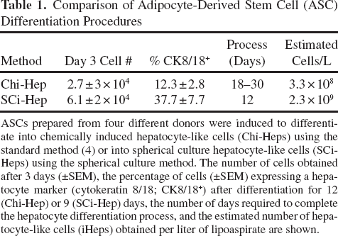
ASCs prepared from four different donors were induced to differentiate into chemically induced hepatocyte-like cells (Chi-Heps) using the standard method (4) or into spherical culture hepatocyte-like cells (SCi-Heps) using the spherical culture method. The number of cells obtained after 3 days (±SEM), the percentage of cells (±SEM) expressing a hepatocyte marker (cytokeratin 8/18; CK8/18+) after differentiation for 12 (Chi-Hep) or 9 (SCi-Hep) days, the number of days required to complete the hepatocyte differentiation process, and the estimated number of hepatocyte-like cells (iHeps) obtained per liter of lipoaspirate are shown.
We also compared the properties of Chi- and SCi-Heps with iPS-Heps, which are ASCs (obtained from the same donor) that were reprogramed into iPS cells after transfection of four genes [octamer-binding transcription factor 4 (OCT4), SOX2, Kruppel-like factor 4 (KLF4), and c-MYC] (36) and then induced to differentiate into hepatocytes using the Ochiya protocol (4). The iPS-Heps expressed a protein (CK8/18) found on hepatocytes (Fig. 1C), could endocytose LDL, synthesize glycogen (Fig. S3; available at http://med.stanford.edu/peltzlab/SuppInfo/liver-regen/), secrete albumin, produce urea, and had CYP450 activity (Fig. S4; available at http://med.stanford.edu/peltzlab/SuppInfo/liver-regen/). Of importance, SCi-Heps produced albumin and urea after only 3 days of in vitro differentiation, which was 3–6 days before Chi-Heps (Fig. 1E). The SCi-Heps produced maximal amounts of albumin and urea by days 6 and 9, respectively (Fig. 1E), which was 14 or more days before iPS-Heps (Fig. S4; available at http://med.stanford.edu/peltzlab/SuppInfo/liver-regen/) produced comparable amounts of these analytes. ASCs must first be reprogramed into and then exit from the pluripotent state before they can differentiate into iPS-Heps. In contrast, SCi-Heps are produced by direct differentiation of ASCs into endoderm, which explains why they can more quickly produce these analytes. Consistent with the more rapid differentiation process, SCi-Heps expressed mRNAs for endodermal (epithelial cell adhesion molecule, EpCAM) and hepatocyte-specific [albumin, forkhead box a2 (Foxa2)] genes within 3 days after initiation of hepatic differentiation (Fig. 2B). Also, SCi-Heps had the highest level of albumin production (on a per cell basis) among the three types of ASC-derived cells tested. Their increased level of albumin production is consistent with the fluorescenceactivated cell sorting (FACS) results indicating that 37% of SCi-Heps expressed a mature hepatocyte marker, while only ~12% and 20% of Chi-Heps and iPS-Heps, respectively, expressed this marker (Fig. 1C). Thus, the SCi-Heps method produces an increased number of hepatocyte-like cells from a lipo aspirate, and the cells are prepared within a time frame that makes it possible that they could be used in an acute clinical situation, such as would occur after an overdose of acetaminophen.
To further characterize these cells, gene expression profiling was performed in ASCs, Chi-Heps, SCi-Heps, iPS-Heps, and hepatocytes using microarrays, and the data analysis is described in the supplemental information. In brief, multiple comparisons indicated that in vitro differentiation significantly altered the gene expression pattern in ASCs, and that Chi- and SCi-Heps expressed a very large number of hepatocyte-specific genes (Table S2; available at http://med.stanford.edu/peltzlab/SuppInfo/liver-regen/). Moreover, two analyses [a space diagram of the gene expression differences (Fig. 2C) and principal component analysis (Fig. S5; available at http://med.stanford.edu/peltzlab/SuppInfo/liver-regen/)] that are described in the supplement indicate that the Chi- and SCi-Heps had a gene expression profile that was closer to that of hepatocytes than the iPS-Hep profile. As described in the supplemental information, iPS-Heps expressed a larger number of genes that were not expressed in adipocytes or hepatocytes (Table S3; available at http://med.stanford.edu/peltzlab/SuppInfo/liver-regen/). In summary, SCi-Heps have a gene expression pattern that resembles, but it does not fully mirror, that of hepatocytes. Since only 37% of the Sci-Hep cells expressed a mature hepatocyte marker (Fig. 1C), it is possible that the gene expression pattern in fully differentiated SCi-Heps could more closely resemble that of hepatocytes than is suggested by this analysis, since some of the gene expression changes could be masked (diluted) by the preponderance of less differentiated cells in the population. Although SCi-Heps were produced by a different method and were cultured for a much shorter time period than were Chi-Heps, their gene expression profiles were extremely similar: 48,165 of 49,395 probes did not show a significant expression difference (adjusted values of p > 0.01 or fold change <2) between these two types of iHeps. The level of expression of 306 genes (0.6%) had a greater than threefold (adjusted p < 0.01) and only 44 genes (0.08%) had a >10-fold (adjusted p < 0.01) difference in expression in these two cell types (Table S4; available at http://med.stanford.edu/peltzlab/SuppInfo/liver-regen/). Although a few liver-specific genes [e.g., cytochrome P450, family 3, subfamily A, polypeptide 4 (CYP3A4)] were differentially expressed, the vast majority of genes had a similar level of expression in Chi-Heps and SCi-Heps, which indicates that both types of cells have a similar liver-specific gene expression profile.
Human Liver Regeneration
To determine whether SCi-Heps could reconstitute human liver in vivo, 5 × 10⁶ SCi-Heps were transplanted by ultrasound-guided injection directly into the liver of four ganciclovir-conditioned TK-NOG mice (Fig. 3A). We previously demonstrated that the amount of human albumin in the sera of chimeric mice is an indicator of the extent of liver humanization (18), so the serum human albumin level was serially assessed over an 8-week period after SCi-Heps transplantation. All four of these mice produced substantial and increasing amounts of human serum albumin when monitored over an 8-week period after transplantation. They had an average human albumin concentration of 0.29 (±0.09) mg/ml in their sera at 4 weeks, which increased to 0.82 (±0.41) at 8 weeks after SCi-Heps transplantation (Fig. 3B). In contrast, none of the three mice that were transplanted with the same number of undifferentiated ASCs produced detectable human serum albumin.

Transplantation of iHeps into TK-NOG mice. (A) An ultrasound-generated image (in transverse view) of SCi-Heps being injected through a 30-gauge needle directly into the right lobe of the liver of a herpes simplex virus type 1 thymidine kinase (TK) transgene expressing nonobese diabetic/severe combined immunodeficient/interleukin-2 receptor γ chain knockout (NOD/Shi-scid/IL-2Rγnull; NOG) (TK-NOG) mouse. (B) Chimeric mice produce human albumin after SCi-Hep transplantation. The amount of human albumin in plasma was serially measured over an 8-week period after transplantation of SCi-Heps into four ganciclovir-conditioned mice. Each dashed line shows the amount of human albumin measured in a chimeric mouse at the indicated time. In all four chimeric mice, human serum albumin was detectable 4 weeks after transplantation, and the amount increased with time after transplantation. In contrast, albumin was not produced by any of the four mice that were recipients of control, undifferentiated ASCs (red triangles and solid line). (C) Immunofluorescent staining of liver sections (at 100× magnification) obtained from TK-NOG mice 4 weeks after implantation of SCi-Heps (a–c) or undifferentiated ASCs (d–f). The liver sections were stained with human-specific anti-albumin (a, d), anti-CK8/18 (b, e), or anti-asialoglycoprotein receptor 1 (ASGR1) antibodies and counterstained with 4,6-diamidino-2-phenylindole (DAPI) to show the cell nuclei. (D) Immunofluorescent staining of liver sections (200× magnification) obtained from TK-NOG mice 4 weeks after transplantation with SCi-Heps or undifferentiated ASCs. The liver sections were stained with anti-CK8/18, anti-Ki-67, or anti-zonula occludens-1 (ZO-1) antibodies and were then counterstained with DAPI to show the cell nuclei.
Liver histology revealed that the transplanted SCi-Heps integrated into the liver, produced human albumin, and expressed markers found on mature human hepatocytes (ASGR1, CK8/18) (Fig. 3C). Since SCi-Heps did not express ASGR1 in vitro just prior to transplantation (Fig. S6; available at http://med.stanford.edu/peltzlab/SuppInfo/liver-regen/), ASGR1 expression indicates that the SCi-Heps continue to differentiate after liver engraftment. Since it was important to determine if the engrafted SCi-Heps would proliferate in the liver, we investigated whether the SCi-Heps expressed the Ki-67 nuclear protein, which is a specific marker for cellular proliferation (32). A significant percentage of the engrafted SCi-Heps were Ki-67 positive (Fig. 3D), which indicates that they proliferate in situ in the liver. In addition, a significant percentage of the engrafted SCi-Heps cells also expressed tight junction protein ZO-1 (Fig. 3D). This indicates that they have established tight junction interactions with one another, which is required for human bile duct formation (21). In contrast, livers obtained from mice that were transplanted with undifferentiated ASCs did not have any Ki-67- or ZO-1-positive cells (Fig. 3D).
SCi-Heps Do Not Form Tumors
Malignant potential is a critical determinant of whether SCi-Heps could be used for liver regeneration in human subjects, so we examined whether SCi-Heps would form tumors after implantation under the kidney capsule of immunocompromised NOG mice. No tumors were formed over a 2-month observation period after 5 × 10⁴ Chi- or SCi-Heps were implanted into each of five NOG mice. Moreover, analysis of tissue sections indicated that only normal tissue was present in the area of implantation. In contrast, implantation of the same number of iPS-Heps into each of four NOG mice resulted in the formation of multiple tumors within 3 weeks, which could be palpated through the body wall (Fig. 4A). Analysis of the tumor tissue obtained 2 months after iPS-Heps transplantation revealed that the tumors contained tissue derived from all three germ cell layers from all four mice (Fig. 4B).

Tumorgenicity of transplanted iHeps in immunocompromised mice. (A) Sci-Heps do not form tumors in immunocom-promised mice. As early as 3 weeks after 5 × 10⁴ iPS-Heps were implanted under the kidney capsule of NOG mice, palpable tumors were formed in the area of implantation. In contrast, no tumors were detected 2 months after the same number of Chi- or SCi-Heps were implanted (top row). The tumors were visible in tissue sections obtained from the area of iPS-Heps implantation, while only normal tissue was present in the area of Chi-Hep implantation (middle row). The magnification of images in the top and middle rows is 10×. The images in the bottom rows were obtained from the boxed regions of the corresponding image and are shown at 200× magnification. (B) Tumor formation after iPS-Heps implantation. Top row: Images of the tumors formed 3 weeks after iPS-Hep cells were implanted under the kidney capsule. The 300× and 100× images were obtained from the indicated region (dashed box) of the corresponding 40× and 100× images. Bottom row: Two months after the iPS-Hep cells were implanted, the tumors contained cells from all three germ cell layers. These images are at 200× magnification. Scale bars: 50 μm.
Discussion
These results demonstrate that functioning human liver tissue could be generated in vivo after direct implantation of ASC-derived cells into the liver. The SCi-Heps were produced from ASCs by in vitro differentiation for a short period of time in chemically defined media. We certainly can achieve higher levels of human liver reconstitution by intrasplenic injection of mature hepatocytes (18). However, given the limitations of the transplantation method used here, the level of liver reconstitution achieved here is quite remarkable. It corresponds to replacement of 10–20% of the mouse's liver with human liver tissue, which has stable biosynthetic function. The SCi-Heps were transplanted by ultrasound-guided direct injection into the liver because we wanted to use implantation methods that could be used on humans. Moreover, because of the small size of a mouse liver, the cells were injected into a relatively small number of sites. To put this level of human liver reconstitution into perspective, human albumin was not detected in the sera of mice reconstituted with Chi-Heps in three prior studies (4,5,33), and only a minimal (160 ng/ml) amount of human serum albumin was detected in one mouse at 10 weeks in the only other study (9), an amount that is 5,125-fold below that obtained here. Also, the TK-NOG system was set up to model the transplantation of autologous cells in humans, which will not trigger an immune response. The highly immunodeficient mouse strain (NOG) used in our model system has mutations that inactivate the innate (Il2rg-/-) and adaptive (SCID) immune responses, which enables this mouse to accept xenografts of human liver cells. Because of the severe effect that these mutations have on the immune system, the mice do not mount an immune response to the transplanted human cells.
The ability of these readily accessible stem cells— which were obtained and transplanted via procedures that are commonly used in clinical medicine—to efficiently regenerate human liver in vivo raises an important question for regenerative medicine. Could these procedures be scaled to enable autologous liver regeneration in humans? Since there are ~140 × 106 hepatocytes per gram of human or mouse liver tissue (34), a 1,500-g adult human liver has ~2 × 10¹¹ hepatocytes, while a 1.8-g mouse liver has 2.5 × 10⁸ cells. We note that after 1 million human hepatocytes are transplanted into a TK-NOG mouse, we routinely obtain ~50% replacement of the mouse hepatocytes (18), which amounts to ~1.2 × 10⁸ functioning human hepatocytes in the mouse liver. This represents a 120-fold increase (or seven population doublings) in the number of human hepatocytes generated in vivo relative to the number of transplanted cells. If a similar regeneration efficiency could be achieved in humans, 800 million SCi-Heps (1011/120) would have to be transplanted to replace 50% of the cells in a human liver. Of relevance to this question, 1 L (1,000 g) of lipoaspirate can easily be obtained from a single liposuction procedure. This volume is estimated to contain 2–10 × 109 cells (8,39,40), of which 1–10% of these cells are estimated to be true ASCs (26,29). Thus, 1 L of lipoaspirate contains between 2 × 10⁷ and 109 ASCs, which could provide a sufficient number of cells to enable autologous human liver replacement. Here we demonstrate that spherical culture coupled with a chemical differentiation process can increase the yield of SCi-Heps by sevenfold. The spherical culture method helps to ensure that a sufficient number of SCi-Heps can be obtained to enable liver regeneration in a human subject. Moreover, elimination of intraspecies differences makes it likely that the efficiency of liver regeneration may be greater when autologous SCi-Heps are transplanted into human subjects. Mouse hepatocytes underwent twice the number of population doublings (~12–14) after transplantation into autologous murine liver (31) than was noted in this xenogeneic transplant model. The spherical culture method also reduces the culture volume by 12-fold and decreases the time in culture by 40%; these factors dramatically reduce the cost of differentiating ASCs into iHeps. The estimated cost of the medium and growth factors required for producing Chi-Heps is ~$49,000 per liter of lipoaspirate processed, but spherical culture method reduces these costs by over 20-fold (to ~$2,400).
The methods for assessing the potential safety of stem cells used for regenerative therapies have not been fully established (7). Although ASCs are reported to be genetically stable in culture (25), it was essential to assess their malignant potential in vivo to determine whether they could be used for liver regeneration in human subjects. SCi-Heps did not produce tumors after implantation in NOG mice, which are highly immunocompromised because they have the severe combined immunodeficiency (SCID) mutation and the interleukin-2 receptor γ chain gene knockout (20). NOG mice have been shown to readily support the growth of multiple types of human tumors (9–11,16,27,35). In contrast to the results obtained with SCi-Heps, readily palpable tumors were noted within 3 weeks after implantation of iPS-Heps. The rapid tumor onset should not be surprising, since this iPS-derived cell population has a large percentage of undifferentiated cells. However, ~109 cells must be transplanted to regenerate a human liver, but even as few as 100–1,000 cells with malignant potential in this population could cause tumor formation. These numbers create a very significant hurdle for any selective method used to prepare iPS-derived cells for liver regeneration, since it must have an efficiency that enables it to remove one tumor-causing cell in 107 cells. The relatively short period of this assessment, relative to the possibility that a regenerated liver may persist for decades in humans, prevents drawing any firm conclusions about SCi-Heps safety at present. However, the fact that implanted SCi-Heps did not form tumors indicates that these cells have a markedly reduced risk of causing tumors to form. Since ganciclovir-induced liver damage in genetically engineered TK-NOG mice resembles the acute liver failure in humans that is caused by exogenous toxicants or drug overdose, the liver regenerative procedures developed here could someday be used to treat acute liver failure.
Supplemental methods, figures, and tables for this article are available at http://med.stanford.edu/peltzlab/SuppInfo/liver-regen/
Footnotes
Acknowledgments
This work is dedicated to Dr. Tatsuji Nomura, whose vision and leadership enabled this project to proceed. We thank Dr. Robert Lewis for helpful discussions and critical review of this manuscript. D.X., T.N., M.Z., M.W., N.S., H.S., and G.P. were partially supported by funding from a transformative RO1 award (1R01DK0909921) from the NIDDK. The authors declare no conflicts of interest.