
Abstract
Lamellar (component cell) corneal transplantation is replacing penetrating keratoplasty for some corneal disorders in humans, but the relative risks of immunological graft rejection for the two procedures remain uncertain. A model of component endothelial cell keratoplasty (endokeratoplasty) was developed in outbred sheep. Clinical and histological graft outcomes after endokeratoplasty were then compared with contemporaneous penetrating corneal allografts. No topical or systemic immunosuppression was administered to any recipient sheep. Endothelial cell allografts (n = 10) took significantly longer to achieve perfect transparency following surgery than did penetrating corneal grafts (n = 7) (day 10 vs. day 4; p = 0.003; two-tailed Mann–Whitney U test). The median day to rejection of penetrating grafts was postoperative day 18; for endothelial cell grafts, it was day 48 (p = 0.04; two-tailed Mann–Whitney U test). The clinical courses of the two procedures were therefore quite different. Penetrating grafts gained clarity quickly but exhibited rapid graft neovascularization. Clinical rejection was preceded by inflammation in the anterior segment. Endothelial cell grafts exhibited a fluctuating, more indolent course of opacification, although all did eventually fail. Histological analysis confirmed immunological rejection in all failed grafts, but with different patterns of leukocytic infiltration in endokeratoplasties compared with penetrating keratoplasties. Inflammatory cells in endothelial cell grafts were generally fewer in number and were more often found in the posterior stroma. We conclude that, in the absence of immunosuppression, all endothelial cell allografts do undergo immunological rejection, albeit at a slower rate than penetrating grafts.
Keywords
Introduction
Corneal transplantation is performed to improve vision, to reduce pain, and for emergency structural repair in cases of corneal perforation (25). In penetrating corneal transplantation, the full thickness of the cornea is replaced (31). New techniques for lamellar (partial thickness) grafts have evolved over the recent years and are now being applied to cases that used to be treated by penetrating keratoplasty (1,25). Component endothelial cell transplantation, or endokeratoplasty, is a procedure in which the anterior portion of the host cornea, including the ocular surface, is retained, but the posterior section, including Descemet's membrane and the corneal endothelium, is replaced with human donor tissue (20,24). A healthy corneal endothelium, the postmitotic monolayer of cells on the posterior surface of the cornea, is essential for the maintenance of corneal transparency (25). The rationale for endokeratoplasty is that, in conditions in which only the corneal endothelium is dysfunctional or absent, only this component of the cornea needs to be replaced (20). Endokeratoplasty is widely used for bullous keratopathy (corneal edema caused by corneal endothelial cell loss, resulting in pain and poor vision) and for Fuchs' endothelial dystrophy (an adult-onset corneal dystrophy caused by corneal endothelial cell dysfunction). These two indications together represent one third of all indications for corneal transplantation (26).
The benefits claimed for endokeratoplasty in its various forms are reduced postoperative suture complications, inflammation and astigmatism, increased wound strength, and faster visual rehabilitation compared with penetrating keratoplasty (20,24). An important unresolved issue is the extent to which endothelial allografts undergo rejection and therefore the extent to which postoperative topical immunosuppression needs to be administered. Many of the essential elements that initiate an allograft response—for example, blood vessels, lymphatics, and antigen-presenting cells—are located within the corneal stroma (8,25). In penetrating keratoplasty, all of these elements are of donor origin, so that the foreign antigenic load is relatively high. Indeed, the major cause of the failure of penetrating corneal grafts is irreversible rejection (6,26). The anterior chamber, in contrast, is an immune-privileged site in which allogeneic skin grafts are not necessarily rejected (10,16,17). Because endothelial grafts are placed into the anterior chamber, the pattern of rejection may conceivably differ from that seen with penetrating keratoplasty.
The outbred sheep is a suitable outbred preclinical model in which to test the relative rejection rates of penetrating and component endothelial cell corneal grafts. The ovine endothelium is nonreplicative, as in humans, and the anatomy of the eye is akin to that of the human, with a deep anterior chamber. In the absence of topical, local, or systemic immunosuppression, penetrating ovine corneal allografts undergo rejection several weeks after transplantation in a manner that is similar clinically and histologically to corneal allograft rejection in humans (28). We developed a method of endokeratoplasty in the sheep that is similar to the procedure known as Descemet's stripping endothelial keratoplasty (DSEK) used in humans (18). We then performed contemporaneous ovine penetrating and endothelial cell allografts to examine the macroscopic and histological outcomes and, in particular, the relative rates of rejection in the absence of any confounding immunosuppression.
Materials and Methods
Animal Ethics
All experimentation was performed with approval from the Institutional Animal Welfare Committee and conformed to the Association for Research in Vision and Ophthalmology (ARVO) Statement for the Use of Animals in Ophthalmic and Visual Research. Outbred adult crossbred sheep sourced from a local farm were housed indoors, allowed unlimited access to lucerne chaff and water, and were examined daily with a handheld slit lamp (SO-801 Scan Optics, Thebarton, SA, Australia) for a week prior to transplantation to accustom them to being handled. Sheep are herd animals, and so they were always held in groups.
Donor Corneas
Ovine eyes were obtained from a local abattoir within 3 h of death of the donor. Donor sheep were of mixed age and gender and were outbred. One donor eye was used for each corneal graft (n = 17 in total). Eyes were transported on ice to the laboratory, decontaminated in 10% w/v povidone–iodine (Sanofi-Aventis, Virginia, QLD, Australia), and washed twice by immersion in sterile normal saline (0.9% sodium chloride; Baxter Healthcare Pty Ltd., Toongabbie, NSW, Australia).
Penetrating Keratoplasty in Sheep
Recipient sheep were 1- to 2-year-old wether (castrated male) crossbreeds of mixed Merino and Dorset ancestry. Penetrating corneal transplantation was performed essentially as previously described (27). Recipient sheep (n = 7) were fasted overnight. Only one eye per animal was operated on. One hour prior to surgery, the pupil of the eye to be grafted was dilated with topical 1% atropine sulfate (Chauvin Pharmaceuticals, Kingston upon Thames, Surrey, England) and viscous 2.5% phenylephrine hydrochloride (Chauvin Pharmaceuticals). Anesthesia was induced with 25 mg/kg sodium thiopental (Abbott Laboratories, North Chicago, IL, USA) delivered intravenously into the external carotid vein and maintained after intubation of the airway with 1.5% isoflurane (Veterinary Companies Australia, Kings Park, NSW, Australia) in 2:1 air/oxygen. Local anesthesia with topical 5 mg/ml proxymetacaine hydrochloride (Allergan Australia Pty Ltd., Gordon, NSW, Australia) was used as required. A 12-mm-diameter donor central button was transferred to an 11-mm graft bed. Trephines were sourced from Tecfen Corp. (Santa Barbara, CA, USA). The graft was secured by four interrupted 9–0 monofilament nylon cardinal sutures (Alcon Australia, Frenchs Forest, NSW, Australia) and one continuous 9–0 nylon suture. Prophylactic topical 0.5% chloramphenicol (Pfizer Australia Pty. Ltd., West Ryde, NSW, Australia) was administered to the graft once daily for 3 days postoperatively. Postoperatively, grafts were examined each day with the handheld slit lamp and scored for clarity, edema, indices of inflammation, and the degree of neovascularization of the graft using a well-validated proforma. The onset of corneal graft rejection was defined as inflammation, spreading edema, and loss of clarity in a previously thin, clear graft, or the appearance of a corneal epithelial or endothelial rejection line. The day of rejection was defined as the first day that graft opacity reached 2 on a 4-point scale (0 representing transparency, 4 representing a completely opaque graft), such that the iris margins were no longer clearly visible through the graft, or the first day that an endothelial or epithelial rejection line was visible in a previously transparent and quiet graft.
Corneal Endothelial Cell Grafts in Sheep
Recipient sheep were 1- to 2-year-old wethers of mixed Merino and Dorset ancestry. Preoperative treatment and anesthesia of recipient sheep (n = 10) was exactly as for penetrating keratoplasty. The technique used was akin to DSEK in humans (18,20,24). A corneoscleral disc was excised from a donor eye and mounted on an artificial anterior chamber (Barron Precision Instruments, Grand Blanc, MI, USA). A thin lamellar graft of posterior stroma and endothelium was fashioned manually from the corneoscleral disc using a Morlet lamellar knife (Duckworth & Kent Ltd., Baldock, Hertfordshire, England), and a 12-mm-diameter donor graft was cut with a handheld trephine. A 12-mm central disc of Descemet's membrane and endothelium was removed from the recipient sheep by scoring Descemet's membrane with a reverse Sinskey hook (Bausch & Lomb, Macquarie Park, NSW, Australia) and stripping it together with the corneal endothelium. The donor disc endothelium was protected by a small amount of viscoelastic (Abbott Medical Optics, Pymble, NSW, Australia) and pulled with a 10–0 prolene suture (Alcon Australia) into the recipient anterior chamber through a 6-mm limbal wound on a Sheets glide (Beaver-Visitec International, Waltham, MA, USA). The disc was centered and compressed against the defect in the recipient Descemet's membrane for 10 min with a complete air fill of the anterior chamber. Once the graft had adhered, the air was exchanged for ophthalmic balanced salt solution (BSS; Alcon Australia), leaving an 8-mm bubble of air to tamponade the graft. The wound was closed with three interrupted 9–0 nylon sutures (Alcon Australia). Postoperatively, grafted eyes were observed daily with the handheld slit lamp and scored for clarity, edema, indices of inflammation, and the degree of corneal neovascularization, as before.
Endpoint Histology
Recipients with failed (edematous and opaque) corneal grafts were killed by overdose of intravenous sodium pentobarbitone and the eyes removed for histological analysis. Corneal tissues were fixed in buffered formalin, embedded in paraffin wax, cut at 5 μm, and stained with Harris' hematoxylin and eosin Y (Chroma-Gesellschaft Schmid GmbH & Co., Münster, North Rhine-Westphalia, Germany).
Statistical Analyses
Data were analyzed by the two-tailed Mann–Whitney U test and corrected for ties, with p < 0.05 being considered significant.
Results
Clinical Outcomes of Ovine Penetrating Corneal Grafts and Corneal Endothelial Cell Grafts
Penetrating corneal allografts (n = 7) achieved perfect transparency after surgery more quickly than did endothelial cell allografts, at a median of 4 days (range 1–6 days) for penetrating grafts and 10 days (range 4–31 days for endothelial grafts) (p = 0.0034) (Table 1). Six of seven penetrating grafts underwent rejection at a median of 18 days (range 16–29 days) after surgery (Table 1). Graft rejection was accompanied by inflammation and neovascularization of the graft, and graft opacity swiftly reached a score of 3–4. The one graft that was followed for more than 60 days postgraft developed a hyphema (blood in the anterior chamber) at day 16 and was somewhat cloudy from day 25 to day 59, indicative of a rejection episode, although it never developed an opacity score of more than 2 and exhibited a fluctuating course.
Clinical Outcomes of Ovine Penetrating Corneal Allografts and Endokeratoplasties
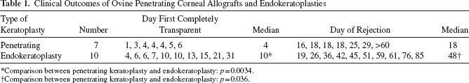
Comparison between penetrating keratoplasty and endokeratoplasty: p = 0.0034.
Comparison between penetrating keratoplasty and endokeratoplasty: p = 0.036.
All ovine endokeratoplasties underwent rejection, as assessed by complete corneal opacification but at a significantly slower pace than did the penetrating corneal grafts, with a median time to rejection of 48 days (range 19–60, p = 0.036) (Table 1).
The rejection process in the penetrating and lamellar graft procedures was different. Eyes with an endothelial cell graft in situ did not become significantly inflamed. The process leading to loss of corneal clarity was often indolent, with fluctuating edema. Keratic precipitates (accumulations of leukocytes) on the graft endothelium and small rejection lines became visible at the slit lamp and subsequently disappeared, only to reappear several days later. However, once corneal clarity reached a score of more than 2, then complete failure over the following few days was inevitable (Fig. 1A, B).

Outcomes of keratoplasty in the sheep. (A, B) Clinical photographs. (A) Transparent endokeratoplasty at day 41 postgraft. (B) Same eye as in (A) at day 48 postgraft: the cornea is opacifying and the graft failing. (C, D) Representative histology (H&E sections) of rejected penetrating keratoplasty and endokeratoplasty (Scale bars: 100 mm). (C) Endothelial rejection in an eye with a penetrating graft. (D) Endothelial rejection in an eye with a corneal endothelial cell graft. (E) Semiorganized lymphoid follicle at limbus in eye with a rejected penetrating corneal graft.
Histological Assessment of Grafted Eyes
Following euthanasia of recipient sheep, fixed and stained corneal sections were examined by a pathologist (SK). Of the seven penetrating grafts, six showed clear evidence of rejection, as evidenced by some or all of the following features: central stromal neovascularization, infiltration of the limbus, stroma, and sometimes epithelium with mononuclear cells, mononuclear cells in the anterior chamber, stromal edema, and missing endothelium (Fig. 1C). The exception was the graft that appeared to undergo a self-limiting rejection episode but that never completely clouded. This graft showed some evidence of corneal neovascularization and a sparse leukocytic infiltrate, but the corneal endothelial monolayer appeared intact.
Of the 10 endokeratoplasties, three showed evidence of an indolent rejection process, marked by deep neovascularization of the stroma, and a slight to moderate infiltrate of mononuclear cells. The endothelial monolayer was mostly intact. In a further five grafts, the evidence of rejection was more pronounced (Fig. 1D), as for the penetrating grafts, with mononuclear cells in the anterior chamber and attached to the corneal endothelium. A leukocytic infiltrate was apparent both in the residual deep posterior stroma of the recipient, as well as in the graft, suggesting infiltration from the limbus. Several of the rejected penetrating grafts exhibited semiorganized accumulations of lymphoid cells that resembled germinal centers at the limbus (Fig. 1E) of the recipient cornea, but these aggregates were not observed in the endokeratoplasties.
Discussion
The early outcomes of penetrating corneal allotransplantation are typically excellent, and graft survival figures of 90% at 1 year after surgery are not uncommon. However, inspection of corneal transplant registry data shows that Kaplan–Meier survival of penetrating corneal allografts at 15 years is 55%, and irreversible rejection is the most important cause of penetrating corneal graft failure (6,8,27). One of the potential benefits of the alternative surgical procedure of endokeratoplasty is that rejection may be less of a concern than for penetrating keratoplasty (2,4). However, we found that, in the absence of any topical or other immunosuppression, all of the ovine endothelial cell grafts in our series underwent rejection, albeit at a slower rate than did contemporaneous penetrating grafts.
Clinical experience with endothelial keratoplasty, as with penetrating keratoplasty, is almost always performed against a background of topical immunosuppression with glucocorticosteroids (6). Under these circumstances, the mean likelihood of a rejection episode (reversible or irreversible) in an endothelial graft is approximately 9% (range >1–36%), depending on comorbidities such as glaucoma, the type of endokeratoplasty, and the length of follow-up (2–4,7,9,12,13,15,21,23,29). Price et al. estimated that the probability of a rejection episode in a DSEK was 7.6% by 1 year and 12% by 2 years postoperatively (19). A review from the American Academy of Ophthalmology in 2009 reported an endothelial graft rejection of 10% (range 0–45%) for DSEKs (14). The corresponding figure for penetrating grafts is approximately 18% for all indications over 20 years (26), but no human endokeratoplasties have as yet been followed for this length of time, and comparative studies in humans have generally been retrospective rather than contemporaneous. Further, in some centers, patients with endothelial grafts are prescribed topical steroids for longer periods than are patients with penetrating grafts (2). It is thus difficult to compare the rejection rates in the two types of procedures.
Animal studies are few, but a recent report compared outcomes of penetrating grafts and endothelial grafts in rabbits over a time frame of 1 month following surgery (11). There were no rejection reactions in the endothelial grafts over this time, whereas half of the penetrating grafts suffered a rejection episode, and the authors of the study concluded that the former exhibited a lower rate of rejection than the latter. The work we report here in the sheep suggests that, in the absence of topical immunosuppression, the rate of rejection may differ between the two procedures, but the final incidence of rejection, of severity sufficient to cause graft failure, is the same. The lesser antigen load associated with endokeratoplasty does not prevent sensitization from occurring, and once sensitization has occurred, the graft will fail.
The clinicopathologic correlates and signs of immunological rejection in ovine endokeratoplasties included corneal neovascularization, diffuse stromal edema, keratic precipitates on the endothelium, and in some cases Khodadoust lines, similar to the pattern seen in rejecting human endokeratoplasties (12). At a histopathological level, failed human component cell grafts have been reported to exhibit evidence of stromal inflammation (22) and corneal endothelial cell loss (30), but specimens are rarely available during or immediately following an irreversible rejection episode. Here we showed that decompensated ovine corneas collected shortly after onset of rejection displayed deep corneal neovascularization together with a variable degree of leukocytic infiltrate in the posterior stroma and the anterior chamber. As expected from previous work, rejected penetrating grafts showed a more substantial infiltrate throughout the cornea, including in the epithelium, and neovascularization was more evenly distributed throughout the stroma (28). Of note, in several instances, we observed organizing lymphoid aggregates with the appearance of a germinal center at the limbus, reminiscent of conjunctiva-associated lymphoid tissue. Similar findings have previously been reported by others in the rat (5), but are unlikely to be seen in humans because peripheral tissue is not available for histology, even after graft failure.
In the absence of immunosuppressive cover, both penetrating and endothelial ovine allografts undergo rejection, albeit with different kinetics. However, once the rejection process has reached a particular stage or tipping point, roughly correlating with corneal opacity of a degree that renders the iris margins indistinct through the graft, then the effector arm of the immune response swiftly leads to decompensation, irrespective of the type of graft. We speculate that the different rates of the initial rejection responses may result from delayed sensitization, in the case of the component cell grafts, perhaps reflecting slower neovascularization of the graft, or less alloantigen finding its way in cell-bound or cell-free form to the secondary lymphoid tissue or to conjunctiva-associated lymphoid tissue.
What are the implications for human corneal grafts? Sensitization to corneal endothelial cell-derived foreign alloantigen is very likely to occur eventually, especially if the recipient is at moderate to high risk of rejection by virtue of comorbidity such as corneal neovascularization. Topical immunosuppression may need to be continued in the longer term.
Footnotes
Acknowledgments
The authors thank Ms. Madison Helm and Animal House staff for expert animal husbandry. This work was funded by the Ophthalmic Research Institute of Australia. KAW is supported by the Australian National Health and Medical Research Council (NHMRC). The authors declare no conflict of interest.