
Abstract
Disorders of the central nervous system (CNS) as a result of trauma or ischemic or neurodegenerative processes still pose a challenge for modern medicine. Due to the complexity of the CNS, and in spite of the advances in the knowledge of its anatomy, pharmacology, and molecular and cellular biology, treatments for these diseases are still limited. The development of cell lines as a source for transplantation into the damaged CNS (cell therapy), and more recently their genetic modification to favor the expression and delivery of molecules with therapeutic potential (ex vivo gene therapy), are some of the techniques used in search of novel restorative strategies. This article reviews the different approaches that have been used and perfected during the last decade to generate cell lines and their use in experimental models of neuronal damage, and evaluates the prospects of applying these methods to treat CNS disorders.
Introduction
Knowledge accumulated in the past several decades has led to a better understanding of the biochemical, molecular, and genetic mechanisms involved in pathologies of the central nervous system (CNS), such as epilepsy and Parkinson's, Alzheimer's, and Huntington's diseases. Nevertheless, therapeutic options remain limited, prompting a search for alternatives, including the use of cellular vehicles for cell therapy, and viral and nonviral vectors for gene therapy for the treatment of neurodegenerative diseases as detailed in the following reviews (68,75,118,130).
Damage caused by CNS degeneration could be restored in vivo by cellular transplants directed to affected regions; cells for transplantation can be derived from stable cell lines, primary cultures, or fetal tissue, and more recently from stem cells [for reviews see (20,210)]. The first studies on intracerebral transplants as a clinical tool for the treatment of nervous system pathologies in humans utilized autologous tissue, for example, from the suprarenal gland (63,132), or human fetal or porcine tissue (62,132). For technical, scientific, and ethical reasons, alternative sources of tissue have been sought, including cell lines cultured in vitro with or without genetic modification (75).
The use of cell lines for intracerebral transplants has been considered to offer various advantages, among them: 1) the fact that cells are cultured under conditions that can be controlled and manipulated; 2) due to their proliferative capacity, cells can be produced on a large scale, sufficient for multiple transplants; 3) that cells maintain homogeneous characteristics over time; and 4) the use of cell lines does not raise ethical problems. In the case of neural stem cells, some of these issues remain to be solved, for example the production of a sufficient number of cells for transplantation (108), and depending on the legislation of the country, the use of stem cells derived from human embryos can still be a matter of ethical concern. Cell lines with specific characteristics can be generated by means of genetic engineering (e.g., by insertion of oncogenic genes to immortalize the cells) and promote their continuous proliferation, enabling the establishment of a cell line. Subsequently, these cells can be further modified for therapeutic purposes by insertion of the desired gene (10).
The concept of ex vivo genetic therapy implies that the cells themselves are used as vehicle for introducing molecules of interest into the brain or other organs. Thus, these cells are modified under in vitro conditions and genetic material for the expression of the protein(s) of interest is transferred into them; for example, neurotrophic factors like brain-derived neurotrophic factor (BDNF) (34,115,160,218), neurotrophin-3 (NT-3), neurotrophin-4/5 (NT-4/5) (77), glial cell-derived neurotrophic factor (GDNF) (60,147), and nerve growth factor (NGF) (6,125,126), enzymes for neurotransmitter synthesis such as tyrosine hydroxylase (TH) (46,96,117,168) and glutamine decarboxylase (GAD) (16,45,57,139,162), and metabolic enzymes (124). The resulting genetically modified cells can function as tiny biological pumps, releasing neurotransmitters, neurotrophic factors, or other peptides that directly affect the underlying cause of the disease and can therefore promote functional recovery (16,60,92). On the other hand, in vivo gene therapy, which is not the subject of this review, consists of directly inserting a functional gene into the cells of an individual in order to correct a specific genetic defect or to endow the cells with a new function. Another alternative involves altering the expression of an endogenous gene in the cells of the individual, and this is achieved by transferring a short piece of genetic material (143).
To transfer genetic information into a cell, delivery systems called vectors, either viral or nonviral, are used. In the case of ex vivo gene therapy, in addition to selecting the best vector for the specific application, other issues need to be considered prior to using modified cells. Among them are the expression level of the gene coding for the protein of interest, and the survival of the cells after implantation, which depends, among other factors, on the implantation mechanism, adequate vascularization (159), and the host's immune response to the cells or to the gene products they express (13). The following sections describe the strategies that have been used to produce cell lines for ex vivo cell therapy in the nervous system.
Strategies for the Development of Cell Lines to be used as Transplants
Advances in molecular and cell biology have allowed scientists to develop and expand cell lines and to refine methods for their implantation; as a result the survival, integration, and functionality of intracerebral transplants have improved (28,32,51,67,117,125,183,187). There are two general strategies for obtaining cell lines that can be used for transplants: the first is to fuse somatic cells of primary cultures to tumor cell lines, and the second is to introduce transforming agents.
Cell Lines Immortalized by Somatic Fusion with Tumor Cells
To obtain cell lines for somatic fusion with tumor cells, polymers like polyethylenimine, poly-L-lysine are used; their cationic properties allow them to condense and form complexes with nucleic acids (19). Somatic fusion has been applied to generate immortalized cell lines of septal, hippocampal, mesencephalic, and striatal origin as well as cell lines from the spinal cord (30,42,50,80,103). The great neurochemical variety of the resulting cells makes them useful in animal models of neural degeneration. It must be emphasized that with this technique it is not necessary for the cells to be in an active mitotic state; thus, it is possible to immortalize various types of neuronal cells in any state of development. However, a major drawback is that the cells obtained by this technique can generate tumors when transplanted into the CNS (200).
There are a variety of cell lines derived from tumor cells, including the cell line N18TG2 derived from a murine neuroblastoma (50), the cell line PC12 obtained from a rat adrenal medulla tumor (76), hNT from a human neuroteratocarcinoma (164), the glial cell line C6 cloned from a rat glial tumor (18), the cell line HCN-1A derived from human cortical tissue (206), and the cell line IMR-32 obtained from a human neuroblastoma (191). Some of these cell lines have been transplanted to evaluate their effect on animal models of brain damage. SH-SY5Y cells derived from human neuroblastoma and transplanted into the lesioned striatum showed integration into the host tissue and improved the behavioral condition in rats (25,135); SMS-LHN and LA-N-6 derived from human neuroblastoma decreased rotational behavior in a Parkinson rat model (121). Somatic fusion is also used for reprogramming of somatic cells. An example is the study of Cowan and colleagues, which showed that human embryonic stem cells fused with human fibroblasts resulted in hybrid cells that showed the morphology, growth rate, and antigen expression of embryonic cells (49).
Cell Lines Generated by Insertion of a Transforming Agent
This strategy employs techniques of molecular biology to introduce a DNA fragment (complementary DNA or cDNA) that codes for a growth factor, neuropeptide, or some biosynthetic or metabolic enzyme (16,41,45,57,64,65,85,123,124,126,162). These cells are first immortalized, usually by incorporating an oncogenic gene like myc, c-mycERTAM, neu, p53, the large T antigen, of either polyoma or simian virus 40 (SV40) (36,89,124,177). One of the most studied genes is the A58 temperature sensitive allele of the SV40 large T antigen (tsA58). The expression of this allele gives the cells the ability to proliferate when they are kept at the permissive temperature of 33°C, and reduces this ability at the nonpermissive temperature of 39.5°C. Cells thus transfected can be kept in constant proliferation in cell culture at low temperatures; after being transplanted they may differentiate and develop into mature neurons in the adult brain.
The latter gene was used, for example, to construct the cell line M213–2O; these cells of striatal origin were immortalized with the tsA58 allele of the SV40 large T antigen (69,70). Other cell lines that were immortalized by the SV40 large T antigen have been derived from cells of the medullary raphe and hippocampus (154,208,211). Whittemore et al. (209) and Renfranz et al. (154) have shown that immortalized embryonic neural cells may develop a complex neuronal phenotype that is determined, in part, by the area of the brain into which they are transplanted, independent of the area from which they originate.
As a way to reduce the tumorogenic potential, Harvey et al. (81) developed the cell line RTC4 immortalized with a mutant of the T antigen; after transplantation into the rat cortex in a model of stroke, this cell line showed a reduction in the volume of infarct and, importantly, no tumor formation (81). To prevent dangerous effects derived from the transforming agent, techniques for reversible immortalization in vitro have been used. For example the retroviral recombinant vector SSR#69 that simultaneously codes SV40T, hygromycin resistance (HygroR), herpes simplex virus thymidine kinase (HSV-TK) flanked by a pair of lox P sequences; since these lox P sequences are recognized by the Cre/loxP system, they can be used to cut out the gene [for a review about reversible immortalization strategies, see Kobayashi (98)].
For immortalization, it is crucial to consider the characteristics of the brain area of interest as well as the choice of the embryonic stage at which the cells will be harvested (45). The next step is to select the gene and cell line most appropriate to be developed; their selection is generally based on the physiopathological characteristics of the disease being modeled (e.g., a transgene can be chosen that codes for an enzyme, for a trophic or growth factor, or for the synthesis of a specific neurotransmitter). The transgenes most frequently employed have been those with therapeutic potential for the neurodegenerative diseases like Parkinson's, Huntington's, and Alzheimer's disease, amyotrophic lateral sclerosis (ALS), and epilepsy [for a review see (20,130)].
There are reports indicating that, in infected cells, expression of the transgene diminishes with time (140,173). This would negatively impact on the stable, long-term expression and ability to maintain adequate levels of the desired gene product. The expression level of the heterologous protein is crucial and will depend on the transduction efficiency, the transcription efficiency, and on the stability of the protein itself and of its mRNA.
Another factor that needs to be considered is the amount of protein produced, which depends on the promoter system used to express the transgene. Promoters may be grouped in various categories. In this review promoters will be grouped into ubiquitous, tissue-specific, or composite promoters, as shown in Table 1. Among the ubiquitous cell promoters used to drive ectopic gene expression are chicken β-actin (CBA), cytomegalovirus (CMV), dihydrofolate reductase (DHFR), elongation factor-1α (EF1-α), murine stem cell virus (MSCV), phosphoglycerate kinase (PGK), Rous sarcoma virus (RSV), the simian virus 40 early promoter (SV40), and the ubiquitin C promoter. Another category of promoters is that of tissue-specific promoters, which only allow the regulation of the expression of the transfected gene in a particular cell type (167–169); for example, glial fibrillary acidic protein (GFAP), neuron-specific promoters such as neuron specific enolase (NSE), platelet-derived growth factor-β (PDGF-β), preproencephalin, synapsin I (SYN1) (Table 1). In addition, it is possible to have regulatory systems of expression made up by a combination of different promoters and enhancers (e.g., CAGG) (Table 1). The selection of the optimal promoter is crucial to ensure its long-term expression after cells are transplanted into the host. Systematic comparisons among different promoters and their ability to drive gene expression in different cell types have been made to aid in the selection of the promoter, depending on the desired target [e.g., (109,152)].
Promoters/Enhancer Elements

CBA, chicken β-actin; CMV, cytomegalovirus; DHFR, dihydrofolate reductase; EF-1α, elongation factor-1α; MSCV, murine stem cell virus; PGK, phosphoglycerate kinase; RSV, Rous sarcoma virus; SV40, simian virus, early expression region 40; GFAP, glial fibrillary acidic protein; NSE, neuron specific enolase; PDGF-β, platelet-derived growth factor-β; SYN1, synapsin I; CAGG, CMV early enhancer/chicken β-actin (CAG) promoter.
Approaches for Delivery of Transgenes for Gene Therapy
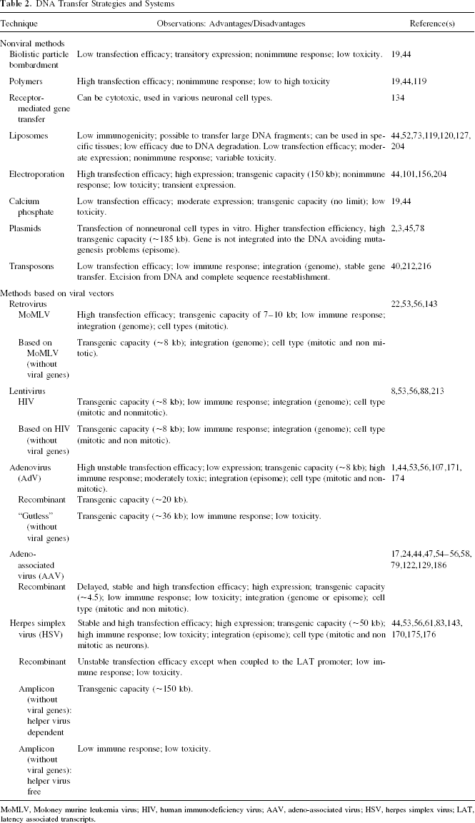
Choosing the appropriate technique to introduce the different types of transgenes into the cells is of fundamental importance; the methods most often used to introduce external genes fall into two groups: nonviral methods that include the biophysical or based on plasmids or transposons, and viral methods that use vectors of diverse types. The vectors used for biophysical transfections contain all the necessary elements to express the inserted transgene. The simplicity of these vectors is an advantage, and they are more reliable and less costly than the viral methods (19,144). On the other hand, these vectors are less efficient than viral vectors for in vivo gene delivery (87). Recent developments, however, are improving the efficiency of nonviral methods, and can now result in both transient and permanent transgene expression when integrating into the genome (73,87,143,144).
Nonviral Methods
Biophysical Methods: Bombardment, Polymers, Liposomes, Electroporation, and Calcium Phosphate
Nanoparticles such as gold particles coated with plasmid DNA can penetrate into the cell through biolistic particle bombardment, and also be used for in vivo gene delivery. Polymers are a biophysical method used mainly because they do not seem to induce an immune response; for target delivery improvement, cations as poly-L-lysine and polyethylenimine have been used with antibodies against specific receptors (119,134) (Table 2).
DNA Transfer Strategies and Systems
MoMLV, Moloney murine leukemia virus; HIV, human immunodeficiency virus; AAV, adeno-associated virus; HSV, herpes simplex virus; LAT, latency associated transcripts.
Other biophysical methods involve the use of liposomes. These consist of hollow spheres or vesicles that are formed by cationic lipids mixed with DNA in water. Lipids form electrostatic complexes with the DNA, and together they adopt an ordered multilamellar structure inside the vesicles. Liposomes are taken up by the cells through pinocytosis or phagocytosis; they are degraded and the DNA must exit into the cytosol, and be imported into the nucleus (73,131) (Table 2). An alternative that is being explored is the use of cationic polymers, called polyplexes for transfection of cells [for reviews see (87,144,189)].
Electroporation-based techniques increase membrane permeability temporarily by applying short electrical pulses of high voltage, forming nanometer-sized pores in the plasma membrane and allowing DNA to pass into the cell (19), and have been used to transfect mature neurons (101) or neural precursor cells (156) (Table 2).
Finally, the calcium phosphate precipitation method is one of the oldest methods used, which leads to the formation of a DNA–calcium phosphate complex that can enter cells by endocytosis, and has been used to transfect dissociated neuronal cultures from the CNS and peripheral nervous system (204) (Table 2).
Plasmids
One method for stabilizing DNA within the cell that enhances its clinical effect in vivo is the use of a plasmid. Plasmids are circular molecules of double helix that occur naturally in bacteria, and they may contain an origin of replication derived from a virus such as the Epstein-Barr or human papilloma virus and antibiotic resistance genes for transfected cells selection, since only those cells that were transfected will survive treatment with that particular antibiotic (185). Plasmid-based vectors have important advantages: they elicit little or no immune response, have few cytopathic effects, and possess high capacity for carrying exogenous DNA, which makes them useful as tools for transferring multiple genes or genomic constructs (185,195). While episomal plasmids do not integrate into the chromosome, some plasmids can integrate into the genome (73,143); these properties then can be exploited for the particular purposes of the experiment.
In contrast to the methods that employ viral vectors (Table 2), these plasmid-based methods, especially Epstein-Barr ori-P sequences, have stable replication and high gene expression (3). These systems must contain all the information necessary to deliver the plasmid DNA that contains a therapeutic gene, the regulatory elements to express the gene of interest, and usually antibiotic resistance genes to facilitate the selection of the cells (see above) (44,73). Since the viral EBNA-1 gene facilitates the extrachromosomal maintenance of these vectors within the nucleus, expression is independent of position effects (44).
The cell lines M213–2O Cl-4 and Cl-2 were transfected with a plasmid based on the Epstein-Barr virus (i.e., the vector pREP-10), which was used to express the cDNA for GAD67h (45) (Table 2). It has been shown that M213–2O Cl-4 cells transplanted into the substantia nigra reticulata are able to modulate kainic acid-induced seizures and reduce the percentage of rats with spontaneous seizures (32,33). Recently, Yu et al. (219) reported on the reprogramming of differentiated human cells (fibroblasts) to induced pluripotent stem cells. They showed that reprogramming could be done by a single transfection with ori-P/Epstein-Barr nuclear antigen-1 (EBNA-1)-based episomal vectors; that is, without integration into the human cell genome, using various factors among them OCT4, SOX 2 (see the Neural Stem section).
Transposons
A new proposal involves the use of transposons (i.e., mobile genetic elements that insert themselves into the DNA through a cut and paste mechanism) (212). Examples of transposons used in mammalian gene transfer are the Tc1-like transposons, Sleeping Beauty (AB) and Frog Prince, and the PiggyBac system (216). These nonviral systems have been tested for genetic modification of a variety of vertebrate cell types, among them human embryonic stem cells; the positive results obtained suggest they could be used as nonviral vectors for transfection of mammalian cells (40,212,216) (Table 2).
Viral Vectors Methods
Retrovirus
Another strategy involves the design of retroviral vectors. These contain 8–10 kb of RNA that, when introduced into the cells, is transcribed into DNA in the cytoplasm of the cell by reverse transcriptase present in the retroviral particle; during cell division the retroviral DNA enters the nucleus and is integrated randomly into the genome of the infected cell (22,35,56) (Table 2). The retroviruses have the advantage of being integrated as a provirus into the genome of the host cell; therefore, they are appropriate for gene transfer to dividing cells because they cannot cross the nuclear membrane (22,170). Currently, there is another class of retroviral vector, the lentiviral vectors, that can transduce postmitotic cells (22).
Packaged retrovirus vectors contain an envelope glycoprotein that determines the types of host cells that can be infected, because the glycoprotein binds to a particular cellular receptor. Ecotropic receptors are only found on rat and mouse cells, whereas amphotropic receptors can be found on human, mouse, and chicken cells, among others; other receptors can also be used for retroviral entry (22,37). Most retroviral vectors are replication defective; thus, once transferred into a cell, infectious particles are not produced because they do not encode the structural genes needed to produce the viral particle (37,143). To produce infectious viral particles, the viral structural proteins are supplied in trans by packaging cell lines (e.g., human 293 cells, primate COS cells, among others) [see (37,143) for a list of packaging cells].
The retroviral vector thus contains in cis the genomic sequences necessary for transcription and packaging of RNA (Ψ), for reverse transcription of the RNA, and for integration into the host cell chromosome (143). The viral coding sequences can be removed, and replaced with the desired gene, and it is useful to include a drug resistance or reporter gene to select the cells (22,37). The available vectors already contain genes for antibiotic resistance; for example, the gene neo, which confers resistance to Geneticine™ (G418) by coding for the enzyme neomycin phosphotransferase, and the gene hygro, which codes for the enzyme hygromycin B phosphotransferase (151,192). These genes allow the infected cells to be selected by maintaining them in the continuous presence of the antibiotic.
Retroviral vectors have been used to generate cell lines that express growth factors for a prolonged period and show good survival after transplantation. Examples are the neural stem cells that express GDNF by means of the retroviral vector F3.GDNF. After transplantation, these cells promoted recovery of motor function in rats with a lesion due to intracerebral hemorrhage (105). In another study, the same group generated the cell line F3. Akt1 from human neural stem cells by using the retroviral vector pLHCX; Akt is a serine/threonine protein kinase implicated in neuronal cell survival. In this study, Lee et al. observed good survival rates of the transplanted cells and found functional recovery in a mouse model of intracerebral hemorrhagic stroke (104).
An alternative to the use of retroviral vectors that cannot infect nonmitotic cells is the use of lentiviral vectors. Lentiviruses contain nuclear localization signals that allow the DNA to be transported to the nucleus without the need of nuclear membrane breakdown (143,215) (Table 2), and have a modest packaging capacity of approximately 8 kb (53). To improve safety when using lentiviral vectors, a number of strategies have been used. The third generation of lentiviral vectors uses four separate plasmids with minimal sequence homology, thus reducing the risk of wild-type virus production through recombination (22). These vectors have been modified so that the entire viral coding regions are removed from the viral genome, and are known as minimal self-inactivating vectors (SIN) (213).
Lentiviral vectors include those derived from the human immunodeficiency virus (HIV) (53), simian immunodeficiency virus (SIV) (138), equine infectious anemia virus (EAIV) (133), and feline immunodeficiency virus (FIV) (149), and have been used for in vivo and ex vivo transduction in the CNS. Astrocytes were transduced with a lentiviral vector containing GAD, the enzyme that synthesizes GABA; this resulted in a 10-fold increase in GAD activity in the rat striatum compared with an intact rat striatum (60). In another study it was found that transplanted neural stem cells modified to express GDNF were indeed able to express this neurotrophic factor and that their survival was enhanced in brains lesioned with 6-hydroxydopamine (6-OHDA), a neurotoxin for catecholaminergic cells, or quinolinic acid, a glutamate analogue (15). With respect to GDNF, a qualitative study was carried out in which cells modified with a GDNF-expressing lentivirus were transplanted into the striatum and substantia nigra of primates treated with 1-methyl-4-phenyl-1,2,3,6-tetrahydropyridine (MPTP) a toxin for dopaminergic neurons; after 3 months, the presence of TH (the synthetic enzyme for catecholamines)-positive cells was demonstrated (59). Gajavelli et al. (66) described cells modified using a lentivirus to express the analgesic peptide [Ser1]-histogranin; after transplantation, the cells were able to secrete the peptide, resulting in an analgesic effect.
A recent study (109) showed that expression of transgenes using lentiviral vectors depends on the choice of the promoter and cell type to be transduced. They measured the expression of enhanced green fluorescence protein (eGFP) under the control of various promoters in primary neocortical and cerebellar neurons, rat oligodendrocytes, and neuroblastoma SH-SY5Y cultures. eGFP expression was more robust with the ubiquitin C promoter and PGK in neurons; CMV promoter showed much lower activity that other ubiquitous promoters in cortical neurons, and the synthetic promoter based on modified Moloney murine leukemia virus (MND) led to the least number of reporter gene-positive neurons. In contrast, all promoters supported robust expression in astrocytes in the primary cultures. The MBP promoter directed eGFP expression in oligodendrocytes, and CMV promoter showed the highest levels of GFP expression for neuroblastoma cells. In another study, the human elongation factor-1α (EF-1α) promoter and the chicken β-actin promoter coupled with CMV early enhancer (CAGG) showed the highest expression levels of GFP in several cell types (mouse fibroblasts, mouse myoblasts, rat mesenchymal stem cells, human fibroblasts, among others), compared with the CMV promoter that showed the greatest variability according to cell type (152).
Adenovirus Vectors
The adenovirus (AdV) has a genome with double-stranded DNA of about 36 kb that undergoes extrachromosomal replication. However, AdVs are pathogenic at high titers, are relatively difficult to construct, and can cause severe immune reactions (53,174) (Table 2). AdV vectors have been used to transfect genes into mitotic and postmitotic cells, and they could infect microglial cells, astrocytes, and neurons in vivo (41,107,171).
Adenoviruses have also been used for ex vivo cell therapy, Corti et al. (46) reported on the construction of a single adenovirus for the regulated expression of human TH under the control of the tetracycline-response regulatory system (222) in human neural stem cells. [This is an externally regulated transgenic system. There are two variants, the tetracycline-controlled transcriptional activator, tTA (“Tet-Off”), and the reverse tetracycline-controlled transcriptional activator, rtTA (“TetOn”). In the case of tTA transcription of the target gene stops in the presence of doxycycline, in the case of rtTA, the responsive elements are activated in the presence of doxycycline.] They observed that transplants of these cells into the rat brain expressed TH, and that this expression was abolished when the animals were treated with doxycycline. Others [e.g., (194)] have used this approach for solid grafts of rat fetal suprachiasmatic nucleus tissue.
Adeno-associated Virus
Vectors based on adeno-associated virus (AAV) can transfer genes into dividing and nondividing cells (47) (Table 2). The AAV genome is a linear single-stranded DNA. In the presence of adenovirus, it is replicated in the nucleus, where it may integrate randomly into DNA; alternatively it may remain as an extrachromosomal element (53). AAV are good candidates for therapeutic use because they have shown low cytotoxicity. Furthermore, they can integrate at specific sites of the host genome. The primary advantage of adeno-associated vectors is their ability to transfer efficiently into many different cells and tissues.
For example, the serotype AAV2 of approximately 4.7 kb has a preference for neuronal cells, although it does not infect all neuronal cells to the same extent (53). In general it has been found that AAV2 has a higher infection efficiency for transformation of retinal cones, serotype AAV4 is better for ependymal cells, and AAV5 is better for Purkinje cells in the brain (4,54,217). One disadvantage is that AVV vectors have a delayed expression; it can take from days to weeks (129), and the size of the insert that it can carry is only 5 kb (24). This approach is being used primarily for cell transfection in vivo [e.g., (54,79)].
Herpes Simplex Virus
Vectors based on herpes simplex virus type 1 (HSV-1) and their derivatives have the following useful characteristics: the ability to infect both dividing and nondividing cells (53), a transgenic capacity of about 150 kb, which allows the insertion of multiple transcription units and of other viral elements; a highly stable virion that yields high viral titers; the virus exists as an episome (i.e., separate from the host DNA); neurons are a natural host for HSV-1 (170) (Table 2). On the other hand, just as with adenoviral vectors, HSV-is known to cause cytotoxic effects as well as an immune response in the host, both of which are problematic for therapeutic use. Therefore, vectors have been developed that lack the cytotoxicity-inducing genes and give rise to infected cells that have greater viability after transplantation and elicit a lower immune response from the host (74) (Table 2).
The amplicons of HSV-1 comprise multiple repetitions of an HSV-1 partial sequence organized in the form of concatamers. The monomeric units contain the origin of replication (oriS o oriL) and a packaging sequence (pac). By cloning these two elements into bacterial plasmids, it was demonstrated that they, together with a helper virus system, enable HSV-1 to be packaged into virions (175,176).
Neural Stem Cells
Stem cells, with their properties of self-renewal and multipotentiality (203), represent another source for transplantation into the nervous system. Pluripotent stem cells (PSC) can generate cells from all three embryonic germ layers: mesoderm, endoderm, and ectoderm. Two types of mammalian PSC have been identified: the embryonic stem (ES) cells derived from the inner cell mass of the blastocyst, and embryonic germ (EG) cells obtained from postimplantation embryos (96). Cell lines have been obtained from human ES (188), becoming a valuable in vitro model for the study of early human development (102). Furthermore, recent studies have identified different means of obtaining desired lineages from these human ES, for example neural stem cells (NSC) (29,155,221), further differentiated into midbrain dopaminergic neurons (145), neural crest lineages (102), and neural progenitor cells from human EG cells (141). Differentiation of mouse stem cells into neural precursors has also been described by various groups, and some of them have found positive functional effects after transplantation [e.g., (11,97,100,157,205,207).]
Among the factors that have been used to promote differentiation from mammalian ES into a neural lineage are the coculture with feeder cells of stromal origin, the use of serum-free media, addition of factors such as fibroblast growth factor (FGF)-2, FGF-8, FGF-20, BDNF, GDNF, transforming growth factor-β3 (TNF-β3), sonic hedgehog (SHH) (a determinant of ventral neural tube), ascorbic acid, and retinoic acid, among others (100,108,142,145,178,179,203). The factors used to differentiate the cells depend on the desired cell type; for example, for obtaining dopaminergic cells SHH and FGF8 appear to be crucial (145), whereas GABAergic cells can be obtained from NSC grown in serum-free culture conditions, followed by expansion of this population with FGF-2 and NT4 (11,207). In addition to the use of soluble factors, genetic manipulation has been used to improve the rate of differentiation, and transcription factors have been overexpressed to favor the generation of dopaminergic neurons, particularly Nurr1, Pitx3, and Lmx1a (86,108). It has also been shown that transient Bcl-XL overexpression directs the fate of immortalized and naive human NSC cells toward neurons while decreasing glia generation (113).
Neural stem cells can also be immortalized to yield homogenous cell lines that can be further modified to express proteins of interest, for example, TH for transplantation in animal models of Parkinson's disease, and, eventually, in patients suffering from this and other neurodegenerative disorders. Human NSC have been immortalized by using vectors carrying v-myc (26,96,198,199). Among these cell lines, some have been shown to express electrophysiological properties of functional neurons and to respond to neurotransmitters in vitro (190), to promote functional recovery in animal models (48,94), and to integrate into the adult intact rat brain (161). Other cell lines, while showing functionality in vitro, have failed to improve behavioral deficits in hemiparkinsonian rats (142). Another alternative that is being considered is the use of parthenogenetic stem cell lines to derive functional dopaminergic neurons; this approach has shown motor recovery in an animal model of Parkinson's disease (163).
Mesenchymal Cells
Another source of PSC, in addition to ES and EG cells, are the precursor cells present in the adult bone marrow in addition to hematopoietic stem cells (7). These nonhematopoietic precursors known as colony-forming unit fibroblasts, mesenchymal stem cells, or marrow stromal cells, can proliferate and differentiate into multiple cell types in vitro, and already a decade ago were considered as potentially useful for cell and gene therapy (7,148,150). Early studies evaluated the effect of direct injection into the brain of mesenchymal stem cells, and showed that they can integrate, migrate, and survive in a manner similar to rat astrocytes (7). Direct injection into the lateral ventricles of neonatal mice of murine stromal cells showed that they could differentiate into astrocytes, and possibly into neurons, and were found throughout the forebrain and cerebellum (99). Similar results were observed when adult rat and human stromal cells were subjected to a differentiation protocol in vitro (21,214). These results should be interpreted with caution, until they are confirmed, and replicated by other groups.
More recently, it has been shown that mesenchymal cells can be differentiated into neural stem cells by expression of the intracellular domain of the Notch1 gene; after transplantation, increase in TH-positive fibers in the 6-OHDA-lesioned host striatum was observed (72); and in a rat model of stroke they observed functional recovery (82). Human stromal cells survive when transplanted into the rat spinal cord with appropriate immune suppression (158). Beneficial effects of mouse mesenchymal cells also have been observed in an animal model of neonatal hypoxic-ischemic brain injury (196); and also in the 6-OHDA animal model, possibly due to the stromal cell-derived factor-1α (201). In an animal model of multiple sclerosis, human mesenchymal cells differentiated into neurotrophic factor-producing cells and, transplanted intracerebroventricularly, resulted in a delay of symptom onset and greater survival (12).
Although mesenchymal cells have been suggested to possess immunosuppressive properties (14), some authors have found that mesenchymal cells appear to induce an immune response in the host's striatum (27). However, a phase I clinical trial showed that transplantation of mesenchymal cells into the spinal cord of patients suffering from ALS was safe, although no beneficial clinical effects were observed, possibly due to the location of the grafts (128).
Reprogramming
So far, we have presented examples of how PSC (ES, EG) or NSC can give rise to a particular cell type suitable for neural transplantation; in all the examples presented above, the researchers started their work using a cell that was not committed to a particular cell lineage. Recently, however, Takahashi and Yamanaka (182) were able to generate PSC, which they named induced pluripotent stem cells (iPSC), directly from mouse embryonic or adult fibroblast cultures by using four transcription factors: OCT3/4, SOX2, KLF4, and c-Myc. The following year, they reported the generation of iPSC from adult human dermal fibroblasts, and human fibroblast-like synoviocytes using the same four factors (181).
From then on, other groups have generated iPSCs from human using OCT4, SOX2, NANOG, and LIN28 (220), and also from monkey and rat somatic sources (43). In 2009, Kaji et al. (91) published a report about reprogramming mouse and human fibroblasts using a nonviral transfection technique to deliver a single multiprotein expression vector that comprised the coding sequences of c-Myc, KLF4, OCT4, and SOX2 linked with 2A peptides. In addition, they removed the exogenous reprogramming factors with subsequent Cre transfection, without disturbing maintenance of the induced pluripotent cell state. The same year, Yu et al. (219) reported the reprogramming of human fibroblasts using an oriP/EBNA1 vector. These plasmids can be transfected without the need for viral packaging, do not require genomic integration, can be removed from the cells by culturing in the absence of drug selection, and can be established as stable episomes in about 1% of the transfected cells. They included the reprogramming factors OCT4, SOX2, KLF4, c-Myc, LIN28, SV40LT, NANOG. The iPSC were similar to human ES cells in terms of expressing the same specific cell surface markers and genes, and differentiated into derivatives of all three germ layers, forming teratomas.
Then, in 2010 Vierbuchen et al. (197) published an article in which they described the direct conversion of fibroblasts to functional neurons, named iN cells, by using three transcription factors (ASCL1, BRN2, and MYT1L). They selected 19 factors and the cDNAs were cloned into lentiviral constructs under the control of the tetracycline operator. The majority of the cells they obtained was excitatory and expressed markers of cortical identity.
Small Regulatory RNAs
In addition to the transcription factors, there is now growing interest into the roles of small regulatory RNAs and their role in determining neuronal identity. The small regulatory RNAs that have been identified are microRNAs, endogenous small interfering RNAs, Piwi-interacting RNAs, promoter-associated short RNAs, and transcription initiation RNAs (111). MicroRNAs were described in 1993 in Caenorhabditis elegans, and currently more than 700 miRNAs have been identified in humans (137). These molecules undergo a series of processing events involving RNase complexes (Drosha, DGCR8, Dicer) before maturing into a functional miRNA–silencer complex, destabilizing and inhibiting translation of mRNAs, thus regulating gene expression posttranscriptionally. These molecules are involved in development (9,71,137), and some groups consider them as regulators of neurogenesis (111), but their function has not been fully determined yet. They are abundantly expressed in embryonic stem cells and embryonic tissues, and there is evidence suggesting that they may contribute to pluripotency, and have important roles in cellular reprogramming. Giraldez (71) suggested that introducing embryonic stem cells microRNAs into differentiated cells could erase the transcriptional landscape of the differentiated cell, thus creating a clear slate where the specific reprogramming transcription factors can return the cell to a pluripotent state.
In addition to miRNAs, small interfering RNAs (siRNA) have been used to suppress gene expression in various organisms, and chemically synthesized siRNA has been proposed as a differentiation method for ES cells to obtain desired cell lineages (180). Their difference with respect to miRNAs is that siRNAs typically induce mRNA cleavage only in a single, specific target in comparison to miRNAs that can target many different mRNAs with similar sequences (146).
Conclusions
Cell therapy for the central nervous system can be conceived as replacement therapy, to substitute for cells lost to injury or disease, and also as regenerative therapy, to induce a response in the host, to reestablish lost connections, and promote cell survival. Transplanted cells could then be viewed as “spare parts” for the brain or as minipumps meant to deliver growth and neurotrophic factors, missing enzymes, and neurotransmitters. Regardless of the particular conception, the main factor is determining the source of cells to be transplanted. Initial studies used embryonic tissue, and autologous transplants; however, these approaches have several limitations, in terms of the number of cells that can be obtained, long-term survival, the immune response of the host, among others, as well as ethical concerns.
Advances in molecular and cell biology led to the development of technical strategies to immortalize neural cells, and to induce the expression of selected molecules, thus providing an unlimited supply of cells tailored to suit the needs of a particular disorder. Among the techniques that have been described in this review are the nonviral and viral methods used to deliver genetic material into the cells. The nonviral methods have the advantage of being efficiently introduced into cells ex vivo, a low tumorogenic potential, and that they cannot recombine to generate replication-competent virus; but they may integrate into the chromosome, and gene expression is transient. In contrast, viral methods have the advantage of a stable gene expression, greater transgenic capacity, and greater transfection efficiency. The disadvantages are the potential for epigenetic changes, including tumorogenic potential, and those particular to the type of vector used, retroviral vectors can only be used with dividing cells, while vectors (e.g., those based on HSV-1) can infect both dividing and nondividing cells but cause an immune response in the host. Vector designs are being constantly modified and improved; thus, it is expected that many of the disadvantages may be reduced with better designs. It is important to stress that the therapeutic results will depend on the correct selection of viral vector, the promoter, the cell type involved, and the experimental model studied.
During the last decade neural stem cells have become an alternative for transplantation into the nervous system. Although unquestionably promising and exciting, there are still some issues to be solved before these cells can be used routinely in the clinic. Several authors have given careful consideration to this matter (5,96,108,112,136,172,178), and thus we will limit ourselves to pointing out their main concerns.
We can divide those concerns into two categories. The first one includes the negative effects of transplantation of stem cells into the brain. These are the immune rejection of the transplant, and the effects of prolonged immunosupression. Alternatives that have been considered are abrogation of MHC molecules; the use of inherent immunosuppressive properties of endogenous cell populations (e.g., mesenchymal cells have immunosuppressive effects, and evoke little immunity when transplanted) (14); the use of genotyped ES, and autologous transplants when possible (108,172). Other negative effects are the potential for tumor formation after differentiated ES are transplanted resulting from residual proliferating ES cells or precursors, or by the epigenetic changes resulting from their manipulation, and the presence of retroviral vectors (95,108). Alternatives include careful selection of the ES cells, and knocking down signaling pathways involved in cell proliferation and survival (108). In the case of cell transplantation in Parkinson's disease there is also the concern for graft-induced dyskinesias (5).
The second category of concerns includes the need to tailor cell therapy to the pathology of interest. There is the question of whether it would be preferable to transplant a homogeneous population (e.g., neural cells) versus transplanting a mixed population including cells of glial lineage in order to favor their integration into the host's nervous system, and functional recovery. In addition to replacing lost or damaged cells, stem cells can also provide host environments rich in trophic and neuroprotective support to promote the recovery of endogenous cells, to mobilize host progenitors, to favor inherent neurogenetic programs within the host, among others (172). One example can be found in the work of Glavaski-Joksimovic et al. (72) described above, where such positive effects were observed in the host TH-positive fibers after mesenchymal cells transplantation. Furthermore, considering the reciprocal stem cell–host interaction (142,172), the host environment may affect stem cell behavior by exposing these cells to new factors, related to the brain pathology of the host, which may confer an invasive phenotype (136). Indeed, NSCs have been shown to migrate to localized and also widespread lesions after transplantation. At least three processes seem to influence this migratory behavior: inflammation, reactive astrocytosis, and angiogenesis (136). This migration of NSCs, in turn, can induce changes in the host. Taken together, these considerations indicate that many challenges remain to be solved before these approaches can become routine in the clinic; also they point out the need to find ways to match the donor cells to the requirements and particularities of the host.
There is no question that all these developments that have taken place during the last decades have significantly increased our understanding about cell biology in general, and about the potential use of cells as vehicles for cell replacement and regeneration in the CNS. The next step will be to improve on these techniques, and to transfer them into the clinical setting, so that they can be used safely and efficiently as therapeutic tools.
Footnotes
Acknowledgments
The authors thank Dorothy Pless and Ataulfo Martínez for his corrections. This work was supported in part by CONACYT (46161-M) and by CONACYT scholarships to C.G.C. (153810) and to J.M.-T. (182421).