
Abstract
BACKGROUND:
Medical images stored in a hospital system are generally confidential and integrated and require strict security. However, medical information stored on digital medical imaging systems, as well as picture archiving and communication systems (PACS), are vulnerable to attack when the data are transferred over wireless or wired communication networks.
OBJECTIVE:
To solve this problem, a watermarking algorithm for medical images is proposed using a bit threshold map based on just noticeable distortion (JND) in the discrete cosine transform (DCT) method.
METHODS:
The low-frequency component comprises a considerable amount of the signal energy for most images. As a result, it has a crucial effect on the image quality. Therefore, in this paper, the proposed algorithm embeds watermarks based on the low-frequency components of the image, such as the DC coefficient of the DCT.
RESULTS:
When watermarks are embedded in a low frequency area, the subjective image quality is often degraded. To compensate for the degradation of the imperceptibility of the watermarking system, which results from embedding watermarks in the low-frequency component, this research considers the human visual system. In addition, the embedding strength of the JND value is used to improve the watermarking imperceptibility.
CONCLUSIONS:
We applied the proposed watermarking algorithm to a variety of medical images using a computer simulation. The algorithm’s performance was verified using a variety of attacks for eliminating watermarks. The simulation results show that the proposed algorithm robustly provides protection against a variety of possible attacks.
Keywords
Introduction
Digital image watermarking is an effective method in copyright protection. In watermarking technology, the robustness of the technology against attacks is key. A digital image watermarking system is generally similar to a communication system composed of three main elements: a watermark embedder, communication channel, and watermark detector. The embedder and detector are typically more attractive to researchers [1]. Researchers additionally consider the design properties of the digital watermarking system, as shown in Eq. (1), which is used to embed the watermark in frequency domain images [2].
where
Today, medical images containing information relating to the health of patients are widely used in patient diagnosis. However, owing to the large data size of medical images, high-quality telemetry communication technology is required for their transfer. Network safety is also significant when transferring medical images. To prevent security problems from occurring, the host medical image can be watermarked with patient information. The cover images used a watermark of encoded patient data that is not detectable before being transferred over the network. The doctor can then check whether the watermark is inserted before proceeding with the diagnosis; moreover, the watermark should not interfere with the image. This application is essential for telemedicine [3, 4].
The significant demand for these technologies has prompted the advancement of watermarking techniques that can be applied to various medical imaging media. Numerous medical image watermarking schemes in the frequency domain have been proposed [5]. More robust methods have been developed for frequency-domain-based watermarking scheme filtering and geometry attacks than for spatial-domain-based watermarking techniques. The fusion-based watermarking technique has also been studied [6, 7]. This technique prevents the distortion of the information contained in the noise added to the original image. Moreover, in 2014, Bose et al. proposed a method for biomedical applications of watermarking algorithms [8] in medical video watermarking systems. In 2014, Eswaraiah et al. presented a fragile region of interest (ROI)-based medical image watermarking technique using tampering detection and recovery [9]. The algorithm stored in the ROI of the fragile watermark information of the least significant bit (LSB) and enhanced ROI is part of the recovery without loss. Furthermore, the run-length coding scheme is used to improve filling capacity.
The most important aspect of the watermarking technique is determining the strength of the watermark insertion. To resist normal signal processing and related attacks, the embedding strength should be as high as possible. However, because the watermark directly affects the host image, the higher the embedding strength, the lower is the quality of the watermarked image. In other words, the robustness and imperceptibility of the watermark are inversely proportional. In addition, when improving invisibility of the embedded watermark, the human visual system (HVS) model must be considered. In this way, the strength of the watermark can be adapted to the features of the host image to guarantee the maximum-possible imperceptibility of the watermark [10, 11]. These technologies are utilized in information security technologies and testing techniques using images in industry, not just in the medical field. In particular, the use of the non-destructive testing technique of C-SCAN using an ultrasound image, and increase in industrial automation, as image processing techniques applied has been applied to security and inspection.
In this paper, we present a medical image watermarking algorithm using DCT and the just noticeable distortion (JND) method based on HVS. To improve the imperceptibility of the watermarking system, a JND mask based on the HVS model is used. In DCT, the domain of the discrete decomposed image has the highest energy in the host image; therefore, it has a crucial effect on the image quality. Conventional DCT watermarking algorithms do not utilize the baseband to retain invisibility. That is, they do not utilize the characteristics of the DC coefficients that participate in the watermark embedding. Therefore, their robustness deteriorates when facing various attacks. Thus, to reduce the limitations of the conventional algorithms, the proposed algorithm uses the DC coefficients in the watermark-embedding step. In addition, because embedding the watermark in the DC coefficients degrades the invisibility, this algorithm includes the JND mask as the embedding strength to compensate for the invisibility degradation.
Overview of proposed method
To embed watermarks in the low-frequency components of the original image, the proposed algorithm uses a 4
The proposed method changes the lower bits of a DC coefficient to embed a watermark, and one watermark bit is embedded in the DC coefficient of each block. To determine the number of lower bits needed for a DC coefficient to embed a watermark, a JND from a self-reference image is used as the embedding strength of the watermark. In the watermark extraction process, this method can extract watermarks using the lower bits of a DC coefficient for each block in the watermarked image. A block diagram of the proposed method is shown in Fig. 1.
Block diagram of the proposed medical image watermarking process: (a) embedding process, (b) detecting process.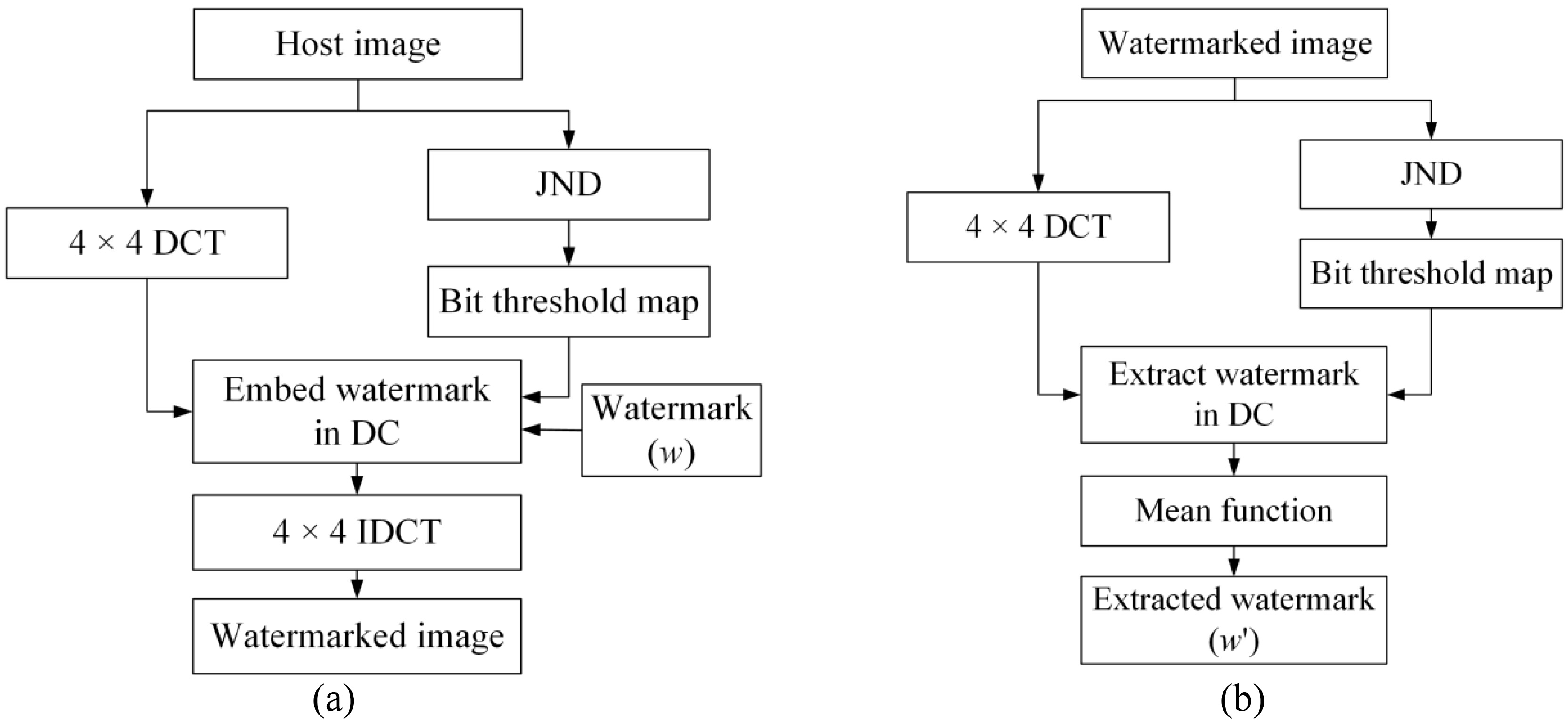
DCT is a widely used transformation technique for image processing of various types. Through DCT, spatial domain data can be transformed into the frequency domain, while frequency domain data can be transformed back to the spatial domain using the inverse DCT (IDCT). The DCT formulas [12] are as follows:
In the above formulas,
HVS is an important aspect of subjective image quality. Therefore, the subjective image quality can be improved by reflecting human visual characteristics. This capability is increasingly being studied [13, 14]. Human visual characteristics can be divided into human visual sensitivity and visual interest. HVS includes various characteristics of luminance adaptive motion and contrast sensitivity. Regarding these characteristics, Chou’s JND model [13] is mathematically constructed as a subjective quality correlation, which is characterized by the highest luminance adjustment.
For this reason, Chou’s JND model was selected for this study to embed the watermarks. JND refers to the maximum distortion that the HVS cannot perceive. It plays an important role in perceptual image processing. Chou’s JND model is employed to quantify the perceptual redundancy and provide each signal being coded with a visibility distortion threshold. Below this threshold, reconstruction errors are rendered imperceptible [15, 16, 17]. This model analyzes the local properties of image signals to incorporate threshold sensitivities due to background luminance and texture-masking effects. The model is estimated by analyzing local properties of image signals.
Bit threshold map
This study uses the JND value and bit threshold map (BTM) for the watermark embedding strength and position. By referencing the bit threshold value of each block, a watermark is embedded or extracted from the lower bits of the DC coefficient. A block diagram of the bit threshold map is shown in Fig. 3. As shown in the block diagram, a BTM is generated by executing Step 4.
Block diagram of bit threshold map. Example of calculating the bit threshold in each block.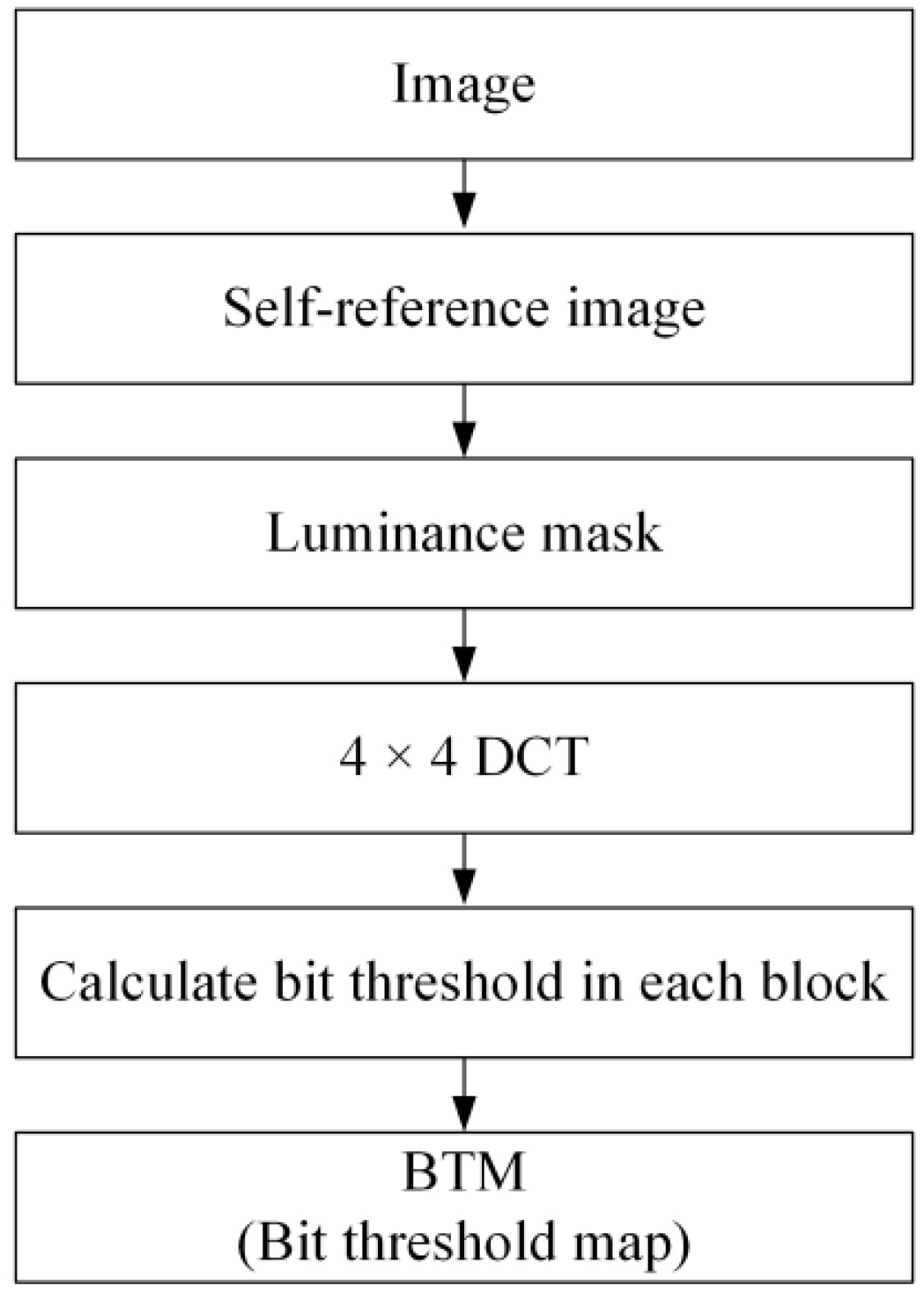
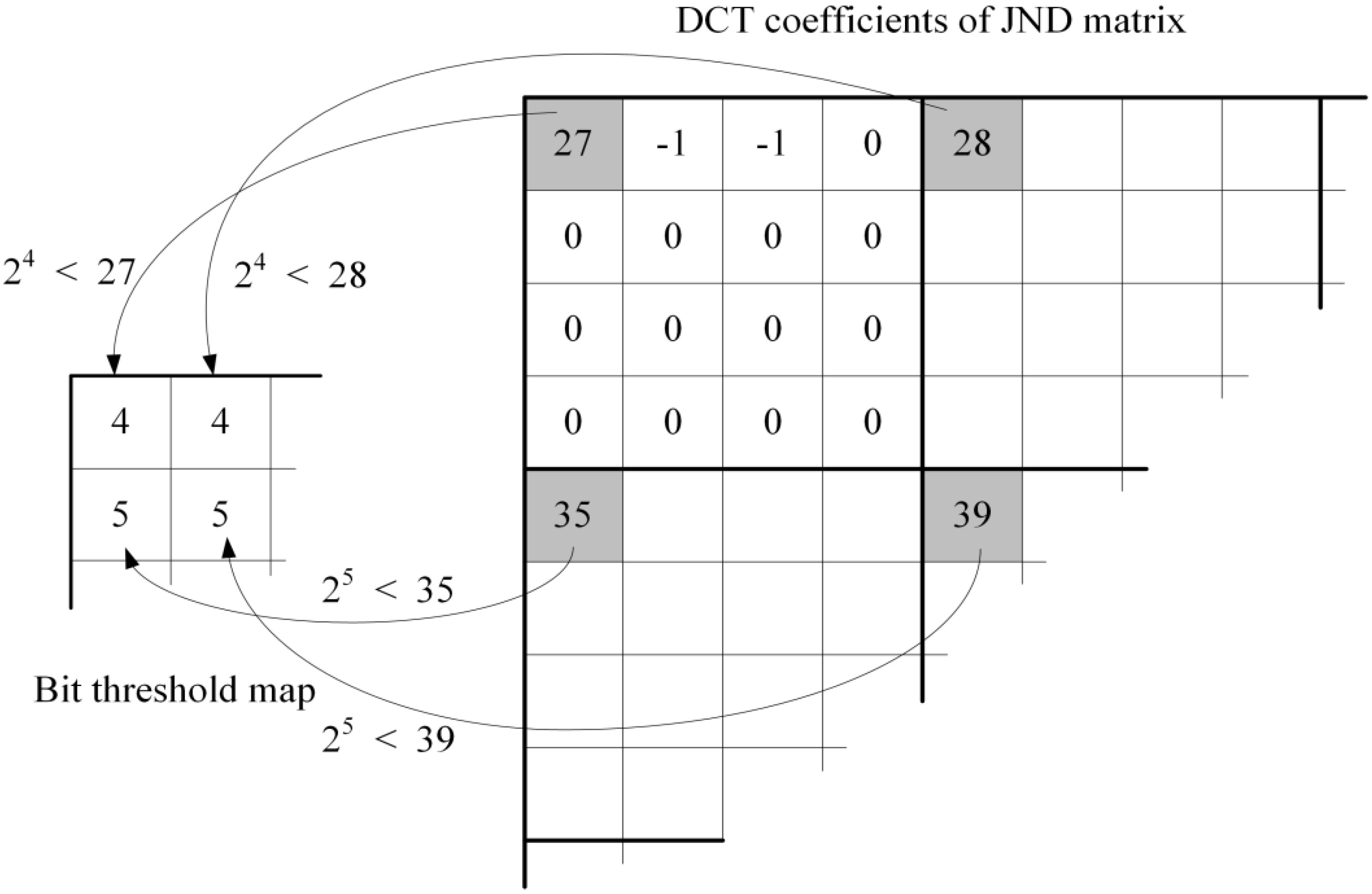
Generate a self-reference image with parts of the low-frequency component in the original image. Apply Chou’s luminance mask to generate the JND matrix for the self-reference image. Decompose the JND matrix into a 4 Use the DC coefficient to generate a BTM. Calculate the bit-threshold value in the DC coefficient using Eq. (4).
where
In the proposed method, a one-bit threshold is generated per block; the bit threshold map is
Example of embedding the watermark in each block.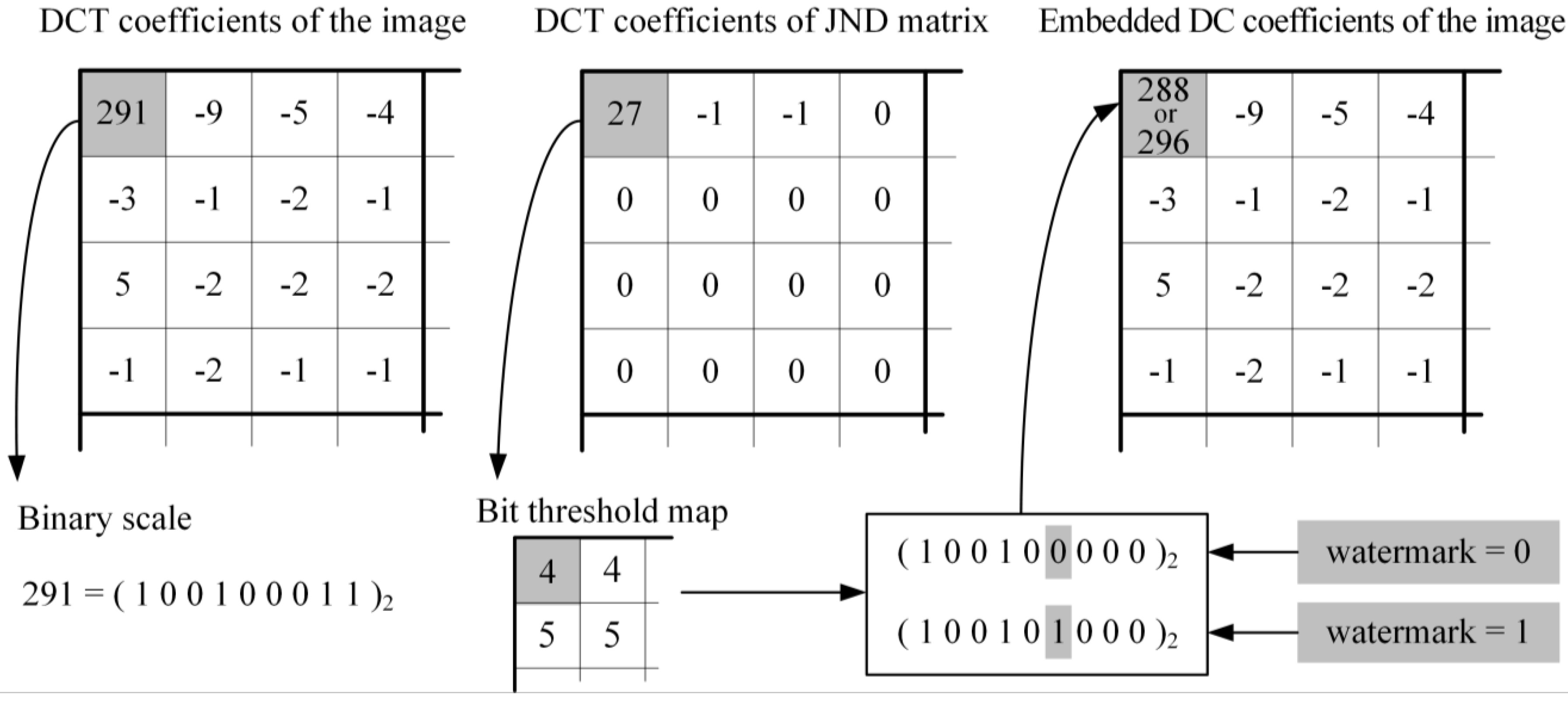
Example of the proposed watermark generation method and selected medical images in the simulation: (a) watermark generation and (b) MRI, (c) CT, and (d) X-ray images.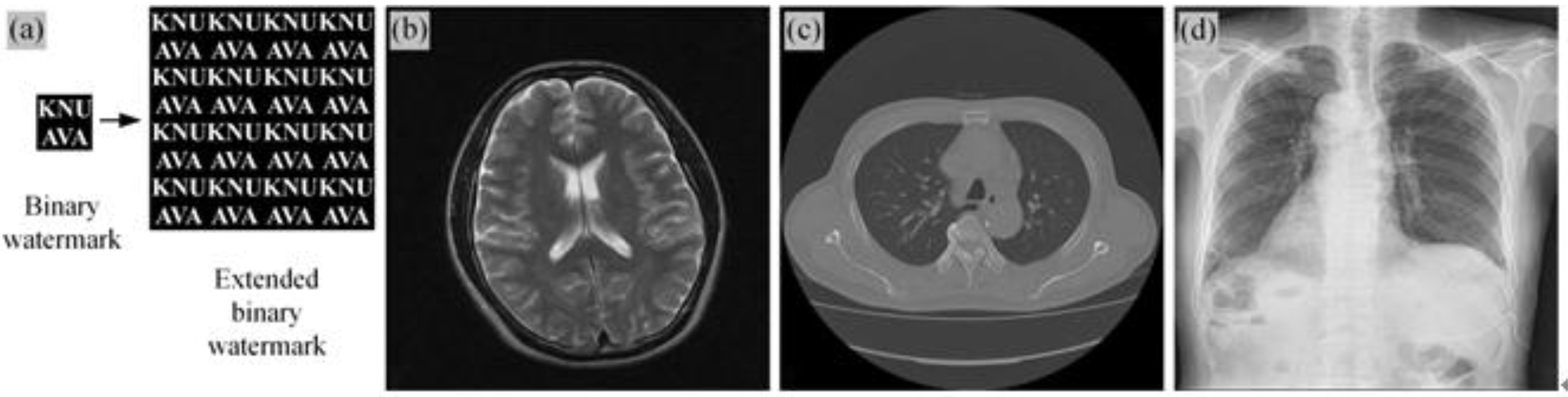
Proposed watermarking algorithm
Watermark embedding algorithm
To embed watermarks in the low-frequency components of the original image, the proposed algorithm uses a 4
Watermark detecting algorithm
In the watermark extraction process, the algorithm can extract watermarks using the lower bits of the DC coefficient of each block in the watermarked image. Medical image watermark extraction is similar to reversing the watermark-embedding process. First, the image is decomposed into non-overlapping 4
Finally, the extended watermark in the watermark bits is obtained for the entire block. A mean function is used in the expanded watermark to extract the final watermark.
The performance of the proposed algorithm was tested on various image types. The test images included medical resonance imaging (MRI), computed tomography (CT), ultrasonic and X-ray images. The images were 512
where
The amount of mismatched data between the embedded watermark and extracted watermark is used to quantify the similarity of the watermarks. The normalized correlation (NC) of the embedded watermark and extracted watermark is defined as follows:
where
A checkmark attack tool was used to evaluate the robustness of the proposed watermarking algorithm. We invoked three types of attacks: JPEG compression, various filters, and a crop method. The simulation results of a comparison of the PSNR value of the proposed method with conventional methods for invisibility evaluation are shown in Table 1. The sample images processed by the proposed method have higher invisibility than those processed by the conventional method. The higher invisibility is assumed to be on account of the HVS based on DC coefficients being changed to prevent quality degradation.
PSNR values of the watermarked images processed by the proposed method and conventional method
PSNR values of the watermarked images processed by the proposed method and conventional method
Attacked watermarked MRI images: (a) Gaussian noise, (b) median filter, (c) Wiener filter, (d) midpoint filter, (e) resampling, (f) sharpening, (g) trimmed mean filtering, (h) salt and pepper noise, (i) crop 25%, (j) crop 50%, (k) JPEG compression 10%, (l) JPEG compression 30%, (m) JPEG compression 50%, and (n) JPEG compression 70%.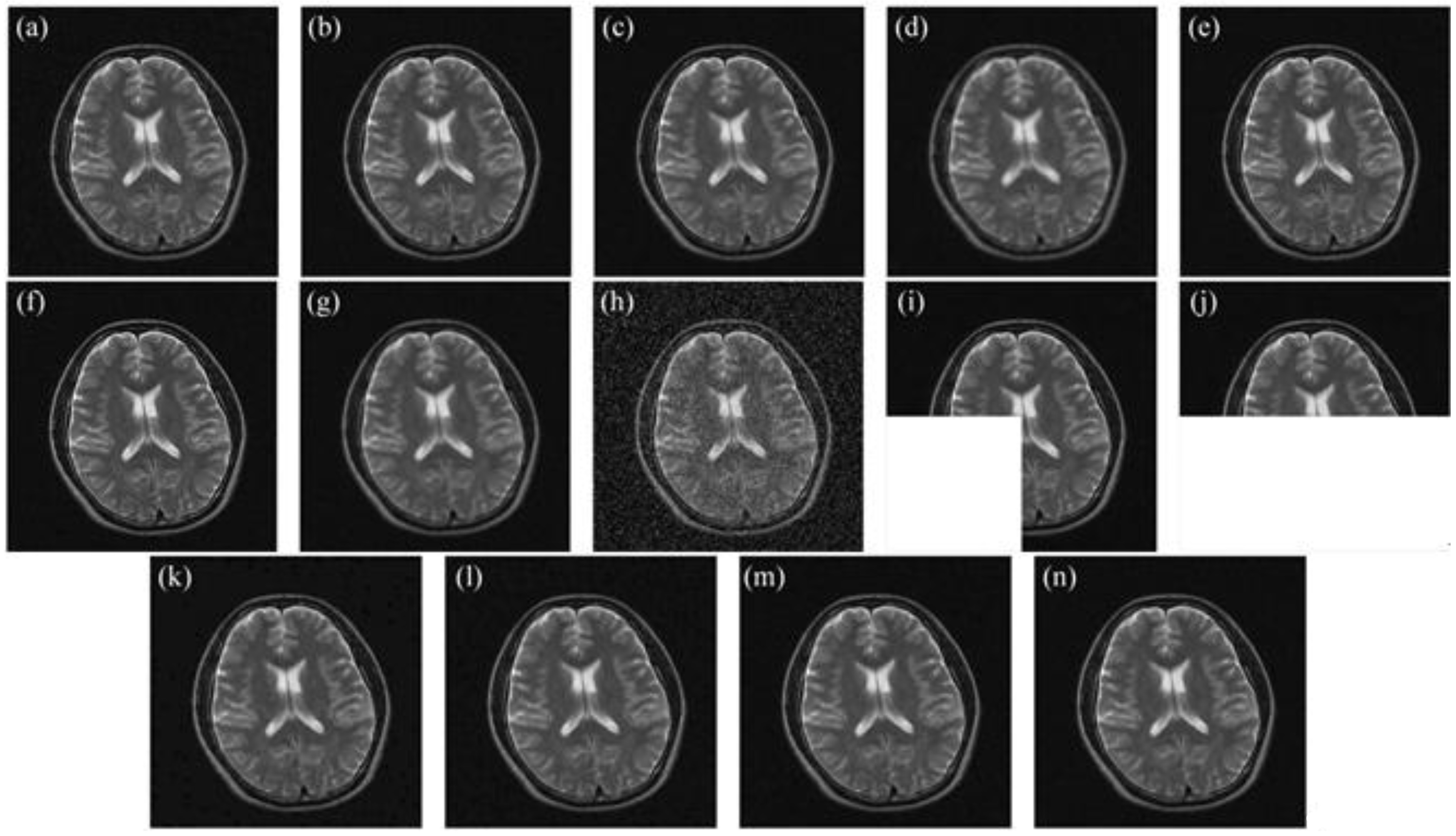
Visual analysis of the attacked watermarked image is shown in Fig. 6. Fourteen types of attack filtering, such as Gaussian noise, median filter, and Wiener filter, were used to evaluate the robustness of the proposed method compared to that of the conventional method. The results show that lower robustness against filters involves an emphasis in high frequency filtering, such as sharpening. However, the NC value is higher in the proposed method than in the conventional method, which indicates that the proposed algorithm has high robustness in protecting against the majority of attack filters.
The extracted watermark images and NC values of the proposed method and conventional method were obtained from the attacked watermarked medical images. These are shown in Table 2.
Extracted watermark images and NC values according to various attacks for test embedded watermark images using the proposed and conventional methods
In this paper, we proposed a medical image watermarking algorithm that employs a bit threshold map based on JND in DCT methods. This technique for embedding and extracting a watermark based on the DC coefficients in the low frequency region of the DCT have been described. Because the low frequency comprises most energy of the original image, it crucially affects the image quality. The proposed algorithm depends on the bit threshold map based on the JND of the image. The watermark is automatically embedded in the DC coefficient. This has a considerable effect on robustness and image quality. Moreover, it provides excellent invisibility because it considers the HVS when embedding the watermark. We performed a simulation evaluation to verify the robustness of the proposed method. The robustness was confirmed by embedding the watermark in the low-frequency component of the DC coefficient. The results show that the proposed approach is robust against a variety of attacks.
In our future work, we intend to develop an ultrasonic scanning non-destructive testing (NDT) technique [19] to enhance the processing performance and reliability.
Conflict of interest
None to report.
Footnotes
Acknowledgments
This research was supported by the Ministry of Trade, Industry & Energy (MOTIE), Korea Institute for advancement of Technology (KIAT) through the Encouragement Program for The Industries of Economic Cooperation Region.