
Abstract
Background
Multiple system atrophy (MSA) and Parkinson's disease (PD) are caused by misfolded α-synuclein spreading throughout the central nervous system. While familial PD is linked to several α-synuclein mutations, no mutations are associated with MSA. We previously showed that the familial PD mutation E46K inhibits replication of MSA prions both in vitro and in vivo, providing key evidence to support the hypothesis that α-synuclein adopts unique strains in patients.
Objective
Here we sought to further interrogate α-synuclein misfolding to identify the structural determinants that contribute to MSA strain biology.
Methods
We engineered a panel of cell lines harbouring both PD-linked and novel mutations designed to identify key residues that facilitate α-synuclein misfolding in MSA. We also used Maestro in silico analyses to predict the effect of each mutation on α-synuclein misfolding into one of the reported MSA cryo-electron microscopy conformations.
Results
In many cases, our modelling accurately identified mutations that facilitated or inhibited MSA replication. However, Maestro was occasionally unable to predict the effect of a mutation, demonstrating the challenge of using computational tools to investigate intrinsically disordered proteins. Finally, we used our cellular models to determine the mechanism underlying the E46K-driven inhibition of MSA replication, finding that the E46/K80 salt bridge is necessary to support α-synuclein misfolding.
Conclusions
Our studies used a structure-based approach to investigate α-synuclein misfolding, resulting in the creation of a powerful panel of cell lines that can be used to interrogate MSA strain biology.
Plain language summary
In patients with Parkinson's disease (PD) and multiple system atrophy (MSA), the protein α-synuclein misfolds into distinct shapes, or strains, causing accumulation of protein aggregates in the brain. Increasing evidence indicates that the shape α-synuclein adopts determines which disease a patient will develop. As a result, it is pivotal that we understand the factors that contribute to protein misfolding in disease. In this study, we used computational modelling to predict the effect of PD-causing and novel mutations on α-synuclein misfolding into the MSA disease conformation. We then tested these predictions in cell lines expressing the same mutations to determine if α-synuclein isolated from MSA patient samples can induce protein aggregation in the presence of each mutation. Using this approach, we not only identified key mutations in the α-synuclein gene that influence the ability of the protein to misfold into the MSA strain, but we also determined the mechanism by which one of these mutations, the PD-causing E46K mutation, exerts its inhibitory effect on α-synuclein misfolding in MSA.
Introduction
Synucleinopathies are a group of neurodegenerative movement disorders, including Parkinson's disease (PD) and multiple system atrophy (MSA), that are defined by distinct clinical and pathological features arising from the accumulation of misfolded α-synuclein proteins in the brain. While PD patients develop abnormal gait, resting tremor, muscle rigidity, and REM sleep behaviour disorder, 1 MSA patients are distinguished by the presence of autonomic failure, which can present as orthostatic hypotension, difficulty controlling urinary movement, and impaired body temperature regulation. 2 Additionally, while PD is more prevalent and patients typically develop disease in their 60 s or later, MSA is a rare disease with an earlier age of onset.1,2 Along with these clinical differences, PD is defined by α-synuclein accumulating into Lewy neurites or Lewy bodies (LB) within neurones, 3 whereas the pathological hallmarks of MSA include glial cytoplasmic inclusions (GCI) in oligodendrocytes and, to a lesser extent, neuronal cytoplasmic inclusions in neurones. 4 Interestingly, while both diseases are defined by the presence of α-synuclein inclusions,5,6 mutations in the α-synuclein gene, SNCA, have only been identified in PD patients, 7 providing strong support for the hypothesis that α-synuclein misfolds into at least two distinct conformations in PD and MSA patients.
Recent advances in cryo-electron microscopy (cryo-EM) enabled resolving the structure of misfolded α-synuclein fibrils from human patient samples, resulting in confirmation that α-synuclein adopts distinct structures in MSA 7 (Figure 1) and PD 8 patients (Supplemental Figure 1A). The α-synuclein fibrils isolated from MSA patient samples are defined by the presence of two asymmetrically arranged protofilaments, both of which adopt a Greek key motif that is stabilized by a salt bridge between residues E46 and K80 (Figure 1). Additionally, several positively charged residues project into a pocket at the protofilament interface that contains a negatively charged non-protein density. 7 It should also be noted that three fibril structures were resolved from MSA patient samples (PDB IDs 6XYO, 6XYP, and 6XYQ), which contain variability in subunit A but a mostly consistent structure in subunit B. In contrast, the α-synuclein Lewy fold contains a single filament with a modified Greek key motif stabilized by a salt bridge between E35 and K80 (Supplemental Figure 1A). The Lewy fold also contains a negatively charged non-protein density; however, in this structure it acts to stabilize the positive charges from four internal facing lysines, none of which include residue E46. 8
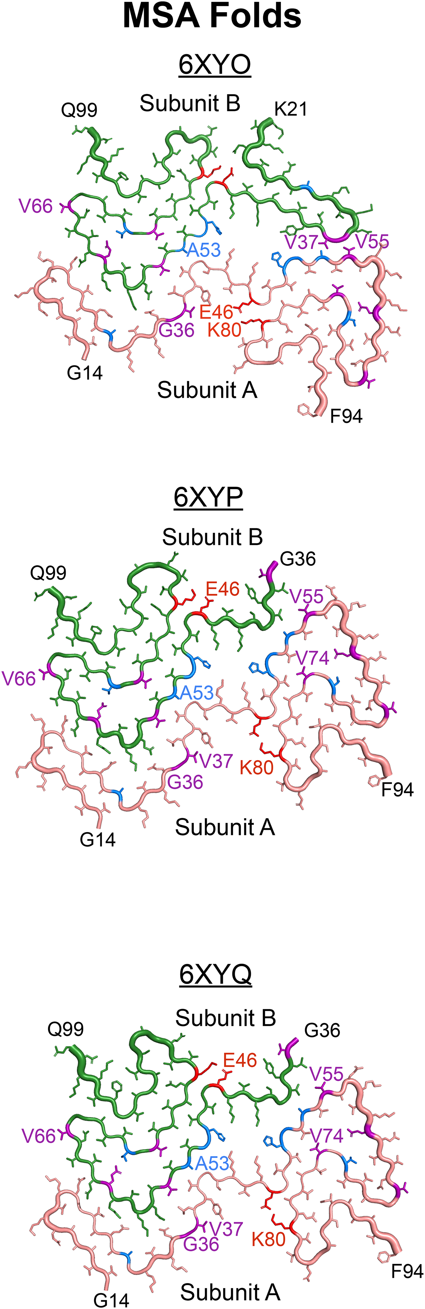
Using the MSA cryo-EM structures as an opportunity to develop hypotheses about how α-synuclein misfolds in disease, here we sought to test the effect of both PD-causing and novel mutations designed to interfere with protein misfolding on MSA propagation in vitro (highlighted in blue and purple in Figure 1, respectively; Supplemental Figure 2). With the goal of identifying computational tools capable of predicting the effect of mutagenesis on α-synuclein misfolding in MSA, we used Maestro software to make in silico predictions about the effects of each mutation on the 6XYQ MSA α-synuclein fibril structure. While total energy changes calculated using Maestro were typically capable of predicting in vitro outcomes, occasional discrepancies between our computational and cellular data demonstrate the challenge of using modelling software to investigate intrinsically disordered proteins that adopt a prion conformation. Additionally, varied effects of some mutations, such as the V55Y and V66F mutations, on MSA prion replication provide initial evidence that previously unreported α-synuclein conformations are likely present in a subset of MSA patients. Finally, to determine the mechanism underlying the inhibitory effect of the E46K mutation on MSA propagation, we showed that disrupting the E46/K80 salt bridge at either residue blocks cellular infection (shown in red in Figure 1), indicating that this salt bridge is necessary to stabilize MSA prions. Unexpectedly, we could not recover MSA propagation in cells expressing the double E46K,K80E mutation, which is likely due to an increase in total energy that occurs when both residues are mutated. Overall, our studies use a structure-based approach to interrogate α-synuclein misfolding in MSA, resulting in the generation of a robust panel of cellular models that can be used more broadly to investigate α-synuclein strain biology.
Materials and methods
Human tissue samples
The Massachusetts Alzheimer's Disease Research Center (MADRC) supplied frozen midbrain tissues from two patients with neuropathologically confirmed MSA and two control samples. The NIH NeuroBioBank supplied tissue from two control patients, and the Sydney Brain Bank supplied frozen samples from three neuropathologically confirmed MSA cases. Demographic information about the patient samples used is included in Supplemental Table 1.
Cell line development
Wild-type (WT) and mutant (E46K, H50Q, G51D, A53E, G36K, V37F, V55Y, E61Q, V66F, V74I, and V74P single mutations and the A30P,A53T double mutation) human α-synuclein cDNA sequences fused to enhanced yellow fluorescent protein by an 18 amino acid flexible linker (EFCSRRYRGPGIHRSPTA) were synthesized and cloned into the pcDNA3.1(+) expression vector by GenScript. The α-syn140-YFP sequences were then subcloned into the pIRESpuro3 vector (Takara) using restriction cloning with EcoRV (5’) and NotI (3’). Gene sequence and insertion were confirmed by Sanger sequencing before subsequent use. Other mutant constructs (A30G, A53V, T72M, K80E, K80N, K80Q, K80W, and the double mutant E46K,K80E) were generated by PCR amplification of the pIRESpuro3 vector containing the WT α-syn140-YFP sequence using primers designed to introduce each mutation, or the E46K mutation for the double mutant. Gene sequence and insertion were confirmed by Sanger sequencing before subsequent use.
HEK293T cells (ATCC) were cultured in Dulbecco's modified Eagle's medium (DMEM; Corning) supplemented with 10% fetal bovine serum (FBS), 100 μg/mL penicillin, and 100 μg/mL streptomycin (ThermoFisher), referred to hereafter as complete media. Cultures were maintained in a humidified atmosphere of 5% CO2 at 37 °C. Cells were plated at a density of 5.7 × 10⁵ cells per well in a 6-well plate overnight in complete media before adding 1 μg of plasmid DNA incubated with 3.5 μL Lipofectamine 2000 (ThermoFisher) for 20 min. Stable cells were selected in complete media containing 1 μg/mL puromycin (ThermoFisher) for 48 h before generating monoclonal lines by limiting dilution of polyclonal cells in 384-well plates. The resulting monoclonal lines were frozen in liquid nitrogen. Lysates from the lines were collected in 1× radioimmunoprecipitation assay (RIPA) buffer containing 50 mM Tris-HCl, pH 7.5 (ThermoFisher), 150 mM NaCl (Sigma), 5 mM EDTA (ThermoFisher), 1% nonidet P-40 (ThermoFisher), 0.5% deoxycholate (ThermoFisher), and 0.1% sodium dodecyl sulfate (SDS; ThermoFisher). Cell lysates in RIPA buffer were frozen, thawed, and clarified using two low-speed spins (500 × g for 5 min followed by 1000 × g for 5 min). Total protein was measured in the supernatants via bicinchoninic acid (BCA) assay (Pierce). To compare the expression of α-syn140-YFP across the clones, a total of 10 μg total protein was prepared in 1× NuPAGE LDS loading buffer and boiled for 10 min. Samples were loaded onto a 10% Bis-Tris gel (ThermoFisher) and SDS-PAGE was performed using the MES buffer system. Protein was transferred to a polyvinylidene fluoride (PVDF) membrane. The membrane was blocked in blocking buffer [5% (wt/vol) nonfat milk in 1× Tris-buffered saline containing 0.05% (vol/vol) Tween 20 (TBST)] for 30 min at room temperature. Blots were incubated with primary antibody for GFP (1:10,000; Abcam) in blocking buffer overnight at 4 °C. Membranes were washed three times with 1× TBST before incubating with horseradish peroxidase-conjugated goat anti-rabbit secondary antibody (1:10,000; Abcam) diluted in blocking buffer for 1 h at 4 °C. After washing blots three times in 1× TBST, membranes were developed using the enhanced chemiluminescent detection system (Pierce) for exposure to X-ray film.
α-Synuclein prion bioassay
Frozen brain samples were used to make a 10% (wt/vol) homogenate in calcium- and magnesium-free 1× Dulbecco's phosphate buffered saline (DPBS) using an Omni Tissue Homogenizer with disposable plastic soft tissue tips (Omni International). Aggregated protein was isolated from the samples using phosphotungstic acid (PTA; Sigma-Aldrich) as described.9,10 Briefly, the brain homogenates were incubated with 2% (vol/vol) sarkosyl (Sigma) and 0.5% (vol/vol) benzonase (Sigma) at 37 °C under constant agitation (850 RPM). Sodium PTA, dissolved in 10% (wt/vol) in double-distilled H2O and brought to pH 7.0, was added to a final concentration of 2% (vol/vol) and incubated overnight as described. Samples were centrifuged at 13,200×g for 30 min, and the supernatants were discarded. The pellets were resuspended in 2% sarkosyl and 10% PTA before incubating for 1 h under agitation as before. Samples were centrifuged at 13,200×g for 30 min, and the supernatants were removed immediately after. The pellets were resuspended in 1× DPBS (Corning) using 10% of the starting volume and were stored at 4 °C until use.
The α-syn140-YFP cell assay was performed as previously described.10,11 Briefly, monoclonal subclones stably expressing WT or mutant α-synuclein were cultured in complete media in a humid 5% CO2 environment at 37 °C. Cells were grown in 384-well plates with black polystyrene walls (Greiner) with growth medium containing 0.012 µg/well of Hoechst 33342 diluted in 1× DPBS (Thermo Fisher) to visualize nuclei. Plates with cells were incubated at 37 °C for 2–4 h. PTA-precipitated patient samples were diluted in 1× DPBS prior to incubation with Lipofectamine 2000 (Thermo Fisher) at room temperature for 1.5 h. Warmed OptiMEM (Thermo Fisher) was added to each sample prior to plating the sample in 6 replicate wells. Optimized assay conditions are reported in Supplemental Table 2. Plates were covered with a membrane to prevent drying during incubation for 4 d at 37 °C in a humidified atmosphere with 5% CO2. The plate was imaged using the MicroXLS (Molecular Devices) collecting a DAPI and FITC image (for Hoechst and YFP, respectively) from 5 areas per well. The images were analyzed with MetaXpress software using parameters developed to detect and quantify intracellular aggregates in live cells using pixel intensity and size thresholds. The measure of aggregation was calculated as the total fluorescence measured across all aggregates per cell in each well (× 106 arbitrary units [A.U.]).
Modeling α-synuclein mutations on the MSA cryo-Em structure
The MSA α-synuclein filament PDB file (6XYQ) was increased to a hetero 12-mer using MOE (Chemical Computing Group) using the build_fibril.svl. From this file, Maestro (Schrödinger) was used to optimize each amino acid side using the Predict Side Chain Panel, then minimized using the OPLS4 force field. Minimization was performed to an RMSD of 10−9 kcal/mol/Å for convergence. From the minimized structure, each mutation was simulated manually and mutations in each heterodimeric sequence were performed independently. To maintain the same ground state energy, the first step in the analysis pipeline was to create a compensating mutation at an amino acid with few neighbouring side chains to serve as a control (shown in Supplemental Table 3). Using the V55Y mutation as an example for the analysis procedure, the minimized structure underwent a computational E83Y mutation on all 6 layers of subunit A in the total fibril structure. Using this control, we computationally made a V55Y mutation and a concomitant Y83V mutation on all 6 layers. After re-predicting the position of all side chains in proximity to both the Y55 and V83 residues using Predict Side Chain panel, both the mutated and control (E83Y) structures were minimized to convergence using the OPLS4 force field. To calculate the relative energy change for each mutation (in kcal/mol), the total calculated ensemble energies for the mutated subunits were subtracted from the structures with only the compensating mutations reported in Supplemental Table 3. For V55Y, the subunit B mutations followed an identical process except that the control remained E83Y on subunit A. In some cases, the E83Q mutation was used. We note that while the E83Q mutation is known to accelerate α-synuclein misfolding kinetics in a cell-free assay, 12 this effect should not impact our structural modelling data.
Statistical analysis
Data are presented as mean ± standard deviation. Cell assay data were analyzed using GraphPad Prism software. Mean fluorescence intensity values and standard deviations were calculated for each cell line by averaging the fluorescence intensity values from the 6 technical replicate wells. Reported p values for each cell line tested are from comparisons between control and MSA patient samples using the average value for each via Welch's two-tailed t-tests with unequal variance. Significance was determined as having a p value <0.05.
Results
Parkinson's mutations exert differential effects on MSA prion replication in vitro
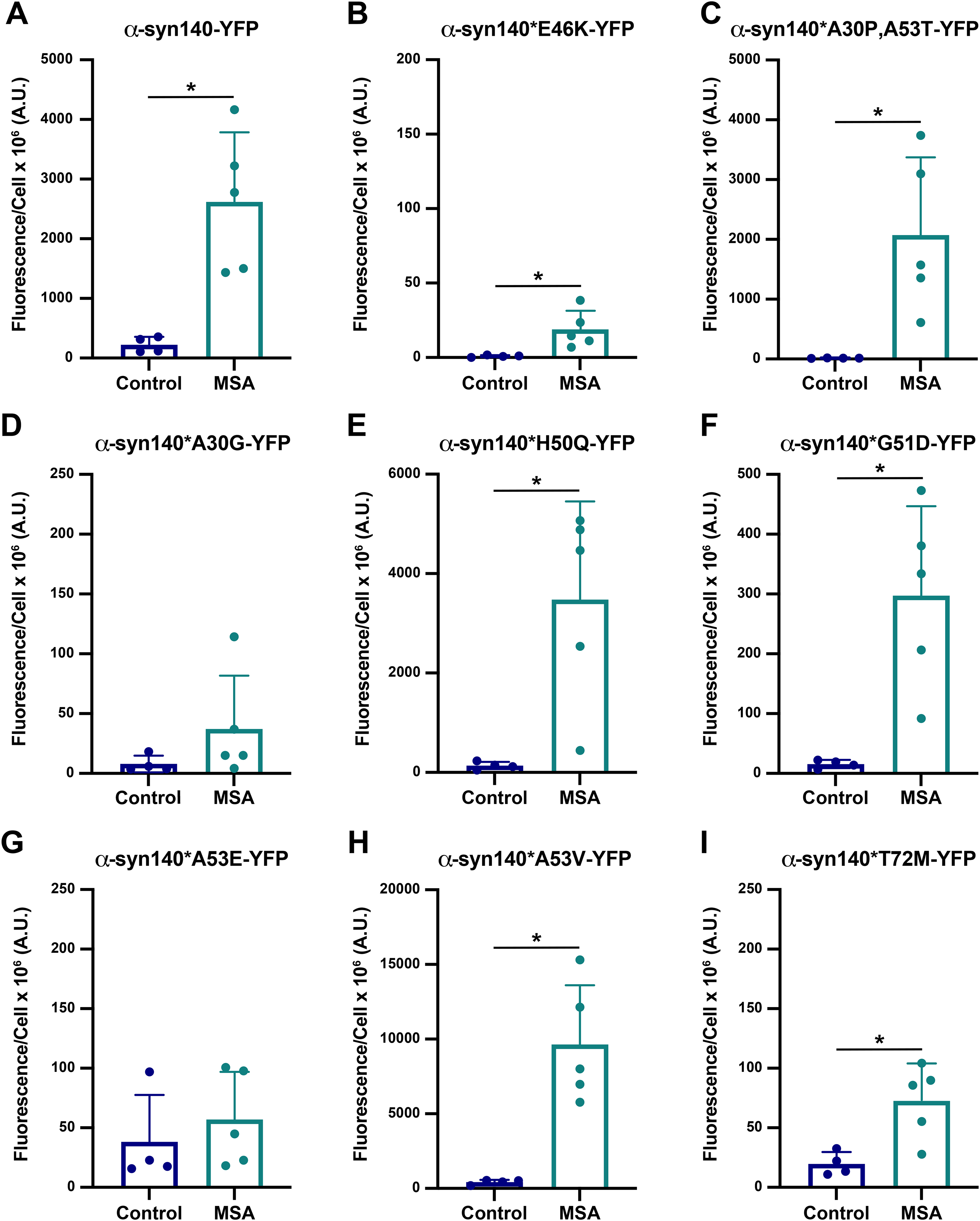
Building on our previous findings that the familial PD mutation E46K 13 prevents MSA prion replication both in vitro and in vivo,11,14 we sought to determine the effect of other known PD mutations on the replicability of MSA prions using a larger panel of α-syn140-YFP cell lines. In this assay, HEK293T cells expressing α-synuclein fused to YFP via a flexible linker are incubated with α-synuclein prions isolated from deceased human patient samples. We compared 4 control samples from patients lacking neuropathological inclusions (C1, C2, C6, and C7) with 5 neuropathologically-confirmed MSA patient samples (MSA2, MSA3, MSA6, MSA7, and MSA12; Supplemental Table 1) to measure α-synuclein prion aggregation in the α-syn140-YFP cells (reported as fluorescence/cell × 106 A.U.). Consistent with our previous results, MSA prions induced aggregation in cells expressing WT α-synuclein (p < 0.05) and the A30P,A53T double mutation (p < 0.05), but replication was inhibited in cells expressing the E46K mutation (Figure 2A-C; p < 0.05). 11 While a statistically significant difference was detected between the control and MSA samples in the E46K cells (Figure 2B), the minimal effect size indicates the lack of a biologically meaningful result. Representative images of WT and E46K cells incubated with patient samples C6 and MSA6 are shown in Supplemental Figure 3.
Our initial studies focused on the first three SNCA mutations identified in Parkinson's cohorts, however, several additional PD-linked mutations have since been reported. These additional mutations include A30G,
15
H50Q,
16
G51D,
17
A53E,
18
A53V,
19
and T72M.
20
To assess the effect of each of these PD-linked mutations on the ability of α-synuclein isolated from MSA patient samples to replicate, we first used Maestro software to measure the relative energy changes (in kcal/mol) of each mutation on the 6XYQ MSA fibril structure (Table 1) resolved via cryo-EM.
7
Energy values below 0 after mutagenesis indicate a favourable energy change, whereas those greater than 0 indicate an unfavourable energy change. It is important to note that residue 30 is outside of the templating region of subunit B in the 6XYQ MSA fibril conformation, limiting our ability to use Maestro to evaluate the effect of the A30G mutation on MSA. However, a slight increase in total energy in subunit A suggested that the A30G mutation would reduce or inhibit MSA replication. In contrast, Maestro predicted the H50Q mutation would decrease the total energy of the MSA structure, increasing the likelihood of replication. Similar predictions were identified for the G51D mutation, though these effects were limited to subunit B as there was essentially no change for subunit A. This latter effect is intriguing given the location of residue 51 at the protofilament interface of the MSA structures. Similarly, residue 53 sits within the protofilament interface, raising the potential that mutations could disrupt replication. Here, mutagenesis of the alanine to either a glutamic acid or a valine resulted in an unfavourable increase in energy in subunit A. Notably, this increase was substantially larger for the A53E mutation compared to A53V. By comparison, the A53E mutation increased total energy for subunit B whereas the A53V mutation yielded a decrease in total energy. It is important to note that the negatively charged non-protein density that fills the pocket between the two protofilament subunits in the MSA cryo-EM structures is absent in the PDB structures. As a result, our initial in silico analysis of the A53E mutation resulted in a salt bridge forming between A53E and K43. In vitro, the presence of that density would prevent this interaction from occurring. To prevent this effect in our modelling data, we inserted a K43A mutation into the control and mutant calculations. Finally, the T72
Total energy change after mutagenesis using PD-causing mutations in MSA structure 6XYQ.
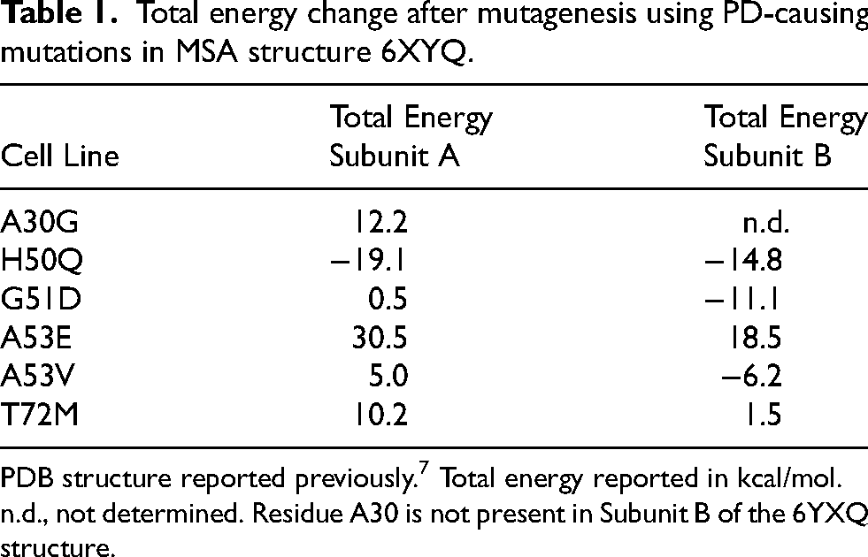
PDB structure reported previously. 7 Total energy reported in kcal/mol.
n.d., not determined. Residue A30 is not present in Subunit B of the 6YXQ structure.
To determine how well Maestro predicted the effect of α-synuclein mutations on MSA replication in vitro, we constructed α-syn140-YFP cell lines expressing each of the PD-linked mutations and found that the A30G mutation largely inhibited MSA replication (Figure 2D; Supplemental Table 4; p = 0.22), which is consistent with the mutagenesis prediction for subunit A. By comparison, the H50Q (Figure 2E) and G51D (Figure 2F) mutations permitted MSA replication in vitro (p < 0.05; Supplemental Table 4), consistent with the favourable predictions from the Maestro analyses. Interestingly, the mutations at A53 resulted in dichotomous effects, with the A53E mutation blocking aggregation (Figure 2G; p = 0.51) while the A53V mutation permitted MSA replication (p < 0.05; Figure 2H; Supplemental Table 4). This is notable given that predictions for the A53V mutation varied by subunit whereas the A53E analyses showed a substantial increase in total energy for both protofilament subunits. Intriguingly, while the T72M mutation did not completely inhibit MSA propagation, we observed a blunted response compared to the α-syn140-YFP cells expressing WT protein, suggesting that the T72M mutation alters replication kinetics in vitro (Figure 2I; Supplemental Table 4; p < 0.05). This is relatively consistent with the modelling data, where the mutation caused a slight increase in total energy.
Overall, the ability of Maestro to predict the effect of PD-causing point mutations on α-synuclein misfolding in MSA was reasonably accurate. Moreover, the cellular results contribute important data to the growing literature showing that the structural differences in α-synuclein fibril conformations in PD 8 and MSA 7 result in unique biological properties between the two strains.11,21–23 Unexpectedly, we found that the effect of mutations at two residues—A30 and A53—are mutation-specific. For example, while the A30P, A53T, and A53V mutations enable MSA replication, the A30G and A53E mutations block this activity. While an explanation for these residue-specific effects is not clear based on the mutagenesis modelling, future studies will focus on investigating this phenomenon.
Structurally informed mutations reveal varied biological activities between MSA patient samples
Preformed fibrils (PFFs) made from recombinant α-synuclein are widely used throughout the literature as a tool to investigate synucleinopathies, including PD and MSA. Previously, we showed that PFFs made from both WT and mutant protein exhibit unique biological and biochemical properties in cell and animal models of disease.11,14,24 These findings are supported by cryo-EM structures of PFFs, which misfold into conformations that are distinct from those found in PD and MSA patient samples (reviewed in 25 ). To further investigate the differences in biological consequences that arise due to differences in MSA versus PFF structure, we developed a library of α-syn140-YFP cell lines expressing novel mutations predicted to interfere with protein misfolding in the ‘rod’ (PDB ID: 6CU7), ‘twister’ (PDB ID: 6CU8),26–28 or MSA fibril structures. 7 Comparing the rod and twister polymorphs (shown in Supplemental Figure 1B, C), 26 we chose to test the effect of mutations at residues E61 and V66 on MSA replication. Our initial assessment of these two structures suggested that the E61Q mutation would alter the stability of the pocket formed by the Greek key motif (which is present in most α-synuclein structures) with a greater disruption to the twister conformation than the rod, where the residues are more tightly packed together. Similarly, mutating the valine at position 66 to a phenylalanine (V66F) would disrupt the intermolecular zipper at the protofilament interface in the twister polymorph without affecting the rod, where V66 projects outward toward solvent. We subsequently used Maestro to predict the effect of these two mutations on the MSA fibril structure and found that the E61Q mutation enhanced the energetic favorability of the MSA fibril while the V66F mutation was neutral (Table 2). Inserting these mutations into the α-syn140-YFP cells, we found that all 5 MSA patient samples tested infected cells expressing the E61Q mutation (Figure 3A; Supplemental Table 5; p < 0.05), whereas the V66F mutation was more selective with some patient samples showing robust replication while others were completely inhibited (p = 0.08; Figure 3B; Supplemental Table 5). Given that the V66F mutation exhibited a neutral effect in our mutagenesis modelling (Table 2) and that the R group projects away from the fibril core in the MSA structures, 7 these findings suggest that previously undetected α-synuclein conformations in MSA patients may exist.
Total energy change after mutagenesis of structurally-informed mutations in MSA structure 6XYQ.
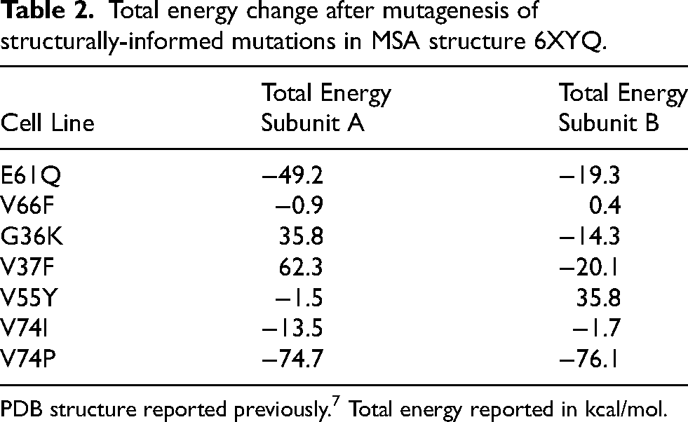
PDB structure reported previously. 7 Total energy reported in kcal/mol.
Analyzing the three MSA fibril structures resolved by cryo-EM, 7 we predicted that mutating residues G36, V37, and V55 would allow us to disrupt the interface between the two protofilaments in the MSA fibrils, with a particularly strong effect on 6XYO. To achieve this goal, we tested the G36K, V37F, and V55Y mutations both in silico and in vitro (Table 2; Supplemental Table 4; Figure 3C-E). Consistent with our design of these mutations, Maestro predicted the G36K and V37F mutations would cause an increase in total energy for subunit A, but a decrease for subunit B (Table 2). By comparison, the V55Y mutation was energetically favourable when we inserted the mutation into subunit A, but caused an increase in total energy for subunit B. In predictions like these where total energy between the two subunits moves in opposite directions, we predict that the disfavoured conformation will exert a stronger effect than the favourable subunit, resulting in an inhibitory effect on protein misfolding. In line with our findings for the V66F mutation, the G36K mutation inhibited or slowed replication of some MSA patient samples while facilitating replication of others (p = 0.06; Figure 3C; Supplemental Table 5). The V37F mutation permitted replication of all MSA samples tested, though the magnitude of change was reduced compared to other cell lines (p < 0.05; Figure 3D; Supplemental Table 5). Lastly, the V55Y mutation inhibited propagation of 2 MSA samples, though only 1 of the 3 positive samples showed robust infection (p = 0.26; Figure 3E; Supplemental Table 5), suggesting residue V55 may play a stronger role in forming the protofilament interface than G36 or V37 in MSA fibrils.
Finally, to disrupt the twister conformation with minimal impact on MSA replication, we mutated the valine at position 74 to either an isoleucine or a proline. Consistent with this objective, Maestro modelling indicated that both the V74I and V74P mutations would reduce the total energy of the α-synuclein conformation, though the effect of the V74P mutation was much stronger, suggesting MSA should be able to propagate (Table 2). However, in vitro studies showed that the V74I mutation blocks MSA replication (p = 0.0499; Figure 3F; Supplemental Table 5). Notably, while we detected a statistical difference between control and MSA samples in this cell line, the minimal effect size between the two groups indicates this statistical change is not biologically relevant. By comparison, the cell assay data showed that the V74P mutation merely blunts MSA replication (p < 0.05; Figure 3G; Supplemental Table 5) compared to the WT cells. This was unexpected given the substantial decrease in total energy predicted by Maestro.
It is worth noting that when we used Maestro to model the effect of mutations that were designed based on protein structures, it variably predicted our in vitro results. For example, while there was largely agreement between the analyses for the E61Q, V66F, and V55Y mutations, we saw less consistency between the predicted data and the biological effects of the G36K, V37F, V74I, and V74P mutations. One possible explanation for the G36K and V37F discrepancy is the fact that these two mutations are located at the end of subunit B, which decreases the reliability of Maestro calculations due to the increased flexibility arising from the smaller number of interactions. However, given that there are sample-to-sample differences in the effect of some mutations, it is possible that the presence of additional α-synuclein conformations in the MSA patient samples confounds our in silico analyses.
The E46K mutation blocks MSA replication by disrupting the E46/K80 salt bridge
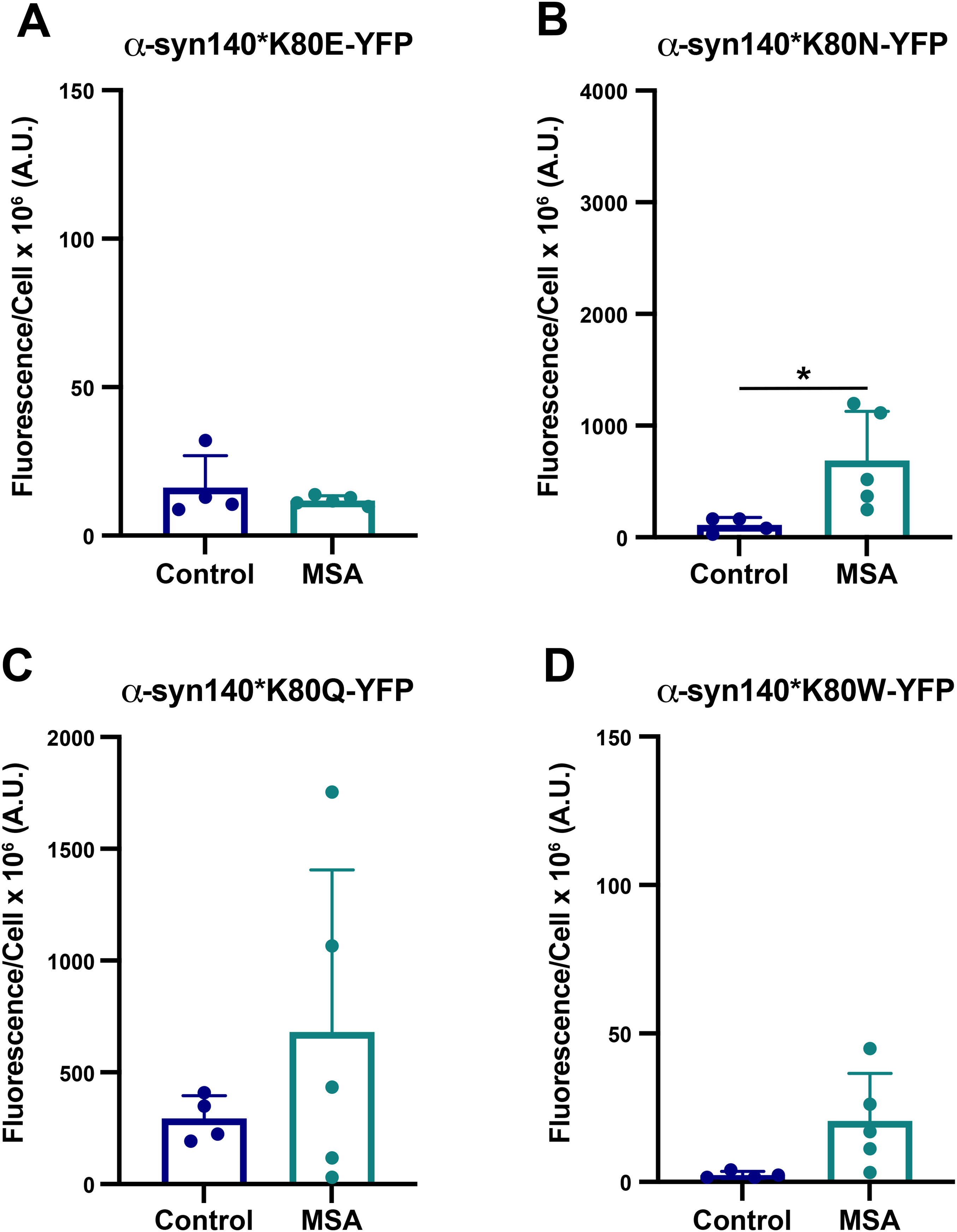
Several of the α-synuclein cryo-EM structures, including the MSA filaments, contain Greek key motifs that are stabilized by an intermolecular salt bridge that typically forms between residues E46 and K80 (reviewed in 25 ). In light of our previous findings that the E46K mutation blocks MSA replication,11,14 we predicted that this effect occurs because the two lysines repel one another when the salt bridge is lost, destabilizing the misfolded conformation. 29 To test this hypothesis, we generated four novel mutations designed to disrupt the E46/K80 salt bridge at position K80. In these mutations, we swapped the positively charged lysine for a negatively charged glutamic acid (K80E), predicting this would create a similar repellant interaction as the E46K mutation. We anticipated that the polar but uncharged asparagine (K80N) and glutamine (K80Q) would allow us to test the role of the positive charge on the lysine in salt bridge formation, given that the asparagine and glutamine have similar R groups to the lysine. And, finally, the bulky hydrophobic tryptophan (K80W) was selected to crowd out the packing of the R groups, preventing α-synuclein from adopting the Greek key motif. Using Maestro, we first confirmed that the E46K mutation is predicted to have a destabilizing effect on both subunits in the MSA fibril structure, with an increase of 70.2 and 51.6 kcal/mol, respectively (Table 3). We then tested the effect of the four K80 mutations using the same approach. While K80E, K80Q, and K80W all showed an increase in total energy, the K80N mutation showed a decrease in total energy for subunit A but an increase for subunit B.
Total energy change after mutagenesis targeting the E46/K80 salt bridge in MSA structure 6XYQ.
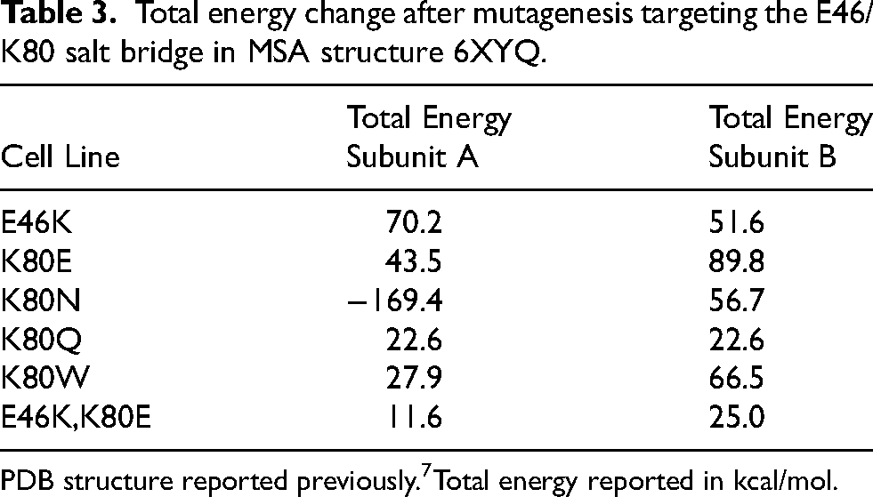
PDB structure reported previously.7Total energy reported in kcal/mol.
Testing these mutations in vitro, we found that the K80E mutation strongly inhibits MSA replication (p = 0.49; Figure 4A; Supplemental Table 6), consistent with our E46K findings and our hypothesis that similar charges (in this case, a negative charge) would force apart the two stabilizing ends of the Greek key. By comparison, the K80N and K80Q mutations exert variable effects on MSA infection, with some MSA patient samples able to replicate using the mutant proteins while others were blocked (Figure 4B, C; Supplemental Table 6). The effect of the K80N mutation on MSA propagation was more consistent (p < 0.05), whereas the MSA samples showed a greater range in the ability to replicate using α-syn*K80Q (p = 0.30). Interestingly, the conflicted effect of the K80N mutation on the two subunits did not result in a completely inhibitory effect. These results suggest that the positive charge on the lysine is important for providing stability through the Greek key, but it is not always necessary for amplification. Additionally, we would expect the longer glutamine to occasionally replace the lysine in the MSA structures. However, the shorter asparagine would lack the length required to form other inter- and intramolecular interactions, leading to reduced stability compared to the K80Q mutation. Finally, the largest amino acid substitution, the K80W mutation, also largely blocked MSA prion replication (p = 0.06; Figure 4D; Supplemental Table 6). We hypothesize this occurs through steric hindrance created by the longer side chain with the large aromatic ring. These data are largely consistent with the predicted effects of each mutation by Maestro, underscoring the importance of the E46/K80 salt bridge in stabilizing the MSA fibril structure.
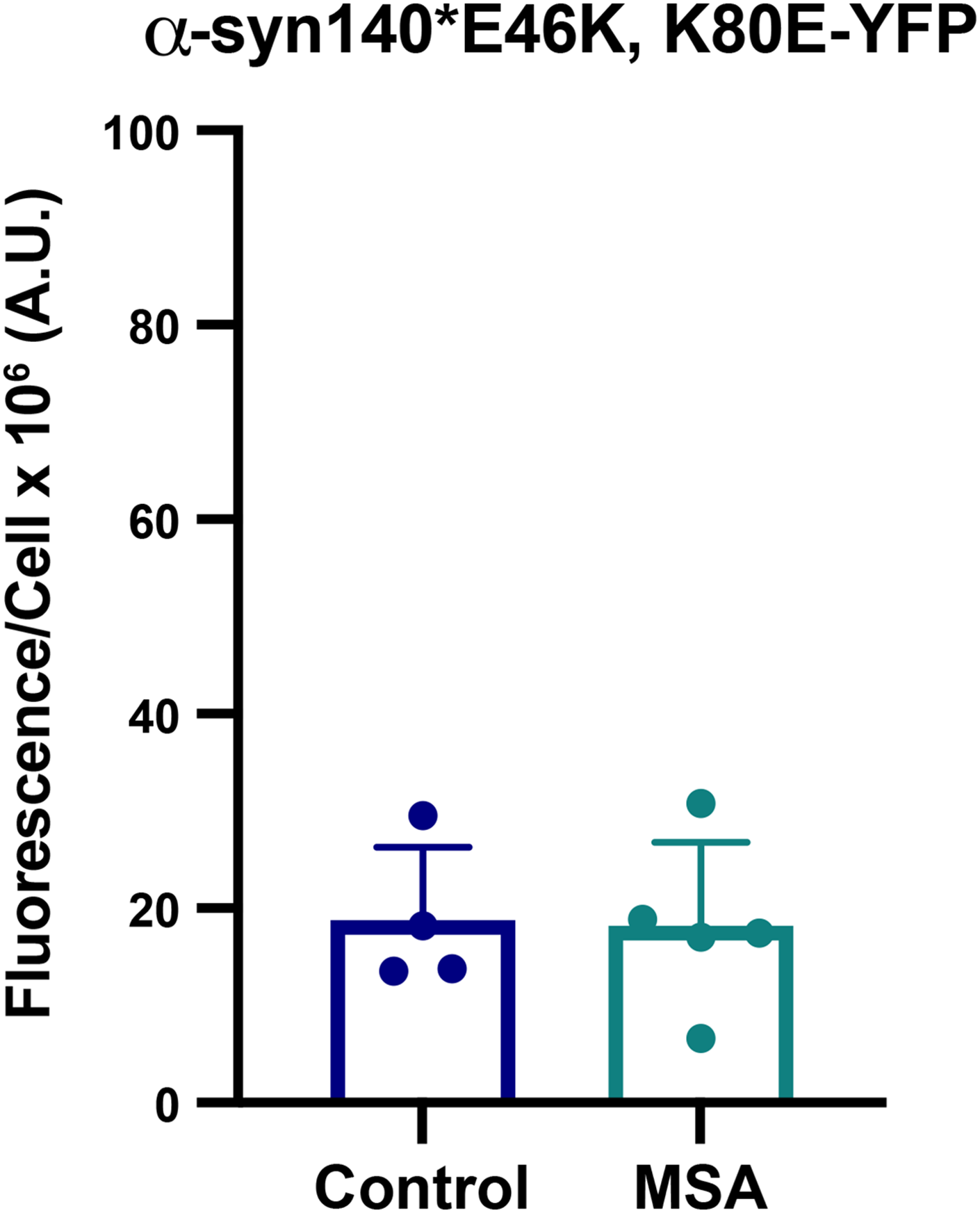
Having established the importance of the E46/K80 salt bridge in MSA prion replication, we hypothesized that swapping the residues would enable salt bridge formation, thereby re-establishing MSA replication in vitro. To test this, we created a cell line expressing both the E46K & K80E mutations (E46K,K80E), which we then incubated with MSA patient samples. Unexpectedly, the E46K,K80E cell line also prevented MSA replication (p = 0.91; Figure 5; Supplemental Table 6), suggesting that the presence of the glutamic acid and lysine alone is not sufficient to enable MSA propagation. To determine what may be contributing to this result, we mutated both residues in the MSA fibril conformations in Maestro and found that the double mutation had a slight increase in total energy compared to the unmutated structure (Table 3). While this overall change is smaller than the individual mutations, the destabilizing effect in both side chains likely contributes to the in vitro measurements.
Discussion
Beginning with the solid-state NMR structure of α-synuclein fibrils reported by Tuttle et al., in 2016, 30 the number of reported structures deposited in Protein Data Bank has grown substantially due to advances in cryo-EM enabling the resolution of fibrillar structures made from recombinant protein26–28,31–38 or isolated from human patient samples.7,8,39 However, as our understanding of the conformations that α-synuclein can adopt has increased, the resulting biological consequences of these structural differences have not been elucidated. Here, to start addressing this pivotal question for the α-synuclein conformations in MSA, we paired an in silico analysis of targeted mutagenesis with in vitro assays to experimentally validate the effect of each mutation on the ability of MSA prions to replicate. In addition to finding that our computational analyses using Maestro were typically able to predict the effect of a point mutation on α-synuclein misfolding, we established a panel of cell lines that can be used to define the biological properties of MSA prions. Moreover, the varied ability of each human patient sample to replicate using these cells suggests there may be multiple α-synuclein strains underlying disease, some of which appear to be biologically incompatible with the previously reported structures. 7
Prior to the Goedert and Scheres labs determining the α-synuclein structures in MSA 7 (Figure 1) and LB diseases (Supplemental Figure 1A), 8 we used our cell-based assay to investigate the ability of MSA prions to replicate in vitro using substrate containing the PD-causing mutations A30P, E46K, and A53T. 11 The strong inhibitory effect of the E46K mutation on MSA propagation added to the growing biological and biochemical data indicating that the two diseases are caused by distinct α-synuclein strains,10,21,22,40 but it was the structural determination that provided the most definitive evidence for this hypothesis. In light of these differences, we began using the protein structures as an opportunity to develop hypotheses about the effect of specific mutations on MSA replication (Figure 1). Starting with the growing number of PD-causing mutations, we sought to further understand the biological differences between the MSA and Lewy strains by modelling the effect of the more recently discovered PD mutations on α-synuclein misfolding in MSA (Table 1; Figure 2). While our Maestro analyses indicated that the A30G, H50Q, and G51D mutations should be well tolerated (noting that we had limited data for A30G given its inconsistent presence in the templating region), we found that the A30G mutation largely inhibited MSA replication whereas it was mostly permissive in the presence of the H50Q and G51D mutations. It is important to note that both H50 and G51 are part of the asymmetrically arranged protofilament pocket containing an unknown density, or co-factor. The Maestro minimizations were conducted on the published structures with no external molecules to balance the positive charge in this region. As a result, we have low confidence in the H50Q and G51D predictions. By comparison, both the A53E and A53V mutations were predicted to be energetically unfavourable in subunit A, but had opposite effects on subunit B, with the A53V mutation reducing total energy. In line with the subunit B analyses, only the A53E mutation inhibited MSA propagation. Finally, the effect of the T72M mutation was more disfavoured, which is consistent with the dampened in vitro replication observed.
We were particularly interested in evaluating the effect of the G51D and A53E mutations on MSA replication due to reports showing that patients with these mutations develop both LB and glial pathology, the latter of which resembles the GCIs in MSA patients.17,18 Anticipating these mutations would exert a similar effect on MSA replication, we were surprised to find that the A53E mutation completely inhibited α-synuclein misfolding in vitro (Figure 2G). Transmission studies using tissue from patients with the A53E mutation have not been reported, however, recent data from Lau et al. demonstrated that intracranial injection of patient samples carrying the G51D mutation can induce subclinical replication and pathological accumulation of α-synuclein in the brains of TgM83+/− mice. 41 The lack of observable neurological disease in these studies is consistent with previous efforts to transmit PD patient samples to the same mouse model. 21 Lau et al. also found that the G51D patient samples behaved more like PD than MSA in an RT-QuIC assay optimized to detect either PD or MSA α-synuclein. These findings, combined with our cellular data, suggest that the glial co-pathology in the G51D and A53E patients is caused by a different α-synuclein strain than is present in MSA, and that the A53E mutation is incompatible with MSA replication.
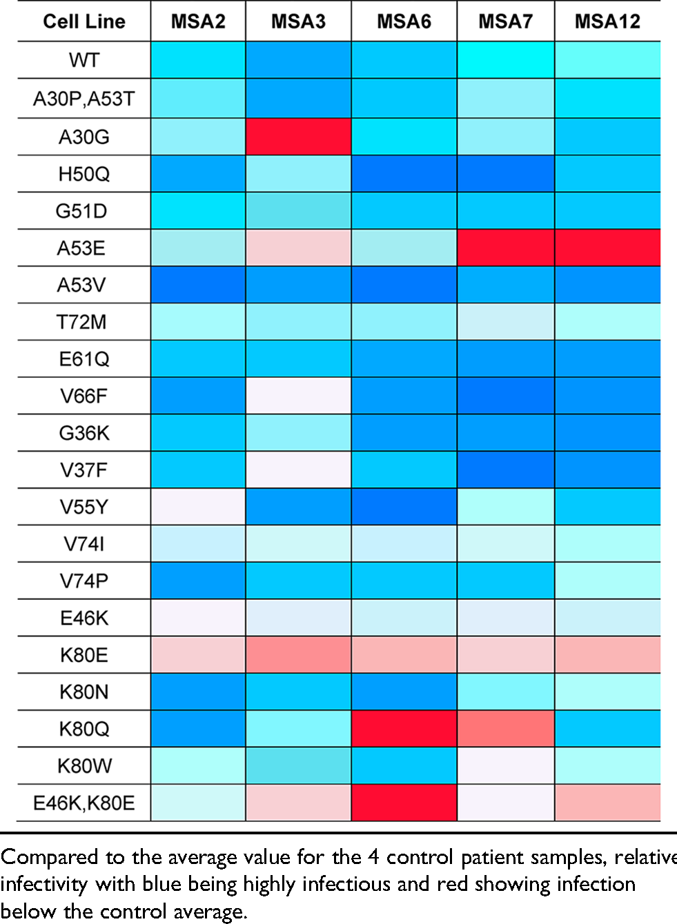
In addition to investigating the effect of PD mutations on MSA propagation, we used the reported MSA and PFF structures to design non-pathogenic mutations with the goal of disrupting α-synuclein misfolding into specific conformations (Table 2; Figure 3). As expected, the E61Q and V66F mutations, which were designed to interfere with the rod and twister PFF conformations (residues noted in purple in Supplemental Figure 1B and C), largely facilitated MSA replication, as did the V74P mutation, though with a blunted response. Unexpectedly, the V74I mutation blocked MSA propagation. This is particularly interesting given that our Maestro analyses suggested that both the V74I and V74P mutations would reduce the total energy of the structure. By comparison, the G36K, V37F, and V55Y mutations were all designed to interfere with the protofilament interface in the MSA structures; however, V55Y exerted the strongest inhibitory effect on MSA patient sample propagation. It should also be noted that residue V55 is located next to a small unfilled pocket in the cryo-EM structure, and it is unclear if that pocket is filled with something exogenous. As a result, we were unable to account for these potential interactions in our calculations. While analyzing our cell assay data, we noticed that in some cell lines, such as V55Y, one of the MSA patient samples appeared as an outlier. We also noticed that there was often a large range in replication ability across the patient samples, as is seen in the V66F and G36K data. To determine if this variability is due to differences in titre, or the amount of replication competent α-synuclein present in each sample, we created a heat map showing cell infection relative to the average of the control samples for each patient sample on every cell line tested (Table 4). We observed that a single patient sample is neither responsible for the lowest nor the highest infection across the cell lines, suggesting that our findings are not due to differences in titre. Instead, these data suggest the potential presence of multiple α-synuclein strains in the MSA patient samples. Given that some mutations tested, for example V66F, should have no impact on MSA replication based on the location of the residue in the reported structures, and others, such as V55Y, should inhibit α-synuclein misfolding, it is highly likely that these unreported strains have conformations that are distinct from reported structures (both residues are highlighted in purple in Figure 1A). One caveat to this interpretation is that it is currently unknown what effect in vitro replication of MSA α-synuclein in the α-syn140*-YFP cell lines has on protein structure, which may contribute to the results reported here.
Heat map of MSA patient sample cell infection relative to control.
Our previous finding that the E46K mutation inhibits MSA replication is consistent with research on the prion protein (PrP), which has shown that a single residue change in the protein sequence can inhibit the conversion of cellular PrPC into pathogenic PrPSc. Notably, a single residue change is responsible for the species barrier creating resistance of the Canidae family to prion diseases,42–44 and for conferring resistance of sheep to scrapie45,46 and humans to Creutzfeldt-Jakob disease. 47 At the time we discovered the effect of the E46K mutation on MSA replication, we relied on the solid-state NMR structure of α-synuclein PFFs 30 to interpret our findings. In this structure, a Greek key motif is stabilized by a salt bridge between residues E46 and K80, leading to our hypothesis that the E46K mutation prevents salt bridge formation. 48 The more recent resolution of α-synuclein fibrils isolated from MSA patient samples confirmed that this salt bridge stabilizes the MSA conformations, 7 supporting our initial hypothesis. To test this, we designed four mutations targeting the second half of the salt bridge (residue K80) and found that the K80E and K80W mutations blocked MSA propagation (Figure 4). With the goal of re-establishing the salt bridge to rescue MSA replication, we then built cells expressing the double E46K,K80E mutations, and were surprised to find that MSA transmission remained inhibited (Figure 5). Modelling the effect of this residue swap using Maestro showed that the double mutation results in a small increase in total energy for both subunits due to an angular salt bridge with minimal orbital overlap, likely resulting in the inhibitory effect observed in vitro. Notably, the E46/K80 salt bridge is an intermolecular interaction, rather than intramolecular, meaning that the salt bridge extends between two layers of the protofilament. Given the strong effect of the E46K and K80E mutations on blocking MSA propagation, this salt bridge may act as a key area on the fibril surface for driving self-templating, serving as an anchoring point to facilitate α-synuclein conversion.
An important finding from this work is that protein modelling software is not always capable of accurately predicting the effect of each mutation on MSA strain biology. For example, as was the case for residues G36, V37, H50, and G51, the location of the residue within the templating region contributes to the reliability of the modelling data. There are three important caveats to our approach that may contribute to these discrepancies. First, our methods used implicit waters in the force field calculations instead of explicit waters. Second, there are pockets in the MSA fibril structures, some of which are known to be occupied by non-protein densities, however, because it is unknown what molecules are present in these pockets, we are unable to account for their interactions with mutated residues. And third, we are using thermodynamic calculations to try to predict a kinetic process. While Maestro can be used to assess the capacity of an established structure to house a mutated residue, it is unable to predict the ability of the mutant substrate to misfold into a specific conformation.
Attempting to address this limitation, our initial approach to modelling the effect of each mutation using Maestro focused on mimicking the experimental design of our in vitro studies by only mutating 3 layers of the protein fibril. The goal was to computationally recapitulate the effect of self-templating on the catalytic surface of the MSA fibril using the mutant α-synuclein expressed in each cell line. However, we quickly discovered that this approach failed to fully capture the consequences of each mutation on the change in total energy, making it difficult to assess the likely effect of each mutation on MSA replication in vitro. Instead, when we expanded our analyses to mutate 6 layers of the protofilament, the effect of each mutation was much more apparent, increasing the applicability of our in silico modelling to interpreting our in vitro data (Table 5). The problem with mutating just 3 layers of the protein fibril may, in part, be an unintended consequence of our control mutation (often E83X, which was chosen to minimize interactions with neighbouring residues). This step was required to keep the amino acids identical for the purpose of comparing total energies. Intramolecular interactions between the sidechains of WT and mutant amino acids were often energetically favourable. The control mutation doubles the energy of these interactions, distorting the energy calculations given that the control mutations are not present in the cell models.
Total energy change after mutagenesis of 3 versus 6 protein layers in MSA structure 6XYQ.
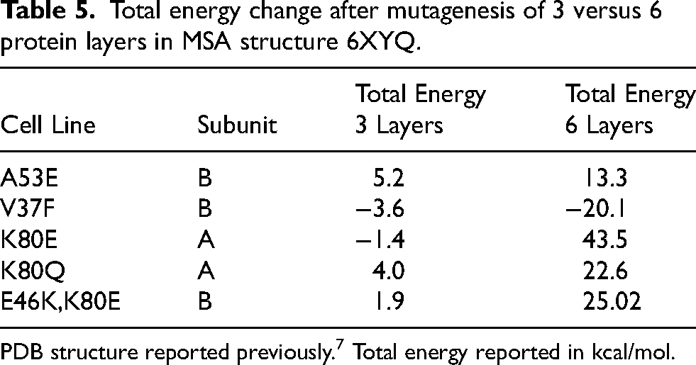
PDB structure reported previously. 7 Total energy reported in kcal/mol.
A final critical limitation to our in silico approach is the inability to predict deformed templating of a prion strain. In cases where Maestro predicted a mutation would increase total energy but templating was still permitted in vitro, such as the V37F mutation, it is possible that a slight alteration in the templating surface occurs to facilitate incorporation of the mutant residue. Deformed templating is thought to be a contributor to prion strain adaptation when strains are successfully transmitted to hosts with a different genotype or across species barriers.49–52 In these cases, future studies should investigate maintenance of strain properties after passaging to determine if deformed templating occurs, and if so, how the altered protein structure impacts prion biology.
The remarkable advances made in structural biology methodology, particularly for prionogenic proteins, have had a tremendous impact on our understanding of protein misfolding and strain biology. The availability of these structures now serves as an opportunity for hypothesis development focused on elucidating the structure-function relationship underlying the ability of each prion strain to exert unique biological consequences in disease. However, the nature of structural techniques limits our understanding of fibril formation to a single snapshot from the brain of a deceased patient, failing to capture the dynamic processes that lead to the final fibrillar product. In this study, we paired structural analysis with in vitro models of α-synuclein misfolding to directly test the effect of targeted mutations on MSA propagation with the goal of understanding how α-synuclein misfolds into distinct conformations. Having shown that MSA prion replication is incompatible with some of the familial PD SNCA mutations, we demonstrated that the mechanism underlying the E46K-driven inhibition is due to disruption of a critical salt bridge between residues E46 and K80. Moreover, using novel mutations designed to perturb protein misfolding, we find evidence suggesting additional unresolved α-synuclein strains are present in MSA patient samples. In addition to furthering our understanding of α-synuclein misfolding in MSA, this work has established a powerful panel of α-syn140-YFP cell lines that can be used to interrogate MSA strain biology.
Supplemental Material
sj-docx-1-pkn-10.3233_JPD-240296 - Supplemental material for Structurally targeted mutagenesis identifies key residues supporting α-synuclein misfolding in multiple system atrophy
Supplemental material, sj-docx-1-pkn-10.3233_JPD-240296 for Structurally targeted mutagenesis identifies key residues supporting α-synuclein misfolding in multiple system atrophy by Patricia M Reis, Sara AM Holec, Chimere Ezeiruaku, Matthew P Frost, Christine K Brown, Samantha L Liu, Steven H Olson and Amanda L. Woerman in Journal of Parkinson's Disease
Footnotes
Acknowledgments
ORCID iDs
Funding
The authors disclosed receipt of the following financial support for the research, authorship, and/or publication of this article: This work was supported by grants from the NIH (R01NS121294 and accompanying supplement and R21NS127002) to A.L.W. Control tissue samples were provided by the NIH NeuroBioBank and by the Massachusetts Alzheimer's Disease Research Center (MADRC), which is supported by 1P30AG062421. Multiple system atrophy patient samples were provided by the MADRC and the Sydney Brain Bank at Neuroscience Research Australia, which is supported by The University of New South Wales and Neuroscience Research Australia.
Declaration of conflicting interests
The authors declared the following potential conflicts of interest with respect to the research, authorship, and/or publication of this article: A.L.W. is an Editorial Board Member of this journal but was not involved in the peer-review process of this article nor had access to any information regarding its peer-review. S.H.O. and A.L.W. are the co-founders of Allagus Therapeutics, which did not contribute financial or any other support to these studies. S.H.O. and A.L.W. are inventors on U.S. Patent Application PCT/US2023/072173, which includes data reported here.
Data availability
The data supporting the findings of this study are available within the article and/or its supplemental material.
Supplemental material
Supplemental material for this article is available online.