
Abstract
Parkinson’s disease (PD) is a movement disorder with many symptoms responsive to treatment with dopamine agonists, anti-cholinergics and the dopamine precursor, levodopa. The cardinal features of PD include tremor, rigidity, bradykinesia, and postural instability. There also are non-motor features that include sleep disorders, cognitive and affective dysfunction, hyposmia, pain and dysautonomia (constipation, bloating, orthostasis, urinary symptoms, sexual dysfunction, dysphagia). Among these non-motor features are signs and symptoms of visual system impairment that range from subtle examination findings to those causing severe disability. In this review we describe common PD-related abnormalities in the visual system, how they present, and potential treatments.
SCOPE OF VISUAL SYSTEM IMPAIRMENT IN PARKINSON’S DISEASE
Pathology in Parkinson’s disease (PD) involving the visual system occur at all levels of visual processing from the ocular environment where light is first detected to cortical areas that mediate interpretation of visual inputs. A recent survey found increased self-reporting of visual impairment in PD patients compared to controls (odds ratio 2.67) [1]. Another study of 848 PD patients and 250 age-matched controls used a detailed 17 question instrument (Visual Impairment in Parkinson’s Disease Questionnaire or VIPD-Q) and found that 82% of patients reported visual symptoms compared to 48% of controls. Symptoms included hallucinations, and other symptoms likely resulting from pathology in the eye, ocular surface, optic nerve and oculomotor function [2]. Visual symptoms interfered with activities of daily living in 68% of patients versus 35% of controls. In addition, people with PD were more likely to see an ophthalmologist, have more ophthalmologic complaints, use a visual aid and report visual aid inadequacy. PD patients had more driving difficulties and more frequent falls.
Overall visual function has been reported to be slower in PD as modeled in a picture identification task (MULES test – Mobile Universal Lexicon Evaluation System) and this correlated with disease severity [3]. In addition, PD patients made more naming errors and seemed to show a larger re-test improvement effect than controls, though a ceiling effect in controls could be responsible. Future studies should compare MULES to other visual assessments in PD and determine its clinical relevance.
EYELID AND OCULAR ABNORMALITIES
Abnormal visual function in PD may involve abnormal eyelid function (usually reduced blink rate, but rarely apraxia of eye opening or reflex blepharospasm), multiple ocular film abnormalities, corneal abnormalities, meibomian gland stenosis/blockage, decreased tear production and seborrheic blepharitis [4–6]).
Dry eye affects 53–87% of patients with PD and is associated with reduced blink rate as low as 1–2 per minute [6, 7]. Decreased blink rate results in increased tear evaporation by reducing the tear film lipid layer, particularly in the presence of associated blepharitis [8]. This in turn may result in increased ocular inflammation and worsening symptoms. Besides reduced blink rate, autonomic dysfunction affecting the lacrimal glands may result in ocular surface abnormalities [6]. Lid abnormalities, including reflex blepharospasm, can lead to added ocular discomfort, vision impairment and light sensitivity. In addition, the PD drug amantadine and other dopaminergic agents have been linked to reversible corneal edema with subsequent vision change [9, 10].
In our experience patients with ocular surface/eyelid abnormalities complain of dry eye, blurred vision, excessive tearing, foreign body sensation, glare, light sensitivity, problems keeping the eye open and general eye discomfort.
Traditional interventions (not PD-specific) to ease symptoms include treatment of blepharitis with lid hygiene, and dry eye with artificial tears, moisture chambers, nutritional supplements, scleral lenses and tear duct occlusion [11, 12] and interested readers are referred to an excellent review detailing diagnosis and treatment of dry eye in general, including level of evidence for particular treatments [13]. Blepharospasm responds to botulinum toxin injection of the eyelids [14].
Future research into dry eye treatment should include testing novel eyedrop formulations such as those containing blood and platelet-derived components and tear proteins (thymosin β4 and lubricin) well as drugs to increase tear production (lactritin, diquafosol, rebamipide) and those to reduce inflammation [15]. There are procedures (meibomian gland probing, intense pulsed light, vector pulsed light therapy) and other physical methods using applied warmth and physical expression of meibum and devices (intranasal lacrimal gland stimulation, scleral lens) in development to protect the eye, treat blepharitis and to unblock meibomian gland ducts [15]. The effectiveness of these newer interventions is unclear in the general population as well as in those with PD thus requiring more study.
The potential severity of eyelid and ocular abnormalities in PD, their high incidence, the generally more advanced age of PD patients and the potential contributions of PD medications on pathology leads us to strongly encourage routine ophthalmological examination. Those having on-going symptoms are referred more urgently. Though no PD-specific guidelines exist, we recommend at least once a year ophthalmological follow up which is only a bit more aggressive than the once every 1–2 years recommended by the National Eye Institute for everyone over age 60 (http://www.nei.nih.gov).
DIPLOPIA AND OCULOMOTOR ABNORMALITIES
Ocular motility abnormalities in PD include saccadic intrusions, vestibulo-ocular reflex abnormalities, increased square-wave jerks associated with fixation, as well as other saccade and pursuit abnormalities [16–18]. Oculomotor abnormalities however are not as prominent as in other Parkinson’s-related disorders, such as progressive supranuclear palsy [19] and their clinical relevance is unclear. It has been shown, however, that saccadic reading assessed by the King-Devick test in PD patients is 20% slower than controls [20] suggesting a potential role for abnormal extraocular movements in clinical impairment.
Diplopia is present in 10–30% of PD patients [7] and occurs more frequently as PD progresses [21]. Convergence insufficiency (CI) is prevalent in PD and often is associated with binocular horizontal diplopia at near. It is experienced during reading, and is exacerbated by dry eyes or ocular surface irritation, particularly with reduction in blink rate [22].
One should differentiate binocular diplopia due to CI or pre-existing strabismus from monocular diplopia generally resulting from refractive errors, corneal pathology, cataract formation or macular disorders that would have more specific ophthalmological treatments (e.g., cataract removal, refraction and spectacle correction, macular and corneal treatments). Selective or cortical diplopia (diplopia of single objects) is a rare phenomenon associated with visual hallucinations [23].
Management of CI by the orthoptist and the ophthalmologist includes single vision reading glasses, base-in prisms or monocular occlusion. We counsel against focal or progressive lenses as they tend to impede binocular vision and prevent the use of prisms in the lower portion of the lens. Patients may benefit from typoscopes, E-tablets and proper lighting to facilitate reading [24].
Intermittent diplopia at near related to CI may improve with prismatic correction and optimization of levodopa (especially if visual complaints are related to waning of dopaminergic medication effect, i.e., wearing off) [25]. In one study of PD patients, nearly 20% reported that levodopa treatment improved diplopia though this was not verified experimentally [26], while convergence did measurably improve in patients in the on-state (90 minutes after taking levodopa) versus prior to their routine levodopa dosing when the medication had started to wear off [27]. The authors suggest that medication-induced fluctuations in convergence ability may make proper prism therapy difficult and this is important as prism placement is likely the most common intervention used in all adults for this condition [28].
RETINAL AND OPTIC NERVE ABNORMALITIES
Optical coherence tomography (OCT) has emerged as a tool in the assessment of retinal changes in PD, often with conflicting findings, reviewed in [29]. Despite the lack of reproducibility in retinal OCT studies, there are likely retina changes in PD with patterns of thinning possibly related to cellular or vascular changes [29]. Abnormal accumulation of retinal alpha-synuclein (whose abnormal accumulation in cells helps to pathologically define PD in the brain) is further evidence of retinal pathology in patients with PD [30, 31]. A recent meta-analysis of 36 studies determined that patients with PD have significantly thinner retinae compared to gender and age-matched controls [32]. This study also noted the similarity of inner retinal layer thinning seen in PD with those of other neurodegenerative disorders.
There is an increased incidence of glaucoma-like changes (optic nerve appearance, cup to disc ratio and glaucoma-like visual field changes) in PD, despite lower average intraocular pressures [4]. Hypotheses linking PD to a glaucoma-like condition have included the role of progressive dopamine depletion and alpha-synuclein induced axonal degeneration [33]. Clinical and pathology similarities and differences between PD-related optic nerve changes and what is commonly referred to as glaucoma require more investigation as does the role of standard glaucoma treatments in PD-related retinal disease.
Symptoms related to retinal pathology include reduced visual acuity, visual field defects, color vision impairment (though central mechanisms may be more important [34]), and reduced contrast sensitivity [35, 36].
Treating retinal and optic nerve pathology involves the optimization of levodopa therapy (found to improve contrast sensitivity and color vision [37, 38]) and ophthalmologic referral for glaucoma evaluation and acuity optimization. New drugs used to treat normal tension glaucoma through mechanisms that rescue nerve fibers may be most applicable to PD and such studies are on-going. For a summary of pathologies and treatments, see Table 1.
Ophthalmic pathology and management in Parkinson’s disease. Treatment recommendations are often not PD-specific and are taken from general ophthalmologic practice, literature review and author’s experience. See text for details
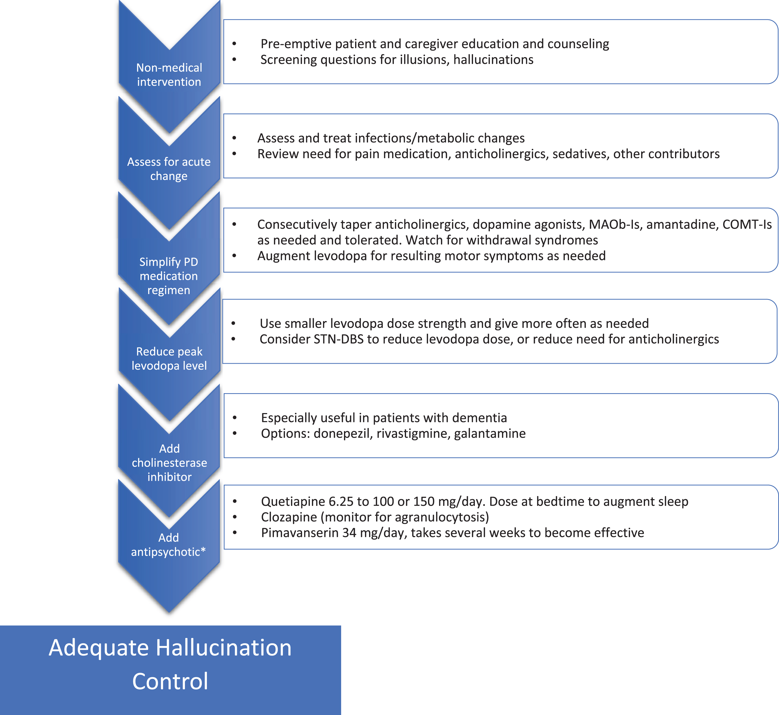
Addressing hallucinations in Parkinson’s disease. Flow chart for stepwise treatment of hallucinations in PD. See text for details and additional warnings. *Use of antipsychotics in patients suffering from dementia must proceed with caution given the black box warning for increased mortality, also QTc should be monitored. MAOb-I, monoamine oxidase B inhibitor; STN-DBS, subthalamic nucleus-deep brain stimulation; COMT-I, catechol-O-methyltransferase inhibitor.
Finally, it is important to educate patients on the benefits of increased levels of lighting and to provide warnings about driving, especially in low contrast environments (fog or twilight) as contrast specificity may be a significant factor in driving fitness [39].
HALLUCINATIONS AND VISUOPERCEPTUAL IMPAIRMENT
Visual hallucinations are reported by about 30% of PD patients, with a lifetime prevalence of up to 60–80% [40, 41]. Often hallucinations are accompanied or preceded by illusions (visual misinterpretations), or a false sense of presence or something moving in the environment. Hallucinations occur with or without insight into their false nature, the latter being more problematic. PD-related hallucinations negatively impact quality of life, ability to live independently and on mortality [11].
Hallucinations and visual misperceptions can be caused by pathology all along the visual pathway from impaired visual sensory input due to ocular changes back through pathological changes in the cortex [42]. Genetic polymorphisms, disease severity, age, sleep disturbances, dementia and certain medications use contribute to hallucination risk [11, 43–45]. Impaired vision due to any cause increases the risk of visual hallucinations (Charles Bonnet syndrome) and causes such as cataracts and macular degeneration should be identified and treated.
The sudden onset or worsening of hallucinations can result from changes in medications or from an infectious/metabolic abnormality. In our experience, dehydration, and urinary tract infections as well as the initiation of pain or urinary (anti-cholinergic) medication are frequent, reversible causes.
Treatment of hallucinations may best begin prior to their development by preparing the patient and caregivers for their emergence. This helps to normalize this symptom and hopefully reduce the stigma and potential distress of their presence. We encourage patients to notify their physician of the emergence of hallucinations and to discuss whether they are causing enough distress to require treatment.
Treating problematic hallucinations involves tapering back contributory medications when possible and this includes every PD medication to varying degrees. The recent publication of guidelines resulting from a workshop on visual hallucinations in a variety of disorders, citing guidelines from the National Institute for Health and Care Excellence from 2017, were incorporated into the strategy for addressing PD-related hallucinations found in Fig. 1 [11]. Levodopa monotherapy for PD symptoms should be the eventual goal.
If hallucinations persist, a reduction of levodopa dose including the use of lower dose strengths, perhaps given more often (to reduce levodopa peak plasma levels) should be attempted. In carefully screened patients who require anticholinergics or levodopa to control symptoms, the dose requirement can be reduced using subthalamic nucleus (STN) deep brain stimulation to mitigate side effects, including hallucinations [46, 47].
If medication adjustment does not improve hallucinations, we consider adding medications to directly target the problem including cholinesterase inhibitors and antipsychotics.
Several studies offer evidence of the potential benefit of cholinesterase inhibitors in PD-related hallucinations, especially in the presence of concomitant dementia [48, 49]. Potential side effects including GI upset/bleeding, sialorrhea, increased tremor, syncope, seizures, and arrhythmia, among others, need to be considered.
Antipsychotics can improve PD hallucinations. The most often employed are quetiapine, clozapine and pimavanserin as others often worsen motor function at therapeutic doses. Other side effects including QTc prolongation and increased mortality must be considered [50, 51]. The use of quetiapine for PD-related hallucinations is common, though clear evidence of benefit is generally lacking [52]. Despite this, the perception by many is that quetiapine is generally well-tolerated when kept at doses at or below 100–150 mg daily, is beneficial for overnight sleep when dosed at bedtime and is more convenient than using Clozapine and pimavanserin (see below).
Pimavanserin is the only medication approved for the treatment of PD psychosis and likely works via serotonin 2a (5-HT2a) receptor inverse agonism. A recent meta-analysis of four relevant studies demonstrated efficacy on a variety of measures and a lack of significant motor side effects [53]. It does require several weeks to take effect and is more expensive than quetiapine.
Clozapine reduces hallucinations in PD patients at daily doses ranging from 12.5 mg to 50 mg without significant effect on motor function [54]. Despite proven efficacy and introduction decades ago, widespread use is limited by concerns regarding granulocytopenia and the frequent laboratory monitoring required for its use.
Importantly, control of hallucinations with additional agents such as clozapine allows levodopa doses to be increased and better optimized, and other anti-Parkinson medications to be used with a reduced risk of hallucinations returning [55].
Addressing visual hallucinations continues to be an unmet need in PD therapy. The presence of hallucinations continues to limit medication options and may continue to be present despite best therapy. Future areas of potential research include options to improve visual acuity through treatment of concurrent condition (such as experimental treatment of macular degeneration) or therapies that may improve PD-related retinal degeneration. A clue to potential therapies comes from case reports addressing hallucinations in Charles Bonnet syndrome where a variety of available medications including gabapentin, serotonergic agents as well as transcranial magnetic stimulation may offer promise (reviewed in [11]). There are on-going trials assessing the potential benefit of ondansetron and novel antipsychotics such as SEP363856 (affecting serotinergic and TAAR1 receptors). Nelotanserin, whose mechanism parallels that of pimvansenin is in clinical trials. Saracatinib, a kinase inhibitor postulated to block 5HT-2a receptor effects via inhibition of the downstream Src kinase, is also under study.
CONCLUSIONS AND FUTURE DIRECTIONS
There is increased awareness of visual dysfunction as a “non-motor” feature of PD. The availability of reporting tools such as the VIPD-Q will lead to increased awareness of visual symptoms in patients with PD and help standardize research in the field. Advances and optimization of OCT will demonstrate the nature of retinal changes that may help in diagnosis, prognosis and treatment not only for visual symptoms, but also for PD in general, as several studies have showed an association between retinal thinning and disease progression and severity. There is a current study examining the association of OCT changes with PD progression and to identify possible PD patients from high risk populations prior to diagnosis. Interventions to treat dry eye and glaucoma may be generalizable to patients with PD and further research is needed to examine this. From a practical standpoint, early recognition and management of visual disorders in PD is important to prevent further deterioration and unnecessary disability. Visual changes impact driving and falling in PD and proper treatment and counseling can reduce these hazards. Physicians are encouraged to ask patients about various symptoms of ocular involvement and refer to ophthalmologists or neuro-ophthalmologists.
CONFLICT OF INTEREST
The authors have no conflict of interest to report.