
Abstract
BACKGROUND:
Blueberry production has generated great commercial expectations, therefore for its agricultural expansion it is necessary to overcome the challenges at the time of mass propagation.
OBJECTIVE:
Evaluate the effect of a set of factors influencing micropropagation, as well as the influence of substrates on the ex vitro morpho-physiological performance of blueberry seedlings.
METHODS:
A set of protocols were developed to optimize all stages of micropropagation (aseptic establishment, multiplication, rooting, and acclimatization) of blueberries.
RESULTS:
Explants immersed in 1.5% NaClO for 8 min and then in 0.1% HgCl2 for 2 min achieved 100% sterility and a viability rate of 86.67% for ‘Biloxi’ and 93.33% for ‘Bluecrop’. At the multiplication stage, the maximum number of shoots of ‘Biloxi’ (3.53) and ‘Bluecrop’ (2.27) were obtained on the medium supplemented with 0.2 and 10 mg L–1 silver nanoparticles (AgNPs), respectively. The percentage of in vitro rooting was significantly improved on media containing activated charcoal, with levels between 80% and 100%. In the acclimatization phase, plants grown in a substrate composed of peat and cocomix® (2:1 ratio) showed greater uniformity and better morpho-physiological behavior.
CONCLUSIONS:
The present results could be successfully used for large-scale commercial production of blueberries of the varieties ‘Biloxi’ and ‘Bluecrop’.
Introduction
The blueberry (Vaccinium corymbosum L.) is a fruit that has gained popularity owing to its multiple health benefits, qualities associated with the content of anthocyanins, flavanol’s and phenolic compounds [1]. These bioactive compounds make blueberries increasingly in demand. Peru is one of the main exporting countries for blueberries in South America [2]. Recent studies have reported the existence of high potential areas to expand the production of blueberry [3]. Nevertheless, agricultural expansion is conditioned by challenges at the time of mass propagation.
Tissue culture is a widely used tool in agriculture to overcome problems related to plant production, as it allows obtaining large numbers of healthy and uniform seedlings in a short period of time [4]. Blueberry micropropagation, which has been extensively studied, always reflects variable responses because there are important factors to be taken into account, such as the genotype introduced [5, 6], the composition of the growing medium and growth regulators [7], and the acclimatization conditions [8].
For the initiation of in vitro propagation, the sterilization of plant material is of crucial importance, especially when the explants come from the field [9]. On the other hand, during the multiplication stage, different growth regulators are usually used to induce the development of new shoots [10–12]. However, in recent years, the addition of nanomaterials has been shown to be a revolutionary alternative to optimize organogenesis protocols [13, 14]. In plant tissue culture, the application of nanoparticles such as silver has been shown to be useful for eliminating microorganisms [15, 16]. In addition, several studies have shown that the use of silver or cobalt nanoparticles can improve the regeneration and growth of explants, as well as the development and quality of seedlings [13, 18], as they play an important role in inhibiting the formation and activity of ethylene gas [13, 14]. However, although the effect on other crops has been positive, the effects of nanoparticles on blueberry micropropagation should be more thoroughly investigated.
In blueberry tissue culture, in vitro rhizogenic development and ex vitro acclimatization are other challenges that need to be addressed. During the in vitro rooting stage, it is important to ensure that seedlings develop an adequate root system and aerial morphology; this is because development with morphological and anatomical abnormalities (absence of cuticle and non-functional stomata) can lead to significant losses during the transition to ex vitro conditions [8]. Therefore, assessing the effect of other factors involved in the process is very important, such as sugar content [19] and activated charcoal [20], which can help improve seedling quality by stimulating pluripotency, and balancing the growth regulators present in the medium [21].
In acclimatization, a delicate stage of micropropagation, the substrate selection represents a vital factor for the survival and development of the new seedlings [22]. Soil-specific requirements such as pH, organic matter content and structure reflect the need to use a suitable support material (substrate) [23], as non-optimal conditions can lead to poor morphological and physiological development of the plants and increased losses due to mortality, limiting their commercial production.
In this context, this study aims to (I) generate a protocol for the establishment of aseptic culture; (II) evaluate the effect of concentrations of silver nanoparticles for in vitro multiplication; (III) evaluate the effect of concentrations and combinations of growth regulators, activated charcoal and sucrose on in vitro rooting and ex vitro hardening, and (IV) evaluate the morpho-physiological characteristics of in vitro propagated seedlings by substrate type.
Materials and methods
In this study, individual experiments were designed to develop and establish methods for successful micropropagation of two blueberry varieties (Vaccinium corymbosum L. var. ‘Biloxi’ and ‘Bluecrop’) using nodal segments, as well as to evaluate acclimation and morpho-physiological behavior of new seedlings (Fig. 1).
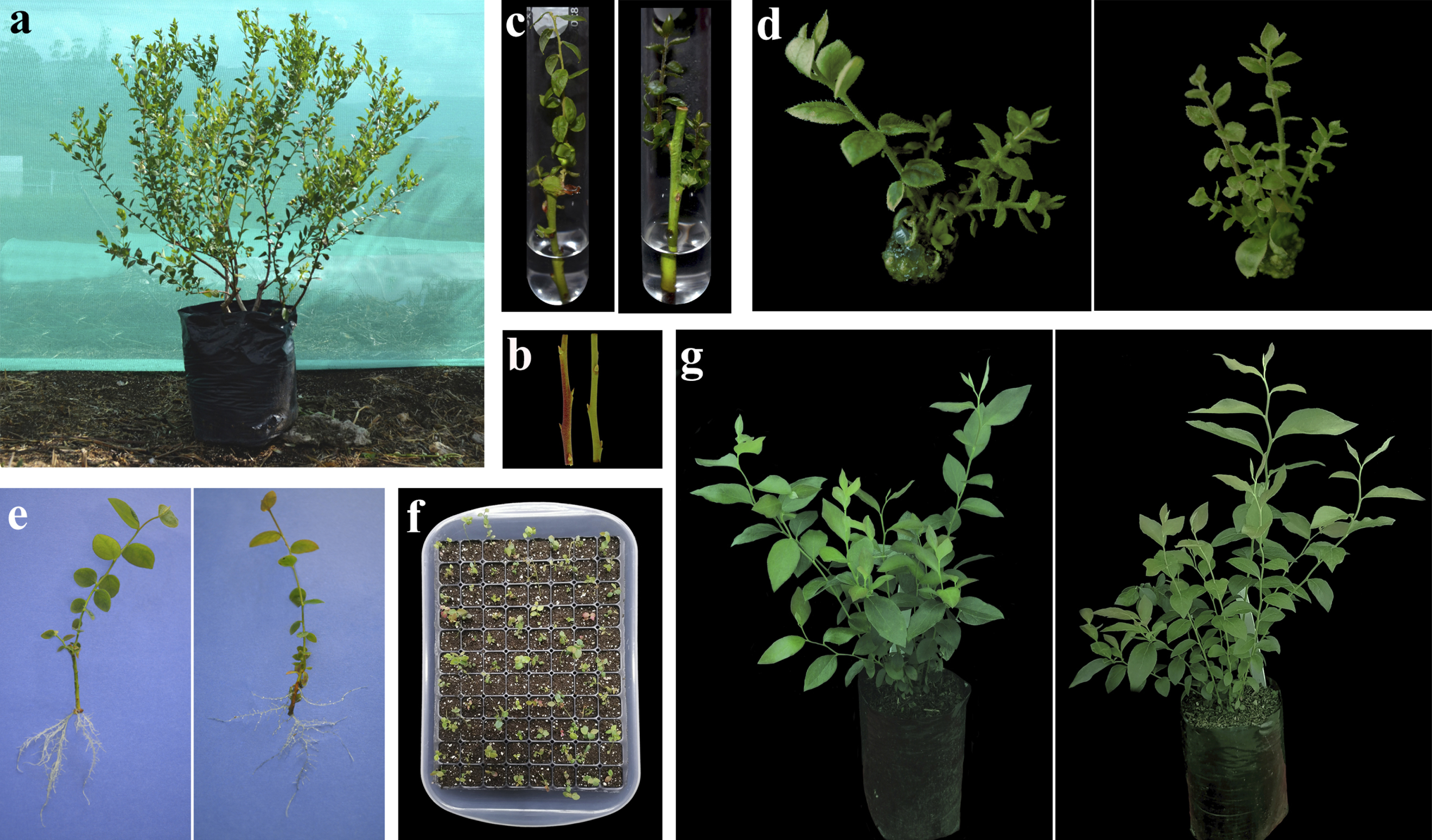
In vitro propagation of Vaccinium corymbosum L. (a) Parent plant, (b) Explants, (c) Establishment, (d) In vitro multiplication, (e) In vitro rooting, (f) Ex vitro acclimatization, (g) Regenerated plant in nursery stage. To the left = ’Bluecrop’, To the Right = ‘Biloxi’.
Semi-woody shoots (15 to 20 cm in length) were collected from healthy and vigorous 5-year-old plants grown at the Experimental Station of the Instituto de Investigación para el Desarrollo Sustentable de Ceja de Selva (6°12’ S and 77°40’ W; 2426 m a.s.l.) in the district of Molinopampa, Chachapoyas, Peru (Fig. 1a).
Culture medium and growth conditions
In this study, we used base culture medium Woody Plant Medium (WPM) with vitamins [24] and 0.15 g L–1 ascorbic acid, supplemented according to the experiment, with different concentrations of silver nanoparticles, growth regulators, sucrose and activated charcoal. The pH was adjusted to 5.2±0.5 (HCl 1N / KOH 1N), 6 g L–l agar was added for solidification (except for in vitro establishment), then autoclaved at 121°C with a pressure of 1.5 Kgf.cm–2 for 20 min. The explants were grown at 25±1°C under 16 h light photoperiod with white fluorescent tubes (3000 lux).
In vitro establishment
The explants were washed with liquid detergent and tap water to remove particles from the shoots; the leaves were then removed and cut into 7 cm segments (Fig. 1b) which were then immersed in 0.5 g L–1 Benomyl for 20 min and thoroughly rinsed under running water. Under aseptic conditions, sterilization of explants started with immersion in 70% (v/v) alcohol for 1 min, followed by immersion in two concentrations of sodium hypochlorite (1.5, 3.0 % NaClO) while shaking for 4 or 8 min, and then treated with two concentrations of mercuric chloride (0.1, 0.2 % HgCl2) at two immersion times (2 and 5 min). Finally, they were rinsed with sterile water and the explant ends were removed. Each explant was cultured in test tube with 15 ml of base medium plus 30 g L–1 sucrose (Fig. 1c). The experiment was conducted under a four-factor design (two concentrations of NaClO at two immersion times, combined with two concentrations of HgCl2 at two immersion times), with three replicates per treatment. Each repetition corresponded to five experimental units (explants). After three weeks, the percentage of oxidized, contaminated and viable explants was recorded.
In vitro multiplication
Nodal segments of 1 cm length with 2 buds were cut and placed on base medium plus 30 g L–l sucrose. All media contained 0.1 mg L–1 indole-3-butyric acid (IBA) and 2 mg L–1 trans-zeatin (used as control) [25], and different concentrations of silver nanoparticles (0.2, 0.5, 1.0, 1.5, 2.0, 5.0, 10.0 mg L–1 AgNPs). The experiment consisted of eight treatments and three replicates. Each repetition corresponded to five experimental units (explants). Five explants were grown per PTL-100 culture flask (volume 370 ml; width 7.5 cm; height 9.8 cm, PhytoTechnology Laboratories, Lenexa, Kansas, USA). After 4 weeks the number of shoots, shoot height and number of leaves were recorded (Fig. 1d).
In vitro rooting and ex vitro hardening
Axillary shoots of 1.5 cm length were grown individually in 3.9” x 11.8” polyethylene bags with base medium at half concentration, supplemented with different types and concentrations of auxins [0.5, 1, 2 mg L–l IBA; 0. 2, 0.5, 1 mg L–1 1-naphthaleneacetic acid (NAA)] which were combined with activated charcoal (0 and 2 g L–1) and sucrose (10 and 20 g L–l). The experiment was conducted under a 6×2×2 factorial design (auxin, activated charcoal and sucrose concentrations), with three replicates. Each repetition corresponded to five experimental units (explants). After 45 days, rooting percentage, number of roots, root length (cm), number of shoots, and shoot length (cm) were recorded (Fig. 1e).
Subsequently, and after the in vitro rooting stage treatments, the seedlings were then inserted into plug trays containing a 2:1 peat + perlite mixture (pH 5.4) (Fig. 1f). The plug trays were placed in trays and covered with a plastic sheet to maintain high humidity. They were placed in a microtunnel (arranged under 70 % shade), with an internal temperature of 22 to 25°C and 80 % relative humidity regulated by a mist irrigation system. The plants were watered regularly and the film was gradually removed in the first two weeks. After three weeks, the percentage of pre-acclimated seedlings (ex vitro hardening) was recorded.
Ex vitro acclimatization
Hardened plants (average height 7 cm and minimum 2 shoots) were transplanted into 6” x 8” nursery bags. Five types of substrates were used (Table 1): sawdust (white wood), cocomix®, raw rice husk, perlite and burnt rice husk, which were combined with peat soil in a 1:2 ratio. Plants (Fig. 1g) were grown in a shaded nursery (50 % shade; light intensity: 635±5 Lux), under photosynthetically active radiation (6:00 am–6:00 pm) of 733.34±93.70μmol m–2 s–1 (mean±SD), average temperature of 16.57±0.53° C and average relative humidity of 80.47±3.96 % (Fig. 2, data recorded by an OREGON Automatic Weather Station model WMR300PU). The plants were regularly irrigated with a solution of ammonium sulphate [0.12 g L–1 (NH4) 2SO4], di-ammonium phosphate [0.06 g L–1 NH4H2PO4], potassium sulphate [0.025 g L–1 MgSO4], magnesium sulphate [0.025 g L–1 MgSO4], zinc sulphate [0.002 g L–1 ZnSO4] and boric acid [0.0002 g L–1 H3BO3]. The experiment consisted of five treatments (substrates), with five replications (plants).
Chemical and physical characteristics of substrates used for ex vitro acclimatization of blueberries
Chemical and physical characteristics of substrates used for ex vitro acclimatization of blueberries
EC: electrical conductivity, CEC: cation exchange capacity, WHC: water holding capacity, EAW: easily available water. Substrate 1: saw dusk + peat; Substrate 2: burn rice husk + peat; Substrate 3: raw rice husk + peat; Substrate 4 = cocomix® + peat; Substrate 5: perlite + peat.
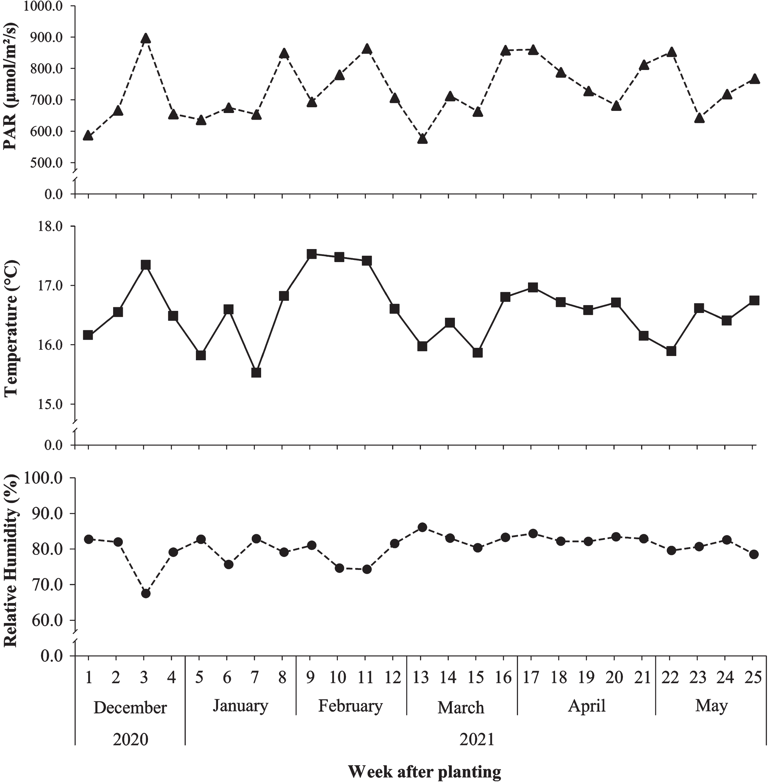
Weekly photosynthetically active radiation (PAR), temperature and relative humidity during the ex vitro acclimatization period of blueberry.
Two weeks after transplanting, the plants were pruned to a height of 4 cm from the root collar. All the morpho-physiological parameters described below were evaluated 25 weeks after the acclimatization stage started.
Morphological measurements were: plant height (every 15 days), number of shoots, leaf area (ImageJ v.1.48 software; http://imagej.nih.gov/ij/), number and length of roots (roots with a length equal to or greater than 1 cm), fresh and dry weights of the cauline and root system. Dry weight was determined after drying the samples in an oven at 80°C to a constant weight.
Physiological characteristics
Chlorophyll content (SPAD index) was measured using a chlorophyll meter SPAD 502 (Konica Minolta, Tokyo, Japan). Stomatic conductance (mmol m–2 s–1) was recorded using a SC-1 leaf porometer (Decagon Devices, Pullman, WA, USA). From each plant, three apical leaves were selected, and the mean of each leaf was the average of three readings, always taken between 8:00 and 11:00 a.m.
Chlorophyll a, b, a + b and carotenoid contents were determined using a Genesys™ 10S UV/Vis spectrophotometer (Thermo Scientific™, USA). Photosynthetic pigments were extracted from 0.2 g of fresh leaf tissue. The samples were crushed, and then 200 mg of magnesium carbonate and 5 ml of 80 % acetone were added. The extracted pigment was transferred to centrifuge tubes (covered with aluminum foil) and centrifuged at 2200 rpm (10–15°C) for 5 minutes. Finally, an aliquot of 1000μl (supernatant) was transferred to spectrophotometer tubes for absorbance readings at 663.2; 646.8 and 470 nm. The concentration of photosynthetic pigments was translated using the following equations [26].
Chlorophyll a (μg/ml): 12.25 A663,2 –2.79 A646,8
Chlorophyll b (μg/ml): 21.50 A646,8 –5.10 A663.2
Total Chlorophyll (μg/ml): Chlorophyll a + Chlorophyll b
Carotenoids (μg/ml): (1000 A470 –1.82 Ca –85.02 Cb) / 198
The relative water content (RWC) was calculated by gravimetry. For this purpose, 3 leaves per plant were selected and 10 discs with a diameter of 8 mm were immediately removed and their fresh weight (FW) was recorded. The samples were then hydrated in Petri dishes with Milli-Q water for 5 hours, surface dried and their turgor weight (TW) was recorded. They were then dried in an oven at 80°C for 48 hours and reweighed to record the dry weight (DW). RWC was calculated using the following equation: RWC = [(FW-DW/TW-DW)×100] [27].
To calculate the stomata index and density, a thin layer of nail varnish was applied evenly on the abaxial surface of the leaflet (middle third free of veins). After a few minutes, the varnish was gently removed, fixed on a slide, stained with methylene blue and observed under a microscope (Leica DM2000 LED, Leica, Germany) equipped with a digital camera (Leica MC170 HD, Leica, Germany). In each optical field (4.5 mm2 area, equivalent to 40x magnification) the number of stomata (NS) and epidermal cells (EC) was counted. The stomata index (SI) was calculated using the following equation: SI = [(NS/(EC+NS)] *100. Stomatal density was determined per mm2 [28].
Experimental design and data analysis
A completely randomized design was used for all experiments. The data were subjected to an analysis of variance, and the means of the variables that were statistically significant were compared using Tukey’s test (P≤0.05). The analysis was performed using the statistical software InfoStat version 2017. Pearson’s correlation test (P≤0.05) was used to analyze the results of all parameters recorded during ex vitro acclimatization.
Results
In vitro establishment
Treatment of explants with 1.5% NaClO for 8 minutes, followed by 0.1% HgCl2 for 2 minutes demonstrated high efficacy for sterilization of the material (both varieties were free of contamination) and achieved the highest viability rate in the in vitro establishment tests of blueberry nodal segments ‘Biloxi’ (86.67%) and ‘Bluecrop’ (93.33%) varieties. In contrast, treatment with 3.0 % NaClO for 8 minutes followed by 0.2 % HgCl2 for 5 minutes resulted in zero percent viable explants, and in the highest oxidation values (Biloxi: 93.33 %; Bluecrop: 100.00 %). The results of treatments differing in both concentration and immersion time in the sterilant used during the establishment of blueberry nodal segments ‘Biloxi’ and ‘Bluecrop’ varieties are detailed in Table 2.
Effect of different sterilization treatments on nodal segments of blueberry varieties ‘Biloxi’ and ‘Bluecrop’ during in vitro establishment
Effect of different sterilization treatments on nodal segments of blueberry varieties ‘Biloxi’ and ‘Bluecrop’ during in vitro establishment
Means within a column followed by the same letter are not significantly different according to Tukey test at P≤0.05.
The number of shoots in vitro showed significant differences for treatments with different concentrations of AgNPs (Table 3). The addition of AgNPs promoted vigorous shoot development and reduced the visual symptoms of vitrification and hyperhydricity. However, the exclusion of this nanomaterial (control treatment) caused an increase in the rate of callus formation.
Effect of silver nanoparticle concentration on in vitro multiplication of blueberry nodal segments ‘Biloxi’ and ‘Bluecrop’ varieties
Effect of silver nanoparticle concentration on in vitro multiplication of blueberry nodal segments ‘Biloxi’ and ‘Bluecrop’ varieties
Means (±standard error) within a column followed by the same letter are not significantly different according to Tukey test at P≤0.05.
The explants of the variety ‘Biloxi’ obtained the highest number of shoots (3.53±0.31) in the treatment with 0.2 mg–1 AgNPs. Regarding the variety ‘Bluecrop’, the highest shoot formation was observed in the treatment with 10 mg L–1 AgNPs; nevertheless, there was no significant difference between the control treatment and the concentrations of 0.5, 1.5, 5.0 and 10.0 mg L–1. In both blueberry varieties, shoot elongation and leaf number of all treatments were statistically similar (Table 3).
The composition of the culture medium had a significant effect on in vitro rhizogenic development and pre-acclimation (Table 4). The best results for the different parameters measured in the in vitro rooting stage were recorded in treatments that had activated charcoal, plus growth regulators and sucrose (regardless of their concentrations). In fact, when explants were grown in the presence of activated charcoal, we observed that seedlings of both blueberry varieties exhibited better root development (number and length of roots) and morphological development (number and length of shoots), which allowed recording preacclimation rates ranging from 80% to 100% (Table 4), while its absence resulted in increased callus formation, which in turn affected regeneration and growth of explants. It should be noted that, regardless of the presence or absence of activated charcoal, the best growth regulator to induce the formation of new roots was IBA. It should be emphasized, however, that the roots formed in a non-activated charcoal medium showed poor growth and, therefore, the seedlings were morphologically underdeveloped.
Effect of IBA, NAA, activated charcoal and sucrose on morphological and rooting characteristics in vitro, and their later influence on ex vitro acclimation of blueberry varieties ‘Biloxi’ and ‘Bluecrop’
Effect of IBA, NAA, activated charcoal and sucrose on morphological and rooting characteristics in vitro, and their later influence on ex vitro acclimation of blueberry varieties ‘Biloxi’ and ‘Bluecrop’
Means (±standard error) within a column followed by the same letter are not significantly different according to Tukey test at P≤0.05.
At the same time, we observed positive correlations (moderate to very high) between root number (‘Bluecrop’: r = 0.91; ‘Biloxi’: r = 0.69), root length (‘Bluecrop’: r = 0.91; ‘Biloxi’: r = 0.87) and the percentage of pre-acclimated plants. From this experiment, the presence of a well-developed system seems to be a fundamental requisite for obtaining successful results in terms of seedlings hardening that could facilitate their direct transplanting into ex vitro environments.
Morphological characteristics
The type of substrate used for vegetative development of the seedlings influenced the morphological characteristics of the blueberry varieties ‘Biloxi’ and ‘Bluecrop’ (Figs. 3, 4). The plant height of the two blueberry varieties was positively correlated with the age of evaluation, i.e., the height, as expected, increased with increasing nursery age (Fig. 3a, b). The most vigorous and tallest plants (‘Bluecrop’: 33.68±0.28 cm; ‘Biloxi’: 29.77±0.85 cm) were observed when they were transplanted in substrate 4, in comparison with plants grown in substrate 1 (‘Bluecrop’: 8.66±0.53 cm; ‘Biloxi’: 8.71±0.48 cm). It was also noted that plants grown on substrate 4 recorded the highest number of shoots (‘Bluecrop’: 7.20±0.84; ‘Biloxi’: 8.00±0.71; Fig. 4a) and leaf area (‘Bluecrop’: 169.43±79.71 cm2; ‘Biloxi’: 107.33±21.57 cm2; Fig. 4b).
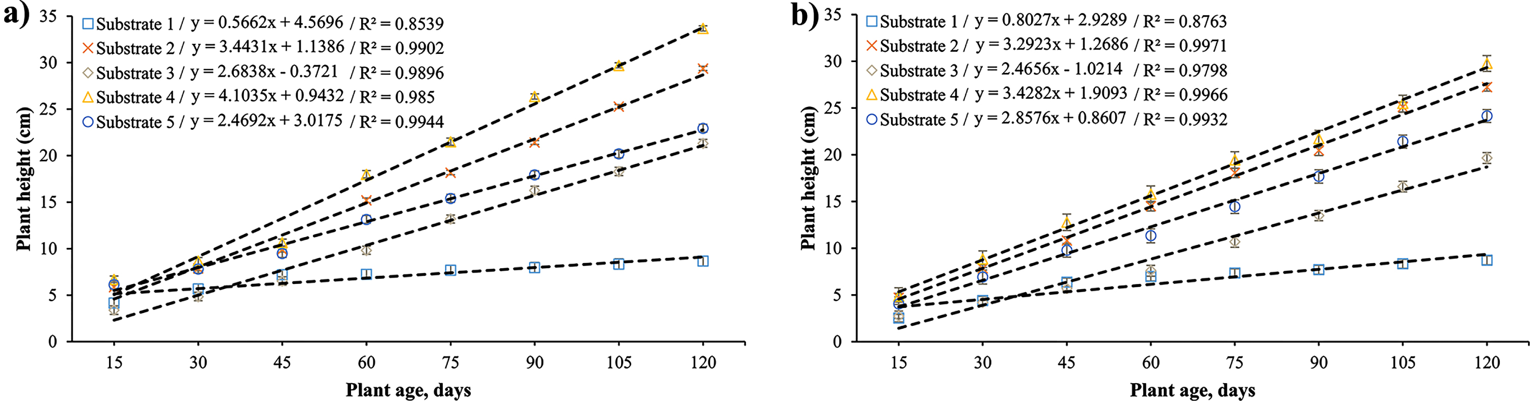
Blueberry plant height on different substrates during ex vitro acclimatization. (a) ‘Bluecrop’ and (b) ‘Biloxi’.
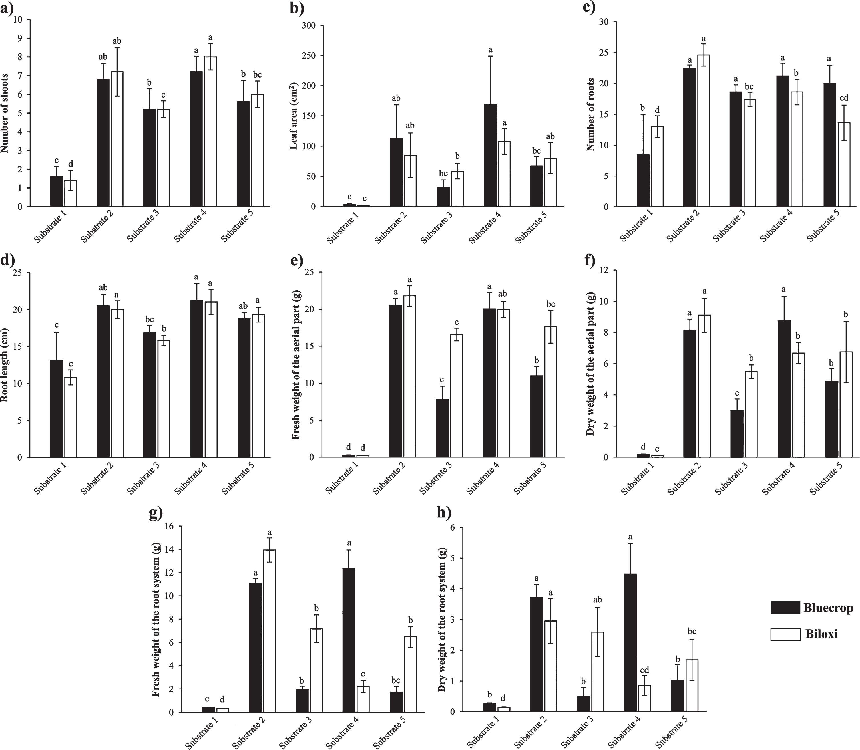
Influence of substrates on morphological parameters of blueberry seedlings (varieties ‘Biloxi’ and ‘Bluecrop’) during ex vitro acclimatization. (a) Number of shoots, (b) Leaf area, (c) Number of roots, (d) Root length, (e) Fresh weight of the aerial part, (f) Dry weight of the aerial part, (g) Fresh weight of the root system, and (h) Dry weight of the root system. Substrate 1: saw dusk + peat; Substrate 2: burn rice husk + peat; Substrate 3: raw rice husk + peat; Substrate 4 = cocomix® + peat; Substrate 5: perlite + peat.
The highest number of roots was observed in plants grown on substrate 2 (‘Bluecrop’: 22.40±0.55; ‘Biloxi’: 24.60±1.82; Fig. 4c); however, the longest roots were recorded in plants transplanted on substrate 4 (‘Bluecrop’: 21.24±2.28 cm; ‘Biloxi’: 21.04±1.70 cm; Fig. 4d). The fresh and dry weight of the cauline system (Fig. 4e, f) and root system (Fig. 4g, h) was directly proportional to the morphological development of the plant, i.e., these parameters reached the highest values in the treatments (substrates) that favored the morphological performance of the plant.
The results showed that the type of substrate influenced the physiological characteristics of the plants (Fig. 5). The highest Chl-a and carotenoid contents in ‘Bluecrop’ leaves were recorded in substrate 2, with values of 30.40±0.05 and 9.35±0.47μg mL–1, respectively (Fig. 5a, d). On the other hand, the highest values of Chl-b and Chl-a+b were observed in plants grown on substrate 3, with values of 28.62±1.65 and 58.68±2.16μg mL–1, respectively (Fig. 5b, c). In ‘Biloxi’ plants, the highest levels of Chl-a, Chl-b and Chl-a+b were observed in substrate 2 and 5, and there was no statistical difference between the values recorded for both substrates (Fig. 5a-c). The highest SPAD value was observed in plants grown on substrate 4 (‘Bluecrop’: 60.58±6.69; ‘Biloxi’: 64.08±3.88; Fig. 5e). Notably, the lowest photosynthetic pigment content and SPAD index were observed in plants grown on substrate 1.
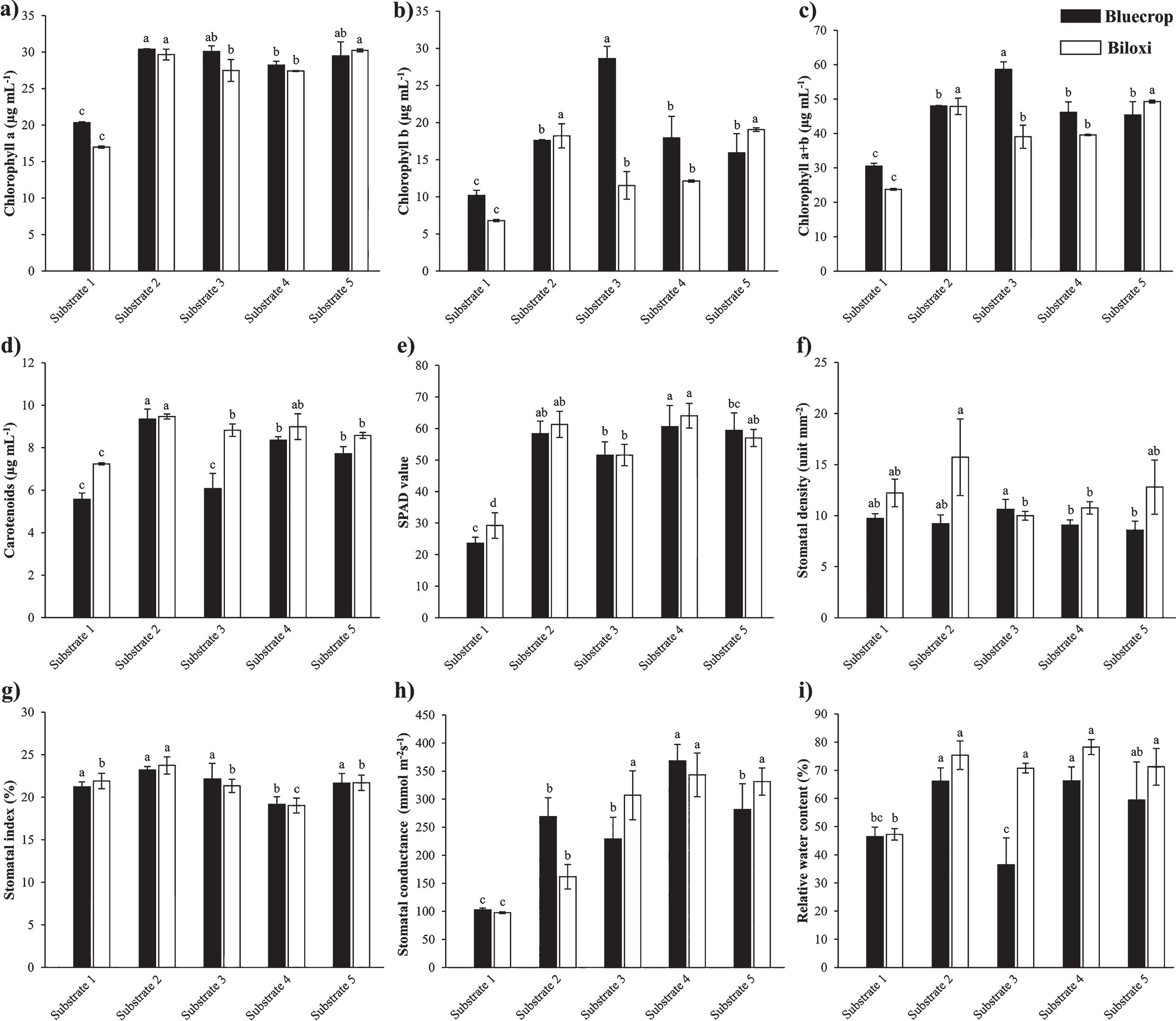
Influence of substrates on physiological parameters of blueberry seedlings (varieties ‘Biloxi’ and ‘Bluecrop’) during ex vitro acclimatization. (a) Chlorophyll a, (b) Chlorophyll b, (c) Chlorophyll a+b, (d) Carotenoids, (e) SPAD index, (f) Stomatal density, (g) Stomatal index, (h) Stomatal conductance, and (i) Relative water content. Substrate 1: saw dusk + peat; Substrate 2: burn rice husk + peat; Substrate 3: raw rice husk + peat; Substrate 4 = cocomix® + peat; Substrate 5: perlite + peat.
Stomatal density (Fig. 5f) ranged from 8.58±0.88 stomata mm–2 to 15.73±3.75 stomata mm–2, which varied by substrate type. ‘Biloxi’ plants grown on burnt rice husk + peat (substrate 2) showed the highest number of stomata (15.73±3.75 stomata mm–2). On the other hand, for ‘Bluecrop’ species showed that plants transplanted in the raw rice husk + peat mixture (substrate 3) had the highest density of stomata (10.62±0.98 stomata mm–2).
The stomatal index (Fig. 5g) recorded in ‘Bluecrop’ plants showed that substrate 4 (19.16±0.89 %) had the lowest value, while no significant effect was observed for other substrates (values obtained between 21.22±0.60 and 23.21±0.41 %). On the other hand, leaves from ‘Biloxi’ showed the highest stomatal index (23.75±0.99 %) among the plants established on a mixture of burnt rice husk + peat (substrate 2).
The stomatal conductance (Fig. 5h) of the two blueberry varieties showed that the highest value was recorded in plants grown on substrate 4 (‘Bluecrop’: 367.98±29.54 mmol m–2s–1; ‘Biloxi’: 343.25±38. 98 mmol m–2s–1, with no statistical difference with substrate 3 and 5), while the lowest reading was recorded in plants transplanted on substrate 1 (‘Bluecrop’: 102.74±2.89 mmol m–2s–1; ‘Biloxi’: 97.55±1.60 mmol m–2s–1).
The relative water content (Fig. 5i) calculated for ‘Biloxi’ shows that there was no significant difference between the evaluations of substrates 2, 3, 4 and 5, with values between 70.80±1.67 and 78.26±2.65 %. On the other hand, the records for ‘Bluecrop’ showed that the relative water content in substrates 2 (66.14±4.76 %) and 4 (66.26±4.93 %) was significantly higher.
Results of the correlation analysis are shown in Fig. 6. In the ‘Bluecrop’ variety (Fig. 5a), plant height had a very high significant correlation with biomass and carotenoid parameters (r = 0.8), followed by leaf area and relative water content (r = 0.7), whereas it had a negative correlation with stomatal density (r = –0.3). In the ‘Biloxi’ variety (Fig. 5b), the results showed a moderate to high positive correlation between morphological parameters and photosynthetic pigment content.
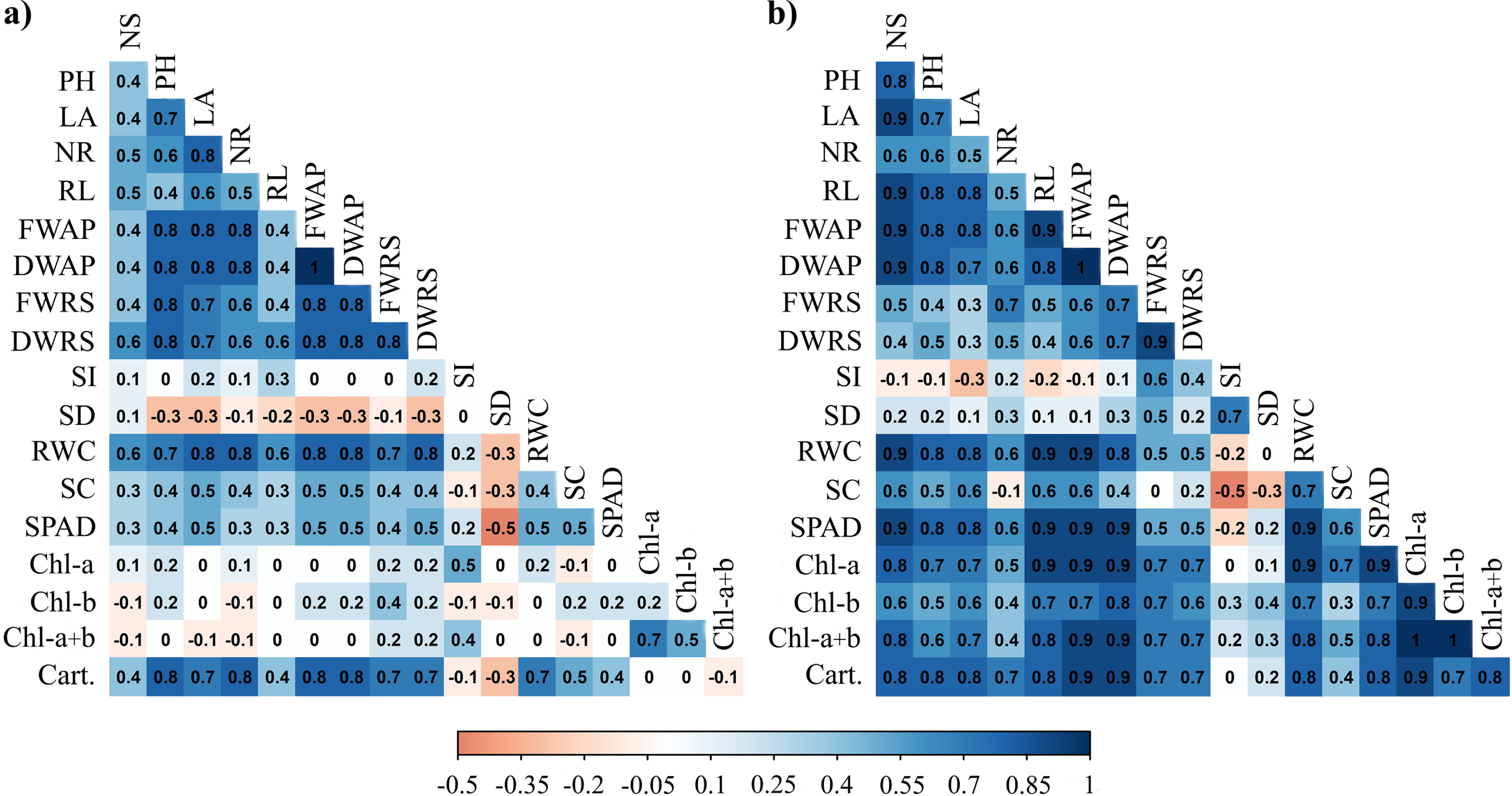
Correlation analysis among studied the morpho-physiological parameters in blueberry seedlings variety (a) ‘Bluecrop’ and (b) ‘Biloxi’ during ex vitro acclimatization. NS: Number of shoots; PH: Plant height; LA: Leaf area; NR: Number of roots; RL: Root length; FWAP: Fresh weight of the aerial part; DWAP: Dry weight of the aerial part; FWRS: Fresh weight of the root system; DWRS: Dry weight of the root system; SI: Stomatic index; SD: Stomatic density; RWC: Relative water content; SC: Stomatic conductance; SPAD: Chlorophyll index; Chl-a: Chlorophyll A; Chl-b: Chlorophyll B; Chl-a+b: Total chlorophyll; Cart.: Carotenoids.
Micropropagation phase
Amazonas region (Northeastern Peru) has favorable agro-climatic conditions for blueberry cultivation [3], generating high expectations as an alternative crop. But, unfortunately, the agricultural expansion of this crop is limited by the scarcity of reproductive material. Therefore, this study aims to evaluate a set of protocols to optimize the micropropagation of blueberry varieties ‘Biloxi’ and ‘Bluecrop’ including all stages (aseptic establishment of plant material, shoot multiplication, rooting and acclimatization).
Sterilization of plant material is a key step in tissue culture, as successful initiation of micropropagation (in vitro establishment) is ensured by the use of explants free of microorganisms that coexist with the donor plant under natural conditions. The results of this study showed that when using 1.5% NaClO for 8 min, and then 0.1% HgCl2 for 2 min for explant surface sterilization, the survival rate (viability) was higher than 85 % (‘Biloxi’: 86.67 %; ‘Bluecrop’: 93.33 %). However, our results agree with Cappai et al. [29], pointing out that it depends on the genotype introduced in the in vitro culture conditions, in fact, modifications of the exposure (in time and concentration) to the disinfectant compound(s) can lead to significant variations in the results.
At the multiplication stage, the concentration of AgNPs had different effects on the formation of new shoots, but did not influence the length of shoots and the number of leaves. One effect observed was a decrease in the rate of callus formation with the use of this nanomaterial. This response is a beneficial effect for in vitro clonal propagation, as shoots growing from callus can show somaclonal variation [30, 31], which in some cases can lead to undesirable consequences for the phenotype [30].
On the other hand, leaves and stems of regenerated shoots in the control treatment (without AgNPs) had a yellowish-reddish coloration, an indicator of nutritional deficiency. The development of yellowish or reddish shoots was also reported by Wang et al. [12] and Li et al. [32], being a characteristic response of shoots grown in WPM due to its low nutrient concentration, particularly nitrogen. Nonetheless, the addition of AgNPs showed a possible hormetic effect on the shoots, as the vitroplants slightly improved their coloring. Although nutrient concentrations in the leaves of the two blueberry varieties were not analyzed in this study, a report by Bello-Bello et al. [33] indicated that the presence of AgNPs in the culture medium allowed for higher N, Mg, and Fe accumulation in sugarcane shoots. In this regard, Ha et al. [13], Mahmoud et al. [34] and Timoteo et al. [35] suggest that AgNPs possibly favor nutrient uptake and assimilation, which may improve the in vitro regeneration process and shoot quality.
Regarding rooting, the results show that the rhizogenic capacity improved when the culture medium contained activated charcoal (in addition to the use of auxins and sucrose), while its exclusion increased the rate of callus formation (between 86.67±11.55 to 100 %). Cüce & Sökmen [25], Nin et al. [36] and Mohamed et al. [37] reported similar results, and when they incorporated activated charcoal (1 g L–l) it was observed that roots of Vaccinium species were more elongated and shoots and leaves developed more vigorously. Similarly, Mohamed et al. [38] reported that the addition of this element (activated charcoal) increased the rooting rate and allowed better acclimatization of seedlings.
The positive changes observed in root development of seedlings grown in activated charcoal medium are often attributed to the fact that it provides a dark environment similar to soil conditions and also adsorbs inhibitory compounds in the growing medium [20, 39]. In this way, it is possible for plants to increase their ability to absorb available nutrients and growth regulators, promoting better development of the root system.
Importantly, blueberry varieties showed natural variation in in vitro responses, which is related to the existence of a large genetic variation in the responses/requirements of the elements that contribute to their development [7].
Acclimatization phase
Seedlings with the best morphological characteristics (plant height, leaf area, number of shoots, root length) were grown on substrate 4, demonstrating that substrate selection is an important factor for plant development in order to reduce plant losses.
In the acclimatization process, it is of utmost importance that the plants have well-developed roots to successfully absorb the elements necessary for their growth. In this study, plants grown in substrate 4 had better root growth, which could be explained by the high porosity and low density of the material, both of which may provide a favorable environment for root development. For blueberry cultivation, it has been pointed out that the right substrate should have sufficient porosity to allow gas exchange and avoid oxygen starvation for root respiration, be resistant to compaction and favor microbial activity in the environment [40]. In this context, de Boodt and Verdonck [41] mention that the porosity in an ideal substrate should be approximately 85 %. In the case of this study, the porosity of all substrates was below the ideal, with values ranging from 59.19 % (substrate 2) to 74.91 % (substrate 5), and there were 8 to 24 roots longer than 10 cm.
According to Bryla et al. [42], Pannunzio et al. [43] and Schuch et al. [44], to allow efficient production of blueberry seedlings, substrates should be characterized by a high organic matter content (> 5%), good moisture retention (> 55%) and acidic pH (4.0 to 5.2). In general, the characteristics described above in almost all substrates were above or very close to optimal, which might suggest that these substrates are ideal for blueberry acclimatization; nevertheless, the evaluations show significant variations in the morphological performance of blueberry plants. Such differences could be explained by the EC and CEC levels of the substrates. In this study, only substrate 4 presented an EC level within the ideal range (0.5 and 1.0 mS cm–1) [45], which represents a beneficial factor for plant development, as it can be nourished without an extra use of energy [46], thus using it to carry out other physiological important processes for plant development. On the other hand, it should be considered that high CEC allows easy storage and release of nutrients [47], consequently a better morphological plant performance.
The supporting medium (substrate) used during ex vitro acclimatization of blueberry plants caused significant changes in photosynthetic pigment content. A similar observation has been reported in Rosa hybrida cv. ‘Classy’ [48]. The assessment of photosynthetic pigments is an important indicator to determine the physiological state of the plant [49], i.e. the quantity and quality of chlorophyll determines photosynthetic activity, thus affecting plant growth and development [50].
The determination of substrates and their characteristics is very important, for instance, considering that the chlorophyll content is related to the uptake of magnesium (Mg+2), which is an important element for the composition of the pigment molecule [51]. The availability of magnesium in the substrates, in this study, was found to be within the normal range. In addition, it is imperative to understand that the amount of photosynthetic pigment also varies according to the environmental conditions by which the plant develops [52, 53]. In effect, the intensity of solar radiation and photoperiod are factors related to chlorophyll content and the rate of photosynthesis, as chlorophyll absorbs the visible part of light and converts it into chemical energy needed for plant development [54].
The assessment of stomatal density is a crucial parameter for determining stress signals [55, 56], which can affect photosynthesis and gas exchange [57, 58]. Importantly, culture conditions play a vital role in minimizing the risk of anatomical, morphological and functional changes that prevent micropropagated seedlings from establishing satisfactorily under ex vitro conditions. For example, substrate characteristics are important to ensure sufficient drainage and water availability to the roots, as stress conditions can affect cell size, stomata conductance and closure, changes in membrane permeability and stomata density, and their cumulative effect can lead to poor growth and reduced plant productivity [59].
In general, it is significant to note that plants grown in the saw dust + peat mixture showed the lowest values for morpho-physiological parameters. These results may be supported by the fact that sawdust-containing substrates can generate phytotoxic effects, leading to nutrient immobilization and/or microbial competition for nutrients, affecting plant development in the early stages of cultivation [60, 61].
Conclusions
An efficient micropropagation protocol has been established for blueberries of the varieties ‘Biloxi’ and ‘Bluecrop’. Both silver nanoparticles and activated charcoal were shown to have beneficial effects on multiplication and in vitro rooting of nodal segments, respectively. When seedlings were grown in a mixture of peat and cocomix®, the ex vitro morpho-physiological characteristics of the seedlings were favored, resulting in a successful transition from laboratory to ex vitro conditions. This protocol can be successfully used for large-scale commercial production of blueberries of the varieties ‘Biloxi’ and ‘Bluecrop’.
Footnotes
Acknowledgments
The authors acknowledge the support of the Soil and Water Research Laboratory.
Funding
The research was funded by SNIP project (312252) / CUI (2252878) “Creación del Servicio de un Laboratorio de Fisiología y Biotecnología Vegetal de la Universidad Nacional Toribio Rodríguez de Mendoza de Amazonas” (Creation of a Plant Physiology and Biotechnology Laboratory Service of the National University Toribio Rodríguez de Mendoza de Amazonas), executed by the Research Institute for the Sustainable Development of Ceja de Selva.
Conflict of interest
The authors have no conflict of interest to report.