
Abstract
Vascular mild cognitive impairment (MCI) is a critical disease. Its prognosis includes not only onset of vascular dementia, but also death by cardiovascular disease. The vascular risk factors for vascular MCI are treatable, and appropriate treatment can prevent or delay the progression to dementia. Therefore, this group is an excellent candidate for secondary prevention. However, community-dwelling older adults with vascular MCI are often undetected and are not clinically identified until they develop frank dementia. Furthermore, older adults with undetected vascular MCI often have decreased ability to follow their medication regimens and this poor medication adherence worsens their vascular comorbidities. This vicious cycle needs to be prevented through community-based interventions. There is evidence that treatment of hypertension or diabetes mellitus could lead to a reduced incidence of vascular MCI and dementia. In this review article, we first explain the background and etiology of vascular MCI. We then summarize phenotype of subcortical vascular dementia which is often unrecognized or “hidden” in the community. Then we introduce the Osaki-Tajiri and Kurihara Projects which have been conducted in Northern Japan, as an example of prevention projects aimed to identify early-stage vascular MCI in the community, reduce the risk factors and facilitate their treatment. Early identification of vascular MCI in the community could lead to a large reduction in the dementia burden worldwide. The outreach efforts presented here could be useful in developing secondary prevention strategies targeted to vascular MCI.
Keywords
INTRODUCTION: VASCULAR MILD COGNITIVE IMPAIRMENT
Historical perspective: mild cognitive impairment
Mild cognitive impairment (MCI), the intermediate condition between normal aging and dementia, should be confirmed as early as possible for therapeutic intervention in order to prevent further deterioration. Two observational scales have been proposed: the Clinical Dementia Rating (CDR) [1, 2] and the Global Deterioration Scale (GDS) [3]. According to Morris et al. [4], CDR 0.5 (questionable dementia) already manifests with specific Alzheimer’s disease (AD) pathology, clearly different from that of normal aging (CDR 0). Also, CDR 0.5 older adults manifest neuropsychological impairments not only in memory but also in psychomotor speed and language. Thus, those with CDR 0.5 are considered to have very mild AD. Morris et al. [5] classified CDR 0.5 into three subgroups, i.e., CDR 0.5/uncertain dementia, CDR 0.5/incipient DAT (Dementia of the Alzheimer’s Type), and CDR 0.5/DAT. Previously we found [6] that those with CDR 0.5/DAT had severely deteriorated cognitive function without subjective memory complaints, suggesting that CDR 0.5 is better conceptualized as very mild clinical AD.
On the contrary, the GDS is based on the assumption that functional degenerative stages in AD appear to reverse the normal human development (the retrogenesis model [7]). The GDS stages of 1 and 2 fall within normal cognition, whereas the term MCI was first used by Reisberg et al. [8] in the description of GDS stage 3.
Petersen et al. re-proposed the concept of MCI [9]. This included normal daily activities and general intelligence, with memory complaints and memory dysfunction. MCI is differentiated from controls and even from very mild AD.
Clinical picture: issues to consider for vascular MCI
Vascular dementia (VaD) has a heterogeneous etiology and clinical course. Hachinski et al. [10] proposed the concept of vascular cognitive impairment (VCI) including a pre-stage of vascular dementia. The term VCI characterizes all forms of cognitive deficits from mild MCI of vascular origin to VaD [11, 12] The mild form of VCI is vascular MCI, and the most severe form of VCI is VaD [11, 12].
VCI could be a consequence of large vessel disease or small vessel disease or both. Large vessel disease (stroke) is more likely to cause dementia while cerebral small vessel disease is the most common cause of vascular MCI [13]. Neuroimaging is important for detecting CVD which is a cause of vascular MCI. Magnetic resonance imaging (MRI) represents the most commonly used neuroimaging technique. MRI markers of small vessels disease (white matter hyperintensities, small subcortical infarcts, enlarged perivascular spaces, and cerebral microbleeds) have been shown to correlate with cognitive impairment [14–18]. Standards for reporting vascular changes in neuroimaging (STRIVE) were proposed by an international working group of experts [19]. STRIVE has provided terms and definitions for lesions visible on MRI in small vessel disease including recent small subcortical infarcts (or lacunar infarcts), lacunes of presumed vascular origin, visible perivascular spaces, white matter hyperintensities of presumed vascular origin and cerebral microbleeds [19].
For further detail, (see Table 1) which summarizes some of the recent systematic reviews with a focus on vascular MCI since 2013 [20–25].
Recent systematic reviews on vascular MCI before 2019
PubMed Date: 29 January 2019.
Erkinjuntti et al. [26] proposed the criteria for subcortical vascular dementia. Among its different subtypes, VCI associated with small vessel disease has an insidious onset due to gradually progressive vascular changes with a slowly progressive cognitive deterioration [26]. The differences in cognitive profiles between VCI and AD in the MCI phase are not immediately obvious. These groups of patients cannot be differentiated on the basis of simple cognitive assessments, such as the Mini-Mental State Examination (MMSE) [27, 28].
There are no significant clinical differences in the course of progression in the pre-dementia phase between patients with vascular MCI and amnestic MCI, even though their clinical courses differ after progressing to dementia [27]. Caution must be exercised before making a conclusion about vascular etiology of cognitive impairment in a particular patient, as cognitive function is highly variable in patients with small vessel disease, and the etiological significance of white matter lesions cannot be determined easily. There is a lack of direct correlation between white matter lesion load and cognitive function [29], and individuals with widespread white matter alterations might exhibit an age-appropriate cognitive profile [30]. Further, white matter changes might be present in patients with a coexisting cognitive disorder, such as Alzheimer’s disease [31]. We herein define vascular MCI as the MCI state of VCI or the CDR 0.5 status with cerebrovascular diseases (CVD).
Prevalence of vascular MCI
No systematic reviews on the prevalence of pure vascular MCI have been reported. However, two reviews of vascular MCI with dementia have reported prevalence rate of 21% by Makin et al. [32] and 30% by van Rooij et al. [20]. According to Harrison et al.’s systematic review [33], the prevalence of vascular MCI (vascular cognitive impairment, no dementia) varied from 24% [34] to 75% [35] in stroke-only populations, and from 4% [36] to 19% [37] in populations where stroke prevalence was low or not reported. There were several studies [38–41] showing the rates for different types which ranged from 3 to 41% depending on the classification, and one study used more than one definition to compare prevalence rates (range 10–50% depending on definition) [42].
Neurobehavioral and neuropsychological characteristics of vascular MCI
Vascular MCI is characterized by executive dysfunction, slowed information processing, memory deficit and mood and personality disorders [43]. The main neuropathological substrate for vascular MCI is a disruption of the fronto-subcortical networks due to white matter lesions. Other vascular lesions could also contribute to the cognitive impairment, damaging white matter and/or subcortical structures (thalamus and basal ganglia) [13].
There are various systematic reviews of the neuropsychological tests for vascular MCI [13, 44]. There were significant differences in all cognitive domains between VCI not demented (VCI-ND) and healthy controls, especially in processing speed, working memory, and visuospatial construction [13]. When compared with non-vascular MCI, subjects with VCI-ND had significantly greater deficits in processing speed and executive function, while those with non-vascular MCI had a greater relative deficit in delayed memory [13].
The most commonly used global cognitive screening instrument, MMSE, has low sensitivity in detecting MCI [45]. Using the MMSE, one study provided information about conversion from MCI to VaD, presenting a sensitivity of 36%, specificity of 80% with incidence of VaD of 6.2% [28]. Other widely used cognitive tests are the Montreal Cognitive Assessment (MoCA) and the Addenbrooke’s Cognitive Examination. Both showed high sensitivity and specificity for MCI [45–47]. The MoCA compares favorably to the MMSE as a screening test that is sensitive to the milder forms of cognitive impairment with cerebrovascular disease, however further longitudinal research is needed in the validity of the MoCA [44].
Other screening instruments have recently been developed specifically for vascular MCI. The Brief Memory and Executive Test includes tasks for executive functioning, processing speed, orientation and memory [48]. The Oxford Cognitive Screen incorporates tests for five cognitive domains: executive function, language, memory, number processing, and praxis [49].
One attempt to standardize the neuropsychological protocol for vascular MCI was the introduction of harmonization standards published by the National Institute of Neurological Disorders and Stroke (NINDS) and Canadian Stroke Network (CSN). These harmonization standards proposed a 60 min neuropsychological test protocol which assesses the following cognitive domains: executive functions (using categorical and letter fluency and WAIS-III Digit Symbol-Coding task), visuospatial functions (Rey-Osterrieth Complex Figure), language (Boston naming test), memory (Hopkins Verbal Learning Test-Revised or California Verbal Learning Test-2) and neuropsychiatric and depressive symptoms (Neuropsychiatric Inventory) [50].
Operational definitions of cognitive impairment (e.g., performance 1 or 1.5 standard deviations below that of an appropriate comparison group) are preferred over qualitative descriptions of cognitive symptoms [11].
Motoric cognitive risk syndrome provides a clinical approach to identify individuals at high risk for dementia, especially vascular dementia [51]. Since slower gait velocity and greater stride time variability are robust markers of mobility decline and falls, changes in these variables may lead to an early identification of vascular MCI and those at risk of falls [24, 53].
VASCULAR RISK FACTORS
Development of vascular MCI
Cardiovascular risk factors play a fundamental role in the development of cognitive impairment [11]. However, the specific mechanism in which cardiovascular risk factors affect cognitive functioning is still unclear. The direction (positive or negative) of the effect may be dependent on the precise time of the personal lifespan. Two of the most important cardiovascular risk factors that play significant roles in the development of vascular MCI is arterial hypertension and blood pressure [54, 55]. Ambulatory blood pressure monitoring and home measured blood pressure (HMBP) are emerging as more reliable measurements than in-clinic blood pressure measurement. They are associated with a better correlation with target organ damage [55] including cognitive impairment.
Blood pressure
Hypertensive patients are at an elevated risk for MCI. A prospective study by Yaneva-Sirakova et al. [56] in hypertensive patients showed a correlation between higher blood pressure and MCI status. A significant, correlation between higher blood pressure variability and lower MoCA and MMSE test scores was also found.
These results are in accord with the conclusions derived from neuroimaging studies [57, 58]. They found that patients with elevated blood pressure variability have higher volume of white matter lesions than those with lower variability. One potential explanation for this association is that microvascular (including cerebrovascular) autoregulation is likely already impaired in hypertensive older adults with concomitant cardiovascular risk factors. Thus, increased blood pressure variability strains microcirculatory compensatory mechanisms beyond their limit. Periods of hypoperfusion are followed by normal or elevated blood flow. This may lead to disruption of the blood-brain barrier, small-vessel dysfunction, and impairment in the neural circuits.
Apart from blood pressure variability, the blood pressure measurements may also be associated with cognitive functioning [56, 59]. Older adults with hypertension, with HMBP <120/70 mmHg or ≥135/85 mmHg, were at an elevated risk for cognitive impairment and scored lower on neuropsychological tests than those with controlled blood pressure. The potential pathophysiological mechanism behind these results is the relative hypoperfusion of the brain after years-long adaptation to high blood pressure values. It is not clear though which is the leading cause and which is the consequence: the low blood pressure or cognitive impairment. Elevated blood pressure may be the reason for impairment in important brain centers, which may lead to deterioration of autonomic functioning and a persistent drop in blood pressure. On the other hand, low blood pressure due to treatment, may have a detrimental effect on target organs’ microvasculature, because of their previous adaptation to high blood pressure values.
Pulse pressure (PP) may be an independent risk factor for target organ damage. Yaneva-Sirakova et al. [60] showed that patients with elevated home PP (measured PP > 55 mmHg) had lower MoCA and MMSE test scores independent of other blood pressure variables. The patients with MCI had significantly higher 24 h, day and night PP values. Elevated PP in sub-optimally controlled hypertensive patients is an important independent factor for persisting target organ damage of the brain- clinically manifested with MCI. It should be evaluated routinely and treated properly in order to reduce their risk for cognitive impairment.
Central aortic pressure is a relatively new method for blood pressure measurement. It is considered a more accurate predictor of cardiovascular risk and mortality than the conventional brachial measurement. Yaneva-Sirakova et al. [61, 62] reported that the mean central pulse pressure values of patients with cognitive impairment were significantly higher than those without cognitive impairment. There was a weak negative correlation between central pulse pressure and the MoCA and MMSE test scores. Central arterial hemodynamics plays an important role in microcirculatory dysfunction and target organ damage in arterial hypertension. Central pulse pressure is a marker of arterial stiffness and a risk factor for target organ damage as well as cognitive impairment.
There are few studies on the correlation between central aortic blood pressure and cognitive impairment. Dias et al. [62] found a significant correlation between central aortic systolic pressure and intima-media thickness, and between cognitive impairment and central aortic systolic pressure. There are some large studies that confirmed the significance of central blood pressure for target organ damage [63], vascular hypertrophy, and extent of atherosclerosis, clinical outcomes and cardiovascular events [64–66]. But their role in cognitive impairment, and which parameter has the leading impact on early brain damage, are not clear so far. With age and cardiovascular risk factors, the arterial wall stiffens and the reflected wave travels at an accelerated speed, thus augmenting central systolic and pulse pressure, rather than diastolic [67]. Left ventricular afterload increases and coronary filling is compromised [68, 69]. Therefore, central hemodynamic parameters are suggested to correspond better with cardiovascular risk than peripheral blood pressure.
Atrial fibrillation
In patients with arterial hypertension non-valvular atrial fibrillation may raise the risk of MCI despite the use of anticoagulation therapy [70]. Patients with arterial hypertension, concomitant cardiovascular risk factors and atrial fibrillation had significantly lower neuropsychological test scores than patients with arterial hypertension, concomitant cardiovascular risk factors, but without atrial fibrillation.
IMPORTANCE OF OBSERVING OLDER RESIDENTS’ DAILY LIVES
The Tajiri & Kurihara Projects
Regarding the prevalence of MCI, unlike a rather clear difference between healthy controls and frank dementia, the more subtle difference between healthy controls and MCI can be affected by the method of sampling or by the neuropsychological tests used. It is important to examine community representative large samples and to perform neuropsychological tests sensitive enough to identify MCI. In the following section, we present the incidence and prevalence of vascular MCI based on the Tajiri & Kurihara Projects. Therefore, we followed our Prevalence Study conducted in 1998 (Prevalence Study 1998) [71, 72] with an Incidence Study in 2003 in Tajiri, Japan (Incidence Study 2003) [73, 74]. The data of the Kurihara Project (Prevalence Study 2008) was also analyzed [75, 76].
Briefly, our Prevalence Study in Tajiri conducted in 1998 included 497 randomly selected participants, including 346 CDR 0, 119 CDR 0.5, and 32 CDR ≥1. The first two groups were targeted for the follow-up Incidence Study in 2003. Based on the database, we reanalyzed the prevalence of subcortical VaD in the subjects with CDR 0.5 (vascular MCI) and VaD, prognosis with CVD, and two types of VaD onset (i.e., subcortical VaD and vascular MCI). The criteria for subcortical VaD were met by 67% of those with vascular dementia and by 7% of those with vascular MCI. In the cognitively normal group, CVD had no effect on prognosis; however, in the CDR 0.5 group, CVD had an effect on death by cardiovascular disease [73, 74].
In our incidence study, we found an incident VaD rate of 18% among adults aged 65 and older. Some cases developed VaD after a stroke (type I), whereas others first met the criteria for subcortical VaD with very mild stage MCI (CDR 0.5) and progressed to VaD (type II). We hypothesized that prognosis of vascular MCI included type II VaD or death due to causes associated with vascular risk factors.
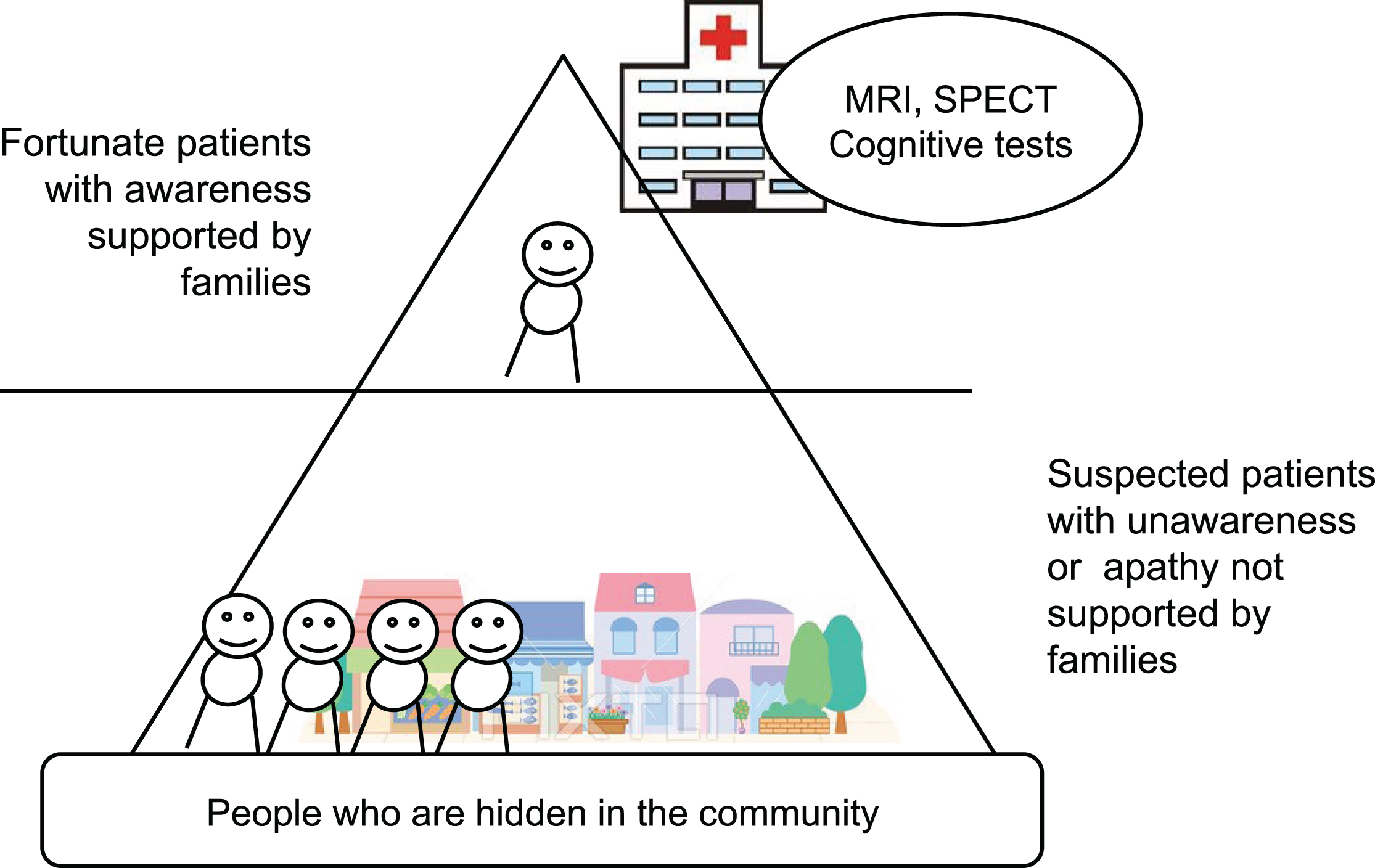
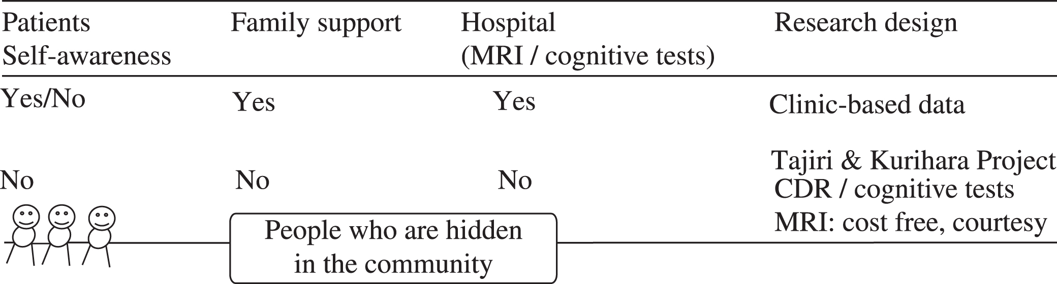
The “hidden in the community” problem
The patient clinic population might be the “tip of the iceberg.” These are the “fortunate” patients with awareness who are supported by families and who visit the outpatient clinic for care. On the contrary, suspected patients with unawareness or apathy not supported by families are easily hidden in the community. The concept is shown in (Fig. 1 and Table 2). Previously, we estimated the prevalence and severity of apathy in vascular MCI, amnestic MCI, and MCI of unknown type, and found that vascular MCI subjects have more severe apathy compared with amnestic MCI subjects on caregiver assessment (discussed detail in [77]). Gaining a full picture of those with vascular MCI requires work in the community. In the following sections, we report details of the techniques we have employed to identify those with vascular MCI “hidden in the communities” in ongoing studies in Japan, the Osaki-Tajiri and Kurihara Projects.
Database of outpatient clinics - The tip of an iceberg?
Clinic-based data involves external validity problem?
CDR assessment focusing on identifying vascular MCI
The CDR is based on memory impairment as the primary symptom since the scale is designed to detect early AD. However, non-memory conditions like frontal executive dysfunction are considered to be important for dementia, especially for the vascular contribution.
To evaluate daily lives of older residents with vascular MCI using the CDR, information from care managers or public health nurses are critical, especially for the CDR domains of Community Affairs, Home & Hobbies, and Personal Care.
The life concern form
The CDR is the international standard for this purpose, but requires long-term training to develop appropriate judgment skills. Based on our experience, we developed a “Life Concern Form” to detect people with vascular MCI in the community [78]. It was developed to assess daily lives of older adults living in the community by specifically observing the following items: Activities they cannot perform in the manner as they used to do in the past; Decreased activity levels at home; Loss of interaction with people other than their family; Need for assistance in taking medications; special attention needs to be paid to older adults taking medications for hypertension or diabetes mellitus with poor medication adherence.
Training of staff members specifically targeted to identify potential subjects with vascular MCI using the above questionnaire and focusing on specific items of CDR are encouraged.
INTERVENTION
Finnish Geriatric Intervention Study to Prevent Cognitive Impairment and Disability (FINGER) [79] examined a multidomain approach to prevent cognitive decline in at-risk elderly people in the community. Findings from their large (intervention 631 versus control 629), long-term, RCT suggest that a multidomain intervention could improve or maintain cognitive functioning in at-risk elderly people from the general population. Recently there is a report that lowering blood pressure will decrease the dementia risk [80]. We have been conducting an intervention study including exercise targeting older adults with vascular MCI in the town of Kurihara, Japan (the Kurihara project) [81]. As discussed earlier, the outpatient clinic population is only a small percentage of those affected by vascular MCI for whom fortunately there is a support network. On the contrary, patients without a support network are likely to be unaware of their disease, apathetic and remain hidden in the community. In the Kurihara Project, free transportation and MRI scans were provided to local residents. By providing free neuropsychological evaluations and MRIs, we aim to identify “hidden” MCI subjects in the community and recruit them into the intervention study. In our prevention study specifically targeted to vascular MCI [81], we examined the efficacy of three intervention approaches (cognitive stimulation or intervention (CI), physical exercise (PA), and group reminiscence approach (GRA)) on cognitive and physical functions and psychological well-being, based on a cluster randomized controlled trial (cluster RCT) design. We recruited 60 participants aged over 75 years with vascular MCI. Vascular MCI was defined as CDR 0.5 with cerebrovascular disease. Participants were randomized into four groups (CI, PA, GRA, and control). The interventions lasted 12 weeks and consisted of weekly sessions (twice per week) and homework. The cognitive and clinical outcomes included MMSE, Trail Making Test part A (TMT-A), Word Fluency, 6-meter-walk time, Geriatric Depression Scale, and quality of life. The CI intervention was found to increase MMSE scores and the PA intervention was found to improve walking speed. TMT-A, Word Fluency, and quality of life improved across all three interventions compared with the control group [81].
CONCLUSION: COMMUNITY-BASED MEASURES FOR MCI AND DEMENTIA
Vascular MCI is a critical disease. Its prognosis includes not only onset of vascular dementia, but also death by cardiovascular disease [82]. The vascular risk factors for vascular MCI are treatable, and appropriate treatment can reduce the incidence of cognitive impairment and dementia due to vascular causes. Prevention and delay of dementia through risk reduction has become increasingly important to reduce the prevalence of dementia worldwide [83]. As highlighted in this review article on vascular MCI, MCI with hypertension and diabetes is often unrecognized in the community and left untreated which further facilitates worsening of the prognosis due to poor management of risk factors and comorbidities. Special outreach efforts specifically targeted to this group include questionnaires focusing on symptoms of vascular MCI, training of staff to accurately assess CDR questions in relation to vascular MCI, and free transportation/access to MRI assessments. These community outreach efforts (as opposed to solely treating those who visit specialty clinics) are critically important for early identification of risk factors and their modifications to delay onset of frank dementia. Some of the efforts might require global initiatives through World Health Organization (WHO) and other entities, especially in low- and middle-income countries which have high prevalence of vascular risk factors and the incidence and prevalence of vascular MCI is likely to rise further. The recent establishment of The International Research Network on Dementia Prevention (IRNDP) initiatives could play a role in facilitating this effort [83].
Footnotes
ACKNOWLEDGMENTS
We are grateful to the discussion at ICVD 2016 conference with Drs. Milica G. Kramberger, Vuk Milosevic, Teodora Yaneva-Sirakova, Berlot Rok, Abantas-Diamla Sharimah, Junko Takada, Latchezar Traykov, Rumiana Tarnovska-Kadreva, and Amos D Korczyn. We are thankful for Ms. Nora Mattek for her superb editing.