
Abstract
BACKGROUND:
Assessment of the plantar flexion (PF) isokinetic performance has been greatly diverse and based on personal preferences rather than standardized guidelines.
OBJECTIVE:
To examine the performance of the plantar flexors under different settings including knee joint angle and subject position.
METHODS:
Thirteen women and 20 men took part in this study. The isokinetic protocol (60
RESULTS:
Knee angle impacted the PF moment (
CONCLUSION:
The 45
Introduction
Assessment of muscular performance is an essential element of the physiotherapeutic practice, especially in the musculoskeletal domain. A valid muscle performance has to ensure that that the tested muscle/s produce maximal moment. This necessitates placing the joints and surrounding muscle/s in optimum position and length, respectively, for maximum exertion. Achieving such optimization could be a challenge, particularly with structurally intricate muscle groups such as the Triceps Surae (TS), which is comprised of two main muscles, the Gastrocnemius (GC) and the Soleus (SOL). Traditionally, both the GC and SOL muscles have been treated as one unit although distinctive functions of these muscles have been reported, particularly during walking and landing activities [1, 2].
However, the assessment of plantarflexion (PF) strength has to a great extent not been standardized. A subject could be assessed in a supine [3, 4], sitting [5, 6, 7], prone [8, 9, 10, 11], or even side lying position [12, 13]. The knee position in these studies ranged from full extension to 90
Isokinetic assessment/rehabilitation of the TS group may be performed in different positions. The sitting position forces the subject to bend the hip joint, which would elongate the Hamstrings that overlaps the origin of the GC muscle. Thus, hip joint position might indirectly affect the activity of the plantarflexors. Additionally, the sitting position is less stable than lying position due to the vertical orientation of the trunk, which might impact the performance of the distal muscles such as the TS. Accordingly, it seems that the subject position during isokinetic assessment/rehabilitation of the TS muscles is significant.
Therefore, the objectives of this study were to examine the isokinetic evaluation of PF strength under different procedural settings including different knee joint angles and subject positions, and explore the positional settings that would result in maximal performance.
Methods
Participants
Thirteen women and 20 men, aged 18–45 years with a BMI of 18–25, took part in the study (Table 1). Participants were excluded if they suffered any musculoskeletal or neurological disorders of the trunk and/or lower extremities, participated in athletic activity on a regular basis for at least a year before joining the study, or were employed in jobs imposing extensive physical exertion on the lower extremities. The study conformed with The Code of Ethics of the World Medical Association (Declaration of Helsinki). All participants signed a consent form approved by the institutional review board (Prince Sultan Military College of Health Sciences, IRB-2018-PT-006) after being informed of the procedures and risks of the investigation.
Participants’ demographic data
Participants’ demographic data
BMI: Body mass index, SD: Standard deviation, CI: Confidence interval.
This study employed a repeated-measures design. The knee joint angles examined were 15
Instrumentation
In this study, a Biodex 4 pro multi-joint dynamometer (Biodex Medical Systems Inc, Shirley, NY, USA) was used. System calibration was performed each session prior to data collection. During the sitting test condition, participants were seated in the Biodex chair with the back support inclined to 70
An eight-channel Delsys Trigno wireless EMG system (Delsys Inc., Natick, Massachusetts, USA) was used to collect the EMG data from the GM, GL and SOL muscles. The recording unit comprised two parallel Silver bar electrodes with a 1 cm inter-electrode distance. Surface EMG data was collected at a sampling rate of 2000 Hz using EMGworks
Procedures
After signing the consent form, the participant’s skin was prepared for placement of EMG electrodes according to the SENIAM recommendations [16]. Electrodes were placed over the SOL, GM, and GL as described before [17]. Signal quality check was performed prior to data collection to detect baseline noise or artifacts.
Participants were instructed to assume the testing position. The trunk and pelvis were stabilized by Velcro straps. The knee joint was placed in the predetermined angle. Finally, the foot was secured to the footplate. The anatomical axis of the ankle joint (Lateral Malleolus) movement was aligned with that of the dynamometer.
The participant assumed the first testing position (supine or sitting) and performed 3–5 familiarization trials. Participants performed a three-second MVIC of the PFs in each test position with the ankle locked in neutral position. This was followed by a five-minute rest. Then, the participant performed the data collection trials. Three repetitions were performed for each test condition with a two-minute rest in-between. Data collection for the different knee angles was completed in the respective participant’s position before moving to the next one. This was followed by a five-minute rest, during which the participant assumed the other testing position and the same procedures were repeated.
Statistical analysis
Data was analysed using SPSS version 21 (SPSS, Chicago, IL). The mean
0pt 0pt
Mean (SE) and 95% CI of the isokinetic data and normalized EMG of male participants as a function of subject position and knee angle main effect
Mean (SE) and 95% CI of the isokinetic data and normalized EMG of male participants as a function of subject position and knee angle main effect
Bold numbers indicate significant difference at
Mean (SE) and 95% CI of the isokinetic data and normalized EMG of female participants as a function of subject position and knee angle main effect
Bold numbers indicate significant difference at
Isokinetic measures
Participants’ demographic data are presented in Table 1. A sample of 33 (13 females) volunteers was recruited for the study. Considering the reported gender-based difference in muscle strength, the data was stratified by gender and reported accordingly. Results revealed that subject position did not significantly influence the isokinetic measures of the PFs in both males and females. However, knee joint angle had a significant main effect on most of the measures including all the moment (
The subject position and knee joint angles did not have an interaction effect on any of the outcome variables (
Isokinetic data for both genders as a function of position and knee angle interaction. The horizontal axis represents knee angles. BW: Body Weight.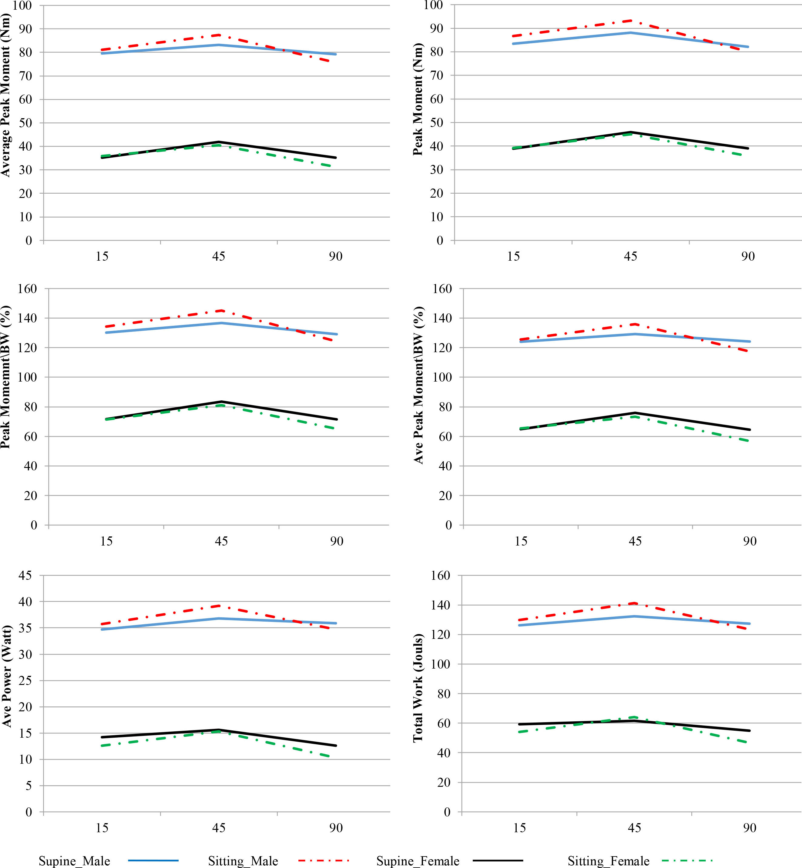
The results showed that the subject position and knee angle main effect and its interaction with knee joint angle did not significantly affect the myoelectric activity of the PF muscles. Only the GL muscle in male participants was significantly affected by the subject position (
Interestingly, the SOL activity showed a consistent pattern for both genders where its activity increased with flexing the knee. This was particularly more noticed among female participants where a difference of 7% was measured between the 15
Normalized EMG data for both genders as a function of position and knee angle interaction. The horizontal axis represents knee angles. GL: Lateral Gastrocnemius, GM: Medial Gastrocnemius, SOL: Soleus.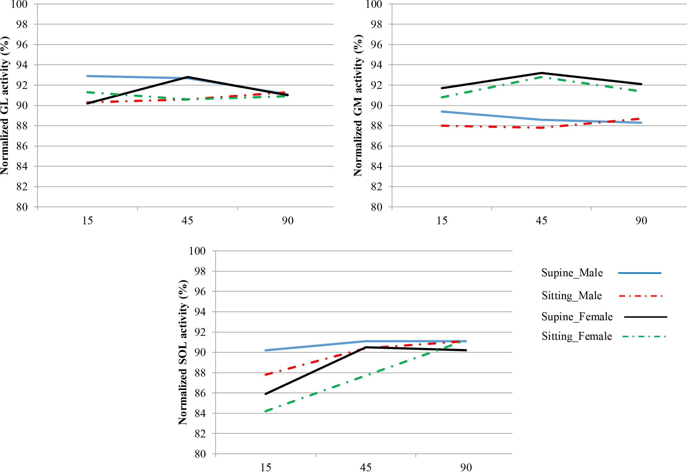
Isokinetic performance measures
Overall, the results showed that the isokinetic moment and work measures of the PFs were mainly affected by the knee angle. The moment and work measures decreased in knee flexion compared with extension. This is in agreement with several previous studies that reported increased PF moment in extended knee position, and vice versa [5, 18, 19, 20, 21, 22, 23]. However, it is important to notice that this relationship was not consistent across all knee angles investigated in the current study. The moment and work values recorded in the 45
The length-tension relationship has been postulated as a key relationship influencing the performance of the PFs. A shorter muscle length, as in flexed knee, would make the PFs mechanically disadvantaged limiting its force productivity. This is further supported by the fact that the two-joint GM and GL of the TS group have been reported to work on the ascending limb of the length-tension relationship curve, which is attributable to the anatomical constraints of the knee and ankle joints [21]. Furthermore, architectural changes including fascicular length and pennation angles could manipulate the muscular performance. Placing the muscle-tendon unit of the PFs in a shortened condition would decrease the fascicle length and increase the pennation angle, which was postulated to decrease the overall force transmitted through the muscle-tendon unit to the calcaneous [24]. Shorter PFs length was found to be associated with decreased moment productivity [22, 25] and inhibited neural drive to the Gastrocnemius [13, 20, 26]. However, evidence showed that the SOL fascicle length is not influenced by knee position as the Gastrocnemius does, despite the changes in moment productivity [12, 24] and muscular activity of the PFs [27]. Accordingly, the reduction of PF moment and work in the flexed (90
To examine the impact of knee position on PF performance, previous studies typically compared flexed (mostly 90
To explain the discordant findings associated with the 45
In the current study, participants were instructed to actively return the foot, as fast as possible, to the starting position (10
Our results showed that the PF isokinetic measures were not influenced by the subject position. However, sitting had slightly higher measures than supine for all the variables in the male participants whereas the findings of the female participants showed the opposite pattern. Previous studies reported variable findings about the influence of position on the PF performance. The PF moment was shown to be greater in the sitting than the prone position [14]. The isometric PF moment was reported to be significantly higher in the supine and prone than the sitting position [11], which disagrees with the current results. Additionally, the superiority of the measured PF performance in the supine and prone positions was instruction-dependent. Previous reports have shown that coactivity of other lower extremity muscles, such as the knee extensors, during a PF exertion could significantly influence the PF moment and work measures [9, 10]. Thus, it seems that several factors could modulate the PFs mechanical performance in different subject positions including instructions, muscular co-activation and joint angle, and probably gender, which could explain the inconsistent findings across the literature.
Plantar flexors electromyography
The current results show that the PFs muscular exertion was not affected by the knee angle or subject position, except the GL muscle in male participants only. Additionally, The SOL activity apparently increased in flexed (45
Some study limitation ought to be considered, though. We implemented an isokinetic assessment protocol only. Other muscle contraction approaches, e.g. eccentric, may provide further valuable insights to illustrate the PF performance under similar assessment procedures. The study sample comprised young and healthy participants, which may limit the generalizability of the findings to other populations as the elderly and those with musculoskeletal conditions of the lower limb. Finally, the detection of the EMG activity of the PF muscle using needle electrodes may have yielded a more distinguished activity of the motor units of each muscle distinctively. Unfortunately, such invasive procedures were inapplicable in the current study.
Conclusion
Considering the above-mentioned elaboration, the 45
Author contributions
CONCEPTION: Ahmed Farrag, Walaa Elsayed and Eidan Alzahrani
PERFORMANCE OF WORK: Walaa Elsayed, Moath Almusallam and Nora Almulhim
INTERPRETATION OR ANALYSIS OF DATA: Ahmed Farrag, Walaa Elsayed and Zaenab Alowa
PREPARATION OF THE MANUSCRIPT: Ahmed Farrag and Walaa Elsayed
REVISION FOR IMPORTANT INTELLECTUAL CONTENT: Ahmed Farrag, Walaa Elsayed, Eidan Alzahrani, Moath Almusallam, Nora Almulhim and Zaenab Alowa
SUPERVISION: Ahmed Farrag
Ethical considerations
This study was approved by the institutional review board of the Prince Sultan Military College of Health Sciences (IRB-2018-PT-006). All participants signed a written informed consent.
Funding
This work was supported by the Prince Sultan Military College of Health Sciences, Saudi Arabia (grant number 2017-PT-003).
Footnotes
Acknowledgments
The authors would like to thank all the participants for their time and effort.
Conflict of interest
The authors report no conflict of interest.