
Abstract
BACKGROUND:
Comparison of knee loads on a Smith machine, which is utilised for maintenance of health and rehabilitation, has not been attempted.
OBJECTIVE:
This study compared lower limb muscle and knee joint forces during front and back squats performed on a Smith Machine.
METHODS:
Eleven participants performed front and back squats with loads at 40%, 60% and 80% of their back squat 1-RMs. Ground reaction forces and three-dimensional full body motion were collected and used for modelling lower limb muscle and knee joint forces.
RESULTS:
Larger loads increased tibiofemoral compressive force during back squat at 80% compared to 40% (
CONCLUSIONS:
Greater external load was associated with increase in gluteus medius and minimus force and with increased tibiofemoral compressive force without effects on tibiofemoral shear force, patellofemoral compressive force or other lower limb muscle forces.
Introduction
Among the various techniques for performing squats, the back squat and the front squat are the most often used for training lower limb and spinal muscles. The key difference between these techniques involves the position of the upper body in relation to the hip and knee joints. During the front squat, it is expected that the vertical projection of the upper body mass and any external load would be closer to the hip than the knee joint [1], assuming that the load lifted is the same between techniques. This is expected to increase the moment-arm for the knee joint and potentially reduce the contribution from the hip joint [2]. Conversely, the back squat should require an increased hip contribution than the knee joint [1]. In parallel, increments in external loads during squats have been shown to increase the contribution from the hip compared to the knee joint [3]. However, it is unclear how this transition in torque and power from the knee to the hip due to changes in technique (front to back) and increments in load affect individual muscle forces. This is critical to allow practitioners to understand the training stimulus that each movement provides.
Internal knee joint loading is important to determine how and when squats should be used clinically [4, 5, 6]. Knee forces during squats include tibiofemoral shear force, tibiofemoral compressive force, and patellofemoral compressive force [7]. Tibiofemoral shear force places stress on the cruciate ligaments and poses a risk of ligament rupture, while tibiofemoral and patellofemoral compressive forces stress the articular cartilage and meniscus [7, 8, 9], which is of concern for patients following a knee injury or surgery. Sahli et al. [10] found that peak compressive and shear force components increased significantly as the external load increased during squats. Hartmann et al. [1] supports these findings by stating that higher loads resulted in increased tibiofemoral and patellofemoral compressive forces when squatting due to an increased knee extensor moment [11]. It was found that joint compressive forces did not differ between the two squat techniques [12]. However, no study compared knee forces during front and back squat taking into account the forces produced by individual muscles (i.e. using musculoskeletal modelling). Moreover, no prior study assessed knee loads or muscles forces on a Smith machine, which could provide valuable evidence to inform exercise prescription for recreational use of strength training, maintenance of health and rehabilitation.
Movement technique in resisted training exercises has been mostly investigated using EMG [13, 14]. Although valuable, EMG only provides information on the electrical activity of the muscles without encompassing responses from length and contractile velocity of the muscles [15, 16, 17]. Musculoskeletal modelling though allows for the physiological (i.e. force-length-velocity) and anatomical (i.e. moment-arms, mass, pennation angles) properties of the muscles to be taken into account when calculating individual muscle forces. This approach potentially provides a more realistic assessment compared to the use of EMG, which only accounts for the muscle excitation [18]. In addition, the use of modelling in strength training ranged from a net-moment driven to the use of musculoskeletal models [19]. However, the use of portable motion tracking instead of optoelectronic systems has been limited to the assessment stair ascent [20] or to the analysis of spinal loads during load handling [21]. Employing portable motion capture systems along with portable force plates would provide a valuable addition when used along with open-access musculoskeletal software (e.g. OpenSim), which should facilitate the analysis of internal loads during a variety of strength training exercises.
Therefore, this study compared lower limb muscle and knee joint forces during front and back squats performed with different external loads on a Smith Machine. It is hypothesized that knee joint and muscle forces would increase when squats are performed with larger external loads and that front squats would increase knee and muscle forces compared to back squats.
Materials and methods
Participants
Eleven male apparently healthy participants without musculoskeletal or neurological diseases volunteered for the study. The sample size was estimated using GPower statistical package for an ANOVA with within-subjects design aiming for an effect size of 0.40 (large effect), alfa of 0.05 and power of the test of 0.80 assuming a minimum of five measures per session. This calculation resulted in nine participants required, which we deemed small and opted for expanding to a sample size similar to prior studies [5, 22]. At the time of the study participants had 22
Procedures
In their first session, after providing consent to participate in the study, anthropometric measures were collected from each participant. Body mass, standing stature, arm span, shoulder and pelvis width, hip, knee and ankle heights to the floor and foot length were measured according to instructions provided by the motion tracking system manufacturer (Xsens, Netherlands) using weighing scales (Model 762; Seca, Germany), portable stadiometer (Model 213; Seca, Germany) and segmometer (MVN 5M/16’; Xsens, Netherlands), as appropriate. These measures were stored to be used in the second session.
Subsequently, participants performed 8-min of walking on a treadmill at 5 km/h of speed for general purpose warm up. They then performed one set of eight repetitions of body weight squats, prior to further warm up on the Smith machine exercise equipment (SCB1000, ProClub Line counter-balanced Smith machine, Body-Solid Inc, IL, USA). This machine had 212 cm of full height of the rail with fore-back displacement of 25 cm, leading to an incline of
After 48-hrs, participants returned to the laboratory for their second session. Following 8-min of walking on a treadmill at 5 km/h of speed, 17 wireless motion sensors (MVN Awinda; Xsens, Netherlands) were attached to pre-defined body segments as described by the manufacturer prior to a static pose calibration. They were then asked to perform one set of eight repetitions of body weight squats for further warm up and for qualitative assessment of their movement using the motion analysis software (MVN Studio 4.4; Xsens, Netherlands). Participants stepped onto a force plate (model 9286A; Kistler Instruments, Switzerland) with their left foot and onto a standard plate of equivalent height with their right foot. They performed one set of 6–8 repetitions with the prescribed load and technique on a Smith machine. For the back squat, the position of the bar and feet followed descriptions from the 1-RM testing. For the front squat, the bar was positioned across the anterior deltoids and clavicles with the shoulders flexed to approximately 90
Illustration of body positions for the back squat (A – upward and B – bottom) and for the front squat (C – upward and D – bottom).
Definition of angles used in this study. Inset A shows the patellofemoral compressive force (PFC) and inset B shows the tibiofemoral compressive (TFC) and shear (TFS) force components. Insets C and D illustrate positions during a back squat in the Smith machine. Inset E shows that feet position on the force plate (left) and on the dummy plate (right).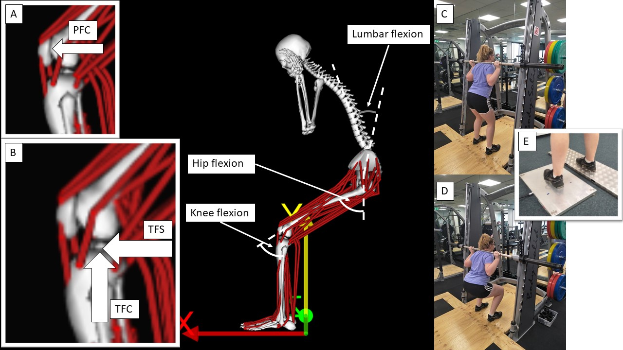
Ground reaction forces and motion data were exported using the manufactures’ software and converted for later use in the muscle modelling software (v.3.3; OpenSim, Stanford University, USA). Static pose calibration data were extracted from the motion tracking data and used for scaling a generic model with 37 degrees of freedom driven by 80 musculotendon actuators across the pelvis and the lower limbs (adapted by Lai et al. 2017). After scaling the model for the participant’s anthropometry, muscle and bone-on-bone forces were calculated. To achieve this inverse kinematics, inverse dynamics, static optimisation and joint reaction analysis were conducted (in this order). This sequence allowed customisation of the model into the body dimensions of each participant and drive the model’s motion using the 3D coordinates gathered from Xsens. During inverse kinematics, inverse dynamics and static optimisation, and joint reaction analysis, motion data was filtered using a zero lag 3
Peak knee joint forces, mean muscle forces, mean angle and range of motion (ROM), mean ground reaction force components (i.e. vertical, anterior-posterior and medio-lateral) and resultant force, and mean sagittal joint moment for the hip, knee and ankle were extracted from the squats and used for statistical analysis. Normality of data distribution were assessed using the Shapiro-Wilk’s test. Patellofemoral and tibiofemoral compressive forces, hip flexors, rectus femoris, gluteus maximus, gluteus medius and minimus, hamstrings, quadriceps, mean lumbar flexion/extension and knee ROM were log transformed as they breached the assumption of normal distribution. ANOVAs were then employed to compare the main effects from load (40% vs. 40% vs. 80% of 1-RM) and technique (i.e. front vs. back squat). Whenever main effects from load or technique were observed, post hoc with Tukey corrections were employed to assess pairwise differences. Magnitude of differences were explored using Cohen’s effect sizes (d) and interpreted according to Rhea [25]. Cohen’s d were calculated as a ratio between the difference in mean values by the pooled standard deviation [26].
All statistical analyses were conducted using JASP software (
Ground reaction force components (vertical – vGRF, anterior-posterior – AP, medio-lateral – ML) and resultant at the three loads and both techniques (Back vs. Front). Total lifted force includes the sum of body weight and external loads
Ground reaction force components (vertical – vGRF, anterior-posterior – AP, medio-lateral – ML) and resultant at the three loads and both techniques (Back vs. Front). Total lifted force includes the sum of body weight and external loads
All force data presented in percentages of body weight. Equal symbols indicate significant differences (
Participants achieved 1-RMs of 124
Illustration of the vertical displacement, velocity and acceleration of the bar.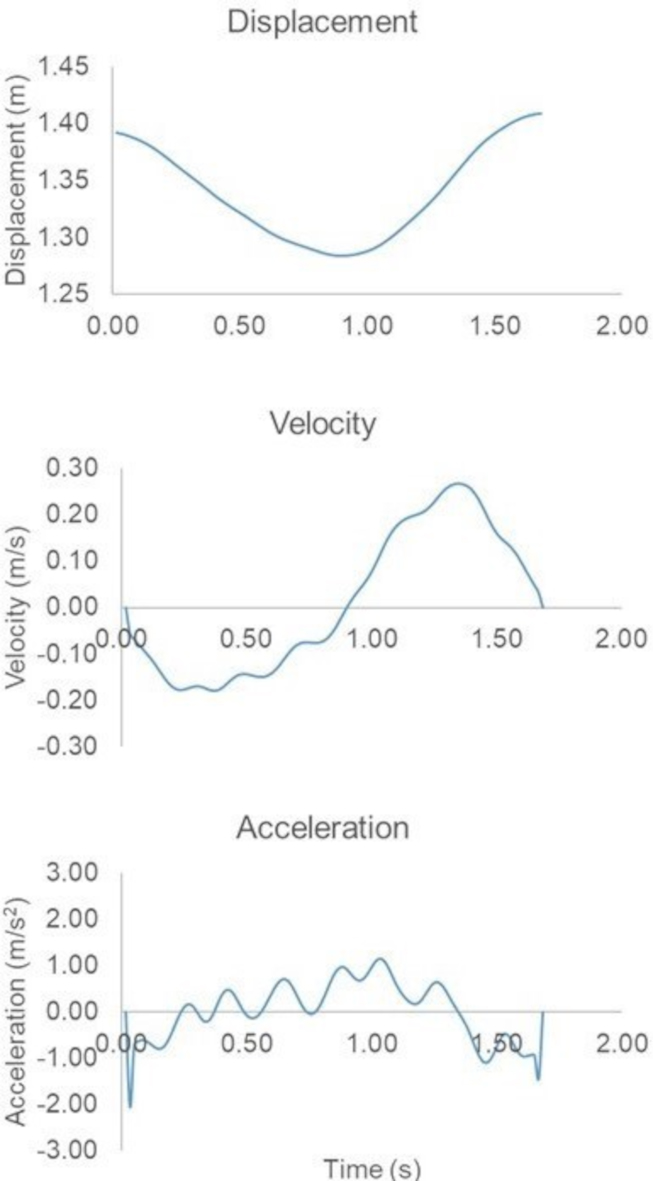
There was no significant difference in patellofemoral compressive force (
Patellofemoral compressive (PFC), tibiofemoral compressive (TFC) and tibiofemoral shear (TFS) forces, along with hip, knee and ankle sagittal moments for each combination of load and technique during squats. Capped arrows indicate pairwise differences (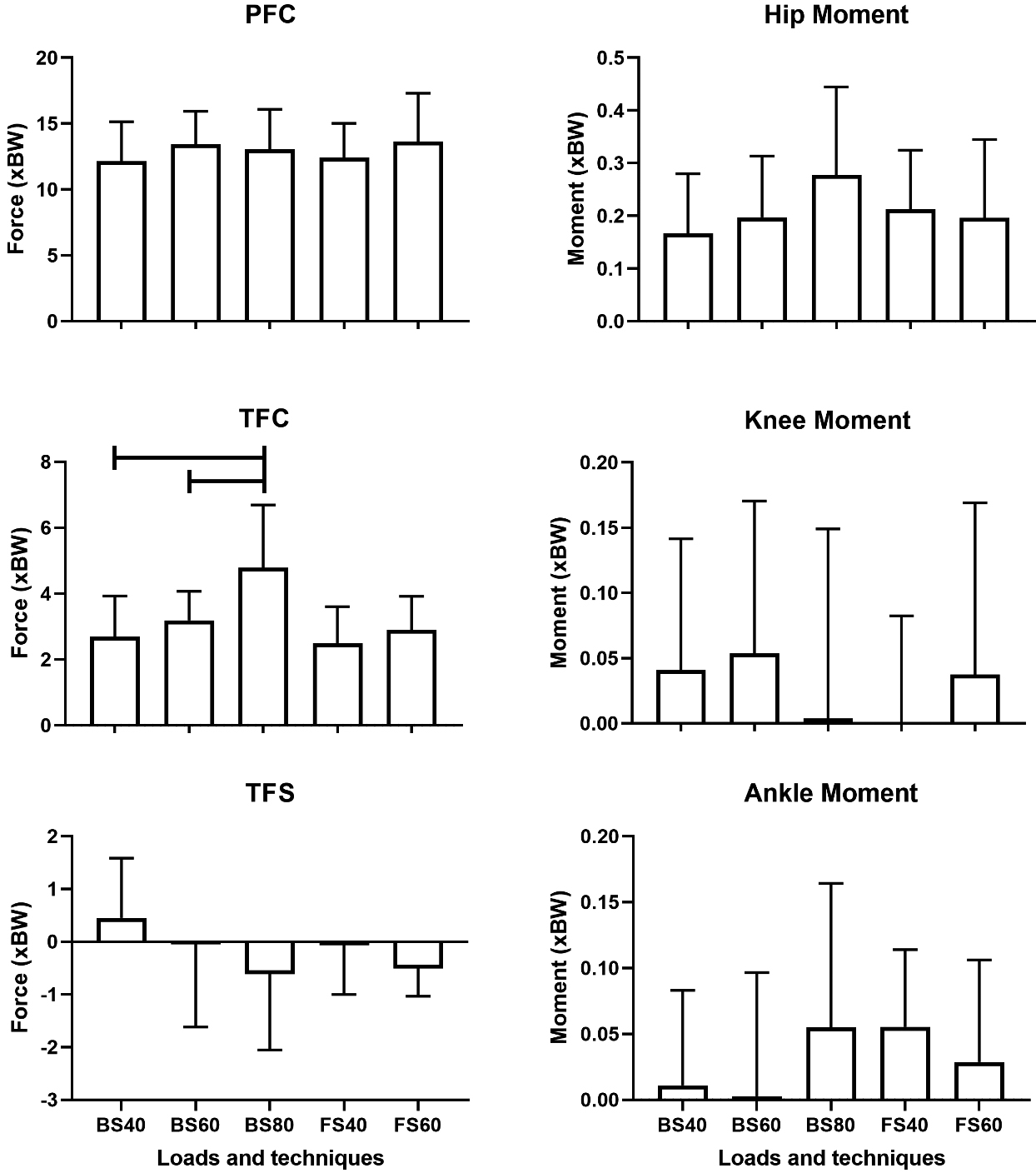
No significant effect from technique (back vs. front squat) was observed for hip flexors (
Hip flexors, rectus femoris, gluteus maximus, gluteus medius and minimus, quadriceps and hamstrings muscle forces for each combination of load and technique during squats. Capped arrows indicate pairwise differences (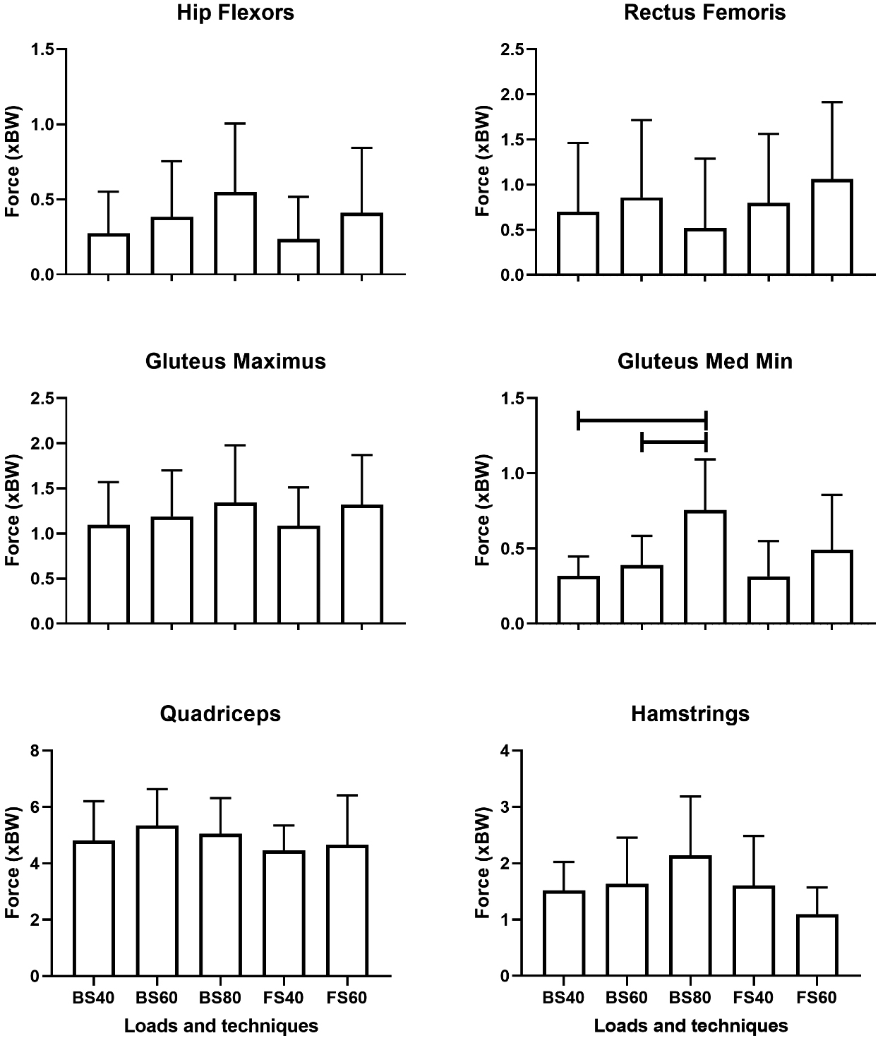
Changes in technique led to moderately greater lumbar extension for the front squat compared to the back squat (
Mean and ROM angles for lumbar flexion/extension, hip and knee flexion for each combination of load and technique during squats. Positive values for lumbar flexion indicate extension. Capped arrows indicate pairwise differences (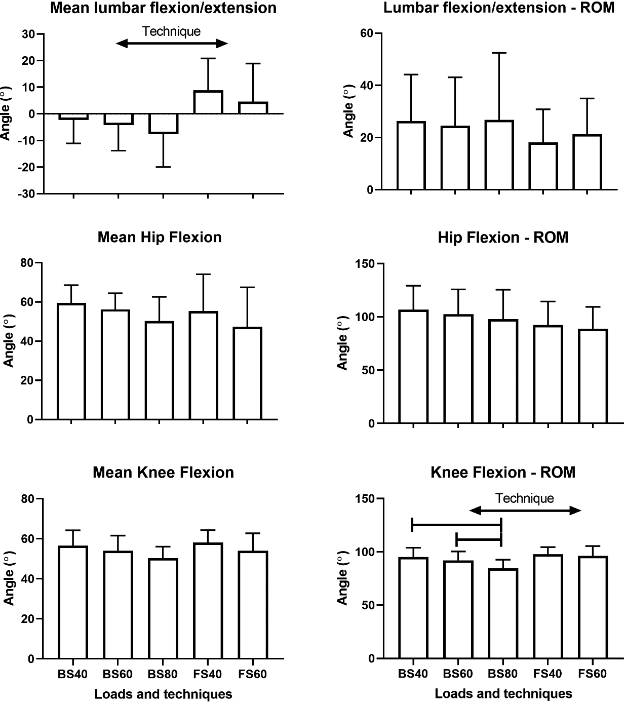
This study assessed the influence from squatting technique (i.e. front vs. back squat) and load (40, 60 and 80% of 1-RM) in knee joint forces and lower limb muscle forces. In line with our hypothesis, tibiofemoral compressive force was somewhat sensitive to increases in external load (i.e. 40 vs. 80% and 60 vs 80% of 1-RM) but other knee joint force components (i.e. patellofemoral and tibiofemoral shear forces) were not affected. Different to initially hypothesized, muscle forces were not largely affected by load or technique, with only gluteus medius and minimus producing more force at 80% compared to 60% and 40%. These findings add to the existing body of knowledge in the literature because no prior study used musculoskeletal modelling to determine joint loads when using loads relative to individuals’ 1-RM. These additions are important because joint loads are more realistically determined when using musculoskeletal modelling compared to inverse dynamics [27] or EMG [18]. In addition, using 1-RM to prescribe the loads provides a more appropriate comparison between participants rather than loads equivalent to body mass [10]. Moreover, the use of a Smith machine adds valuable information to exercise prescription for recreational use of strength training, maintenance of health and rehabilitation due to the access to this equipment in exercise and clinical spaces.
Front vs. Back squats
The lack of differences in joint forces between front and back squat is contrary to a prior study that showed larger compressive forces for the back squat [22], which conflicts with the expected alteration in torque from the knee to the hip [1]. These differences in relation to prior studies may be explained by several elements, including the use of barbells in prior studies and potential differences in position of the upper body and pelvis between studies. The use of barbells in a prior study [22] may have involved different position of the body as these authors did not provide a full description of their instruction to participants. Likewise, Stuart et al. [12] utilized a variation of the front squat with similar spinal flexion than the back squat, which differs from our method.
The only difference in body position between squats (front vs. back) was a larger spinal flexion for the back squat, as expected. This is in line with a prior study [14], which suggests that, during back squats, participants have potentially positioned their pelvis posteriorly in relation to their knees. It is possible though that, in order to maximise differences between techniques, back squats may also need to involve greater anterior pelvic tilt. These combination of positions for the spine and the pelvis could then ensure that hip extensors are longer and engage strongly during a back squat. In addition, a larger spinal flexion would mean that the external load and the trunk centre of mass should be projected more forward and that the pelvis moves backward relation to the hip joint [1]. Because in this study participants were not constrained to a given position of their pelvis, it is likely that they have self-selected their best possible combination for the spine-pelvis-hip complex whilst squatting. This means that, some participants may have opted for less backward movement of the pelvis, which could have resulted in greater anterior displacement for their knees. This is partially supported by the small increase in knee ROM during front squats, when the pelvis is unlikely to move as backward as during back squats. Therefore, further studies are needed to assess if prescribing the position of the pelvis or the knee (i.e. 28) increases the engagement of hip extensors during back squats and potentially affects knee loads.
Effects of loads
A prior study [10] showed that compressive and shear force components of the tibiofemoral joint were larger with increased loads whilst another study [1] also showed that patellofemoral compressive force was greater with larger loads. These results are potentially due to an increased knee extensor moment when load increases [11], which was not observed in the current study. As for the front-back squat comparison, it is possible that large variability in spine-pelvis-hip complex may have reflected into knee moments. This is supported by the potential change in the position of the pelvis, when load was increased [29]. This is likely to have resulted in a further backward position of the pelvis with the purpose of engaging hip extensors to a greater extent, as partially supported by moderate increases in gluteus medius and minimus force.
It is also possible that increases in loads, as observed by larger vertical and resultant ground reaction forces, may have been accommodated across the hip, knee and ankle at different proportions. This is somewhat evident in the visually large standard deviation for knee and ankle moments, and by the moderately larger medio-lateral ground reaction force at 80% compared to 40% of 1-RM Although prior studies suggest that increases in load changes the main demand for torque and power from the knee to the hip joint [1], this could be accomplished by a variety of muscle recruitments. In a prior study [30], it has been shown that load lifting squats (i.e. manual handling) involves an individualized combination of joint angles, regardless of the provision of specific instructions on how the lift should have been performed. This finding reinforces the observation that participants may self-select their muscle recruitment in order to concentrate on optimising their performance during squats.
Modelling and the use of an inclined Smith machine
Findings from Sahli et al. [10] and Hartmann et al. [1] are mostly conflicting to ours because we only observed moderate-to-large increases in tibiofemoral compressive force between 40% and 80% and 60 vs 80% of 1-RM. Differently, our findings align with results from Gullet at al. [22], using a geometrical model of the knee joint for calculating net joint forces. A possible reason for differences between our results and prior findings may have been due to minimum differences in muscle forces (i.e. only changes for gluteus medius and minimus). Schellenberg et al. [19] outlined that there are a variety of methods being used to calculate muscle and joint forces in strength training exercises. Differences between models have been demonstrated to have a major impact in comparison of data [31]. In addition, a small proportion of models have outcomes compared to in vivo data [32], which limits the definition on the levels of reliability of the outcomes. The model used in the current study has been refined in order to allow for realistic outcomes during high knee flexion tasks (i.e. greater than 120
The use of a Smith machine with an inclined rail was a unique feature of this study for measurements of knee loads because prior studies using Smith machines for squats were either limited to the analysis of 1-RM loads [35] or to the assessment of muscle activation [36]. The use of this particular equipment is beneficial as it can help inform exercise prescription for recreational use of strength training, maintenance of health and rehabilitation. For clients or patients that would be introduced to the squats, the use of the Smith machine could be positive as it provides greater stability in relation to free-weights. In addition, the potential for engaging safety lockers allow novice performers to gain instant feedback on the depth of their squat. However, the small incline of this Smith machine provides differential features compared to a Smith machine with no incline. Further studies may be required to determine if the
Limitations
Data from this study was limited to a certain extent. Participants were generally young and although familiar with performing squats on a Smith machine, were recreationally trained. This may have limited their ability to perform the squats consistently, particularly the front squat. In this issue, we then opted for using the 1-RM measured during back squats to determine the loads to be used during front squats. Therefore, it is possible that our outcomes may differ if both techniques are compared at loads relative to their 1-RMs rather than based on the back squats 1-RM. We ensured that they were familiarized properly with the front and back squats using the Smith machine prior to collecting data, in order to minimise learning effects. We were also unable to measure ground reaction forces at the right foot or to use EMG data to refine muscle force calculations from the timings of EMG signals [37]. These data could have provided further information on bilateral asymmetries and could have improved the assessment of individual responses in muscle forces.
Conclusion
In summary, larger external load increased tibiofemoral compressive force without effects on tibiofemoral shear force, patellofemoral compressive force. A moderate increase in gluteus medius and minimus force was observed at greater loads associated with a potential increase in posterior displacement of the pelvis. However, individual’s self-selection for spine-pelvis-hip positions may have had an effect on muscle forces and joint angles during squats, which hinders potential differences between front and back squats in terms of most muscle groups and joint forces.
Author contributions
CONCEPTION: Rodrigo Bini, Megan Lock and Gedd Hommelhoff.
PERFORMANCE OF WORK: Rodrigo Bini, Megan Lock and Gedd Hommelhoff.
INTERPRETATION OR ANALYSIS OF DATA: Rodrigo Bini, Megan Lock and Gedd Hommelhoff.
PREPARATION OF THE MANUSCRIPT: Rodrigo Bini, Megan Lock and Gedd Hommelhoff.
REVISION FOR IMPORTANT INTELLECTUAL CONTENT: Rodrigo Bini.
SUPERVISION: Rodrigo Bini.
Ethical considerations
This study was approved by the ethics committee from La Trobe University (HEC17-028 approved in 18 May 2017). All participants provided signed informed consent.
Funding
The authors report no funding.
Footnotes
Acknowledgments
We would like to thank all participants who volunteered for this study. Authors also acknowledge the support from Dylan D’Souza, Ilena Karas and Kyle Morcombe for support during data collection.
Conflict of interest
The authors have no conflicts of interest to report.