
Abstract
OBJECTIVE:
To examine the effects of fatigue on force reproduction during internal and external rotation of the shoulder using a single group repeated measure design.
METHODS:
Seventeen healthy male subjects who did not regularly compete in overhand sports and had no history of glenohumeral complaints took part in the study. Force reproduction was measured in the dominant shoulder. A target force had to be reproduced in three consecutive trials before and after a fatigue protocol. Maximal voluntary contraction was assessed to determine the target force. Measured data before and after fatigue were analyzed as well as error scores to examine the effect of fatigue.
RESULTS:
Repeated-measures analysis of variance revealed a significant influence only in the testing direction. No difference was found with the target value before or after the fatiguing repetitions. The inter-trial intra-class correlation coefficient showed high reliability.
CONCLUSIONS
: Force reproduction towards external rotation is more accurate than for internal rotation.
Introduction
The shoulder joint, as one of the most mobile joints in the body, has to rely on a dynamic stabilization system apart from its limited osseous and capsular structures. This complex system offers stability through muscles and ligaments [1, 2, 3]. As muscle contractions (output) are involved in this type of stabilization, a very good sense of proprioception (input) is required) [4]. To date, three modalities in the concept of proprioception have been discussed in literature. In addition to joint position sense (JPS) and sense of motion (kinesthesia), force-reproduction (FR) is the third modality in this concept [5, 6, 7, 8, 9]. Several researchers have already stated that FR should be regarded as an important component of proprioception [10, 11]. Affected muscle coordination due to a deficit in proprioception can actually lead to symptomatic shoulder instability [12, 13].
Sherrington defined proprioception as: afferent information from proprioceptors located in the proprioceptive field that contribute to conscious sensation (muscle sense), total posture (postural equilibrium) and segmental posture (joint stability) [14]. It is a specialized modality providing information about the position and direction of movement of a limb [4]. As it includes movement, muscular control is incorporated and vice versa [15]. Therefore, the idea that muscles are crucial for good proprioception has gained attention in scientific literature. For example, whereas JPS is traditionally thought to be initiated from the joint-capsule, some researchers have shown that muscle-receptors are also of great importance. As muscle receptors have the ability to detect an amount of force or tension, researchers can use FR to measure their sensory features [16, 17].
Force-reproduction, or sensation of resistance, is defined by Myers and Lephart as the ability to sense the force produced in a certain joint [18]. A very important mechanism in dynamic stabilization is the muscle contraction to occur preceding a joint movement [3]. This contraction will allow in the adequate amount of muscle-stiffness necessary to obtain joint-stability. When performing certain movements, subjects have to reproduce the correct amount of force to properly stabilize the moving joint. This has been investigated in the knee and ankle by several researchers [19, 20, 21]. There have been few studies concerning this specific subject in the upper limb [11, 22]. Mcnair et al. defined this muscle-stiffness as the ratio of the difference in force to the difference in length [20]. An increase in muscle-stiffness provides resistance to stretch, sensitizes muscle-spindles and lowers the electro-mechanic delay in reflexive stabilization. Muscle-stiffness as a result of a preparatory muscle-contraction can only be obtained in an efficient way due to an efficient FR. Only if a subject is able to accurately reproduce a given force, this results in an adequate amount of preparatory muscle contraction, leading to enough muscle stiffness for joint-stability. In learning how to perform a given movement, the adequate amount of force needed to stabilize the moving joint is perceived by the peripheral sensory system or proprioception, stored and eventually used in planning and executing motor patterns [10]. As a result of this planning, preparatory muscle activity will to occur; the subject will reproduce the adequate amount of force. This results in bracing of the joint before the actual loading with an external force takes place [11].
While using muscles to move a joint, they tend to fatigue. Takahashi et al. described fatigue as a commonly experienced condition that results from a period of intense or prolonged physical activity and is characterized by reduced capacity to exert muscular force. Different mechanisms are involved in this process. Factors proximal to the neuromuscular junction or factors involving the peripheral nervous system and muscle are still not completely understood. Fatigue slows down the muscle fiber conduction velocity, prolongs twitch duration and increases the neural activation required to produce a given force [23]. Fatigue can also affect motor performance skilled activities such as targeted throwing, stoop lift, tennis and balancing on an unstable surface [24, 25, 26, 27].
As the glenohumeral joint mainly relies on a dynamic stabilization, a well-tuned proprioceptive and neuromuscular control system, the rotator cuff is probably an important factor to stabilize this joint [3, 28]. To date, a lot of research on proprioception has been performed with the emphasis on JPS and sense of movement. FR, being the third component of proprioception, has been much less investigated. Several publications have shown that JPS is affected by fatigue [15, 29]. However, there is no consensus on whether or not FR is affected by fatigue.
In this study we hypothesized that 1. a fatiguing protocol would have a significant influence on FR in both internal and external rotation; 2. a difference in FR between both directions of rotation was expected. This research might provide the opportunity to establish a reliable protocol for FR testing in other groups of subjects, such as overhead athletes and subjects with various types of shoulder injuries.
Methods
Participants
The purpose of this study was to determine the effect of muscle fatigue on the FR for internal and external rotation in the glenohumeral joint of healthy subjects.
The study included 17 male subjects aged between 18 and 30 years (age
Procedures and instrumentation
Both the force reproduction test and the fatigue pro- tocol were performed using an isokinetic dynamometer (Biodex system three
All testing was completed at the research laboratory of the department of Rehabilitation Science and Physiotherapy of Ghent University. Before the actual testing procedure started, all subjects had to perform the same warm-up exercises as used in similar studies related to shoulder problems: lifting both arms to forward flexion, abduction, circumductions and push-ups against the wall [31]. Only the dominant side was tested. The dominant arm was identified by asking the subjects which arm they used to throw a ball.
Following the warm-up, the subject was seated on the chair of the Biodex system 3
Testing position.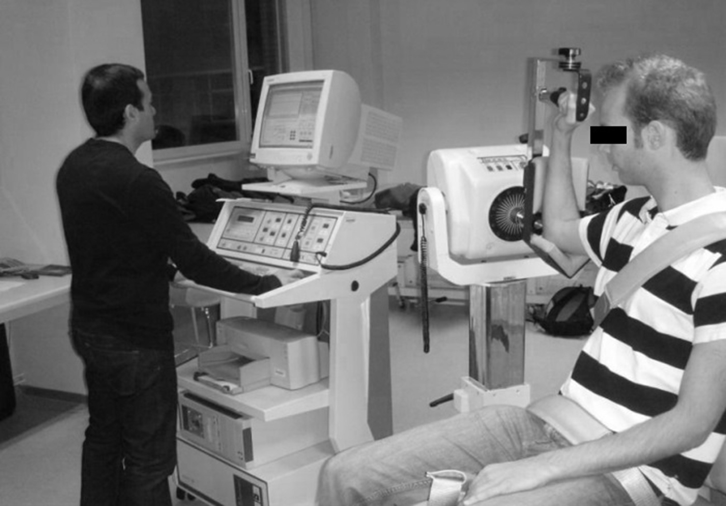
The initial direction of testing (internal or external rotation) was randomly selected. Per subject the sequence of testing stayed the same during all further measurements. The testing position remained the same for the whole testing procedure. The target for force reproduction was set at 50% maximal voluntary contraction (MVC) in accordance with the research of Dover and Power [5].
To determine the MVC for both directions, the subject was asked to perform a maximal isometric contraction against the arm of the dynamometer. Measurements were taken during 3 trials of each 5 seconds with 5 seconds rest between trials. The highest value (peak moment) was used as MVC. Fifty percent (to 1 Nm) of this MVC was used as target-moment for further testing.
After indicating the target-value on the screen, the subject was instructed to: “try to push against the arm of the machine so the line you produce equals the line we have drawn on the screen.” The subject was allowed 3 trial repetitions of 5 seconds each with 5 seconds rest between trials. After this exercise, the same procedure was repeated, but without either visual or auditory feedback. The screen was turned away, out of the subject’s visual field. This was performed for internal as well as for external rotation.
Fatigue protocol
A fatiguing program was selected that consisted of a continuous isokinetic movement with both internal and external rotation at a constant speed of 120
Force reproduction after fatigue
Immediately after the fatigue protocol, within three minutes, the same protocol for isometric force reproduction was applied as before fatigue with the same starting direction.
Mean
SD values for all measured parameters (Nm)
Mean
MVC
All data related to MVC were calculated by the Biodex software and written down on an evaluation sheet. The peak moment was used a representative of the MVC. For reproduction data, the average moment (AM) each individual subject produced over the 3 trials was calculated. Each trial took 5 seconds of contraction, but for data calculation the first and the last second were discarded. For the transformation of the source data to workable data for analysis, Matlab 7.8 (version 7.8.0.347, the Math Works TM, copyright 1984–2009) was used. All moment values are in Nm and presented as “mean
Subsequently, as a measure for FR, error values that the subjects made regarding the target value were calculated in two ways: the “absolute error” is the absolute value of the difference between the produced moment and the target (50%MVC). To allow comparison between the errors for internal and external rotation, the “relative error” was calculated as the absolute error divided by the target value. Type A intra-class correlation coefficients (ICC) using an absolute agreement definition intra observer agreement.
Mean and SD values were calculated for all dependent variables. The Kolmogorov-Smirnov test was used to determine normality of all data. As all data were normally distributed with equal variances, parametric statistics could be used. For analysis of the absolute data, a General Linear Model (GLM) 2-way analysis of variance (ANOVA) for repeated measurements with 1 within-subject factor (time) and 3 levels (50%MVC, before and after fatigue) was used. The analysis of the error data was also performed with a GLM, but with 2 within-subject factors: direction (2 levels: internal and external rotation) and fatigue (2 levels: before and after). Alpha for all statistical analysis was set at 0.05. Post-hoc tests were performed with a paired t-test, with Bonferroni-correction if necessary (Alpha/n; with
Results
The inter-trial ICC showed high reliability between the trials. For internal rotation before the fatigue protocol the ICC was 0.934 and after the fatigue protocol 0.941. The ICC for external rotation was 0.904 before fatigue and 0.958 for the before fatigue and after fatigue, respectively.
The mean values for MVC, target and AM before and after the fatigue protocol are shown in Table 1. The calculated errors are presented in Table 2. The overall force reproduction acuity for internal rotation was higher compared to external rotation (
Error data
Error data
Comparison between target value and AT towards internal rotation
For internal rotation, a significant interaction was found (
While trying to reach the target value, subjects could under- or over-estimate their output force. Before the fatigue protocol 14 out of 17 subjects (82.35%) underestimated the target towards internal rotation, while after the fatigue protocol, 15 subjects underestimated the target (88.24%). Towards external rotation 10 (58.82%) subjects underestimated their target before the fatigue protocol, while after the fatigue protocol only 7 (41.18%) did.
The main goal of this study was to determine whether fatigue affects moment reproduction during internal and external rotation of the shoulder joint.
For external rotation, no significant difference was found in the reproduction before or after the fatigue protocol, nor between the target value and the AM before or after fatigue. Regarding internal rotation, differences were found for the AM with the target force. Subjects were not able to reproduce the target force for internal rotation within an acceptable range (Table 3). They made a similar mistake before as well as after the fatigue protocol (Fig. 2). Based on these results, it seems that fatigue does not influence the accuracy in which a given force can be reproduced. Graphically a difference could be expected (Fig. 3), but no statistical evidence was found. Being a part of proprioception, FR is assumed to be affected in the same way as JPS or movement sense. Other research has already shown different outcomes for proprioception measures [34]. Most studies have shown that fatigue diminishes proprioception [12, 13, 18, 32, 33, 35], although others have shown fatigue to enhance [36] or not to affect [37] proprioception.
Plot of relative error.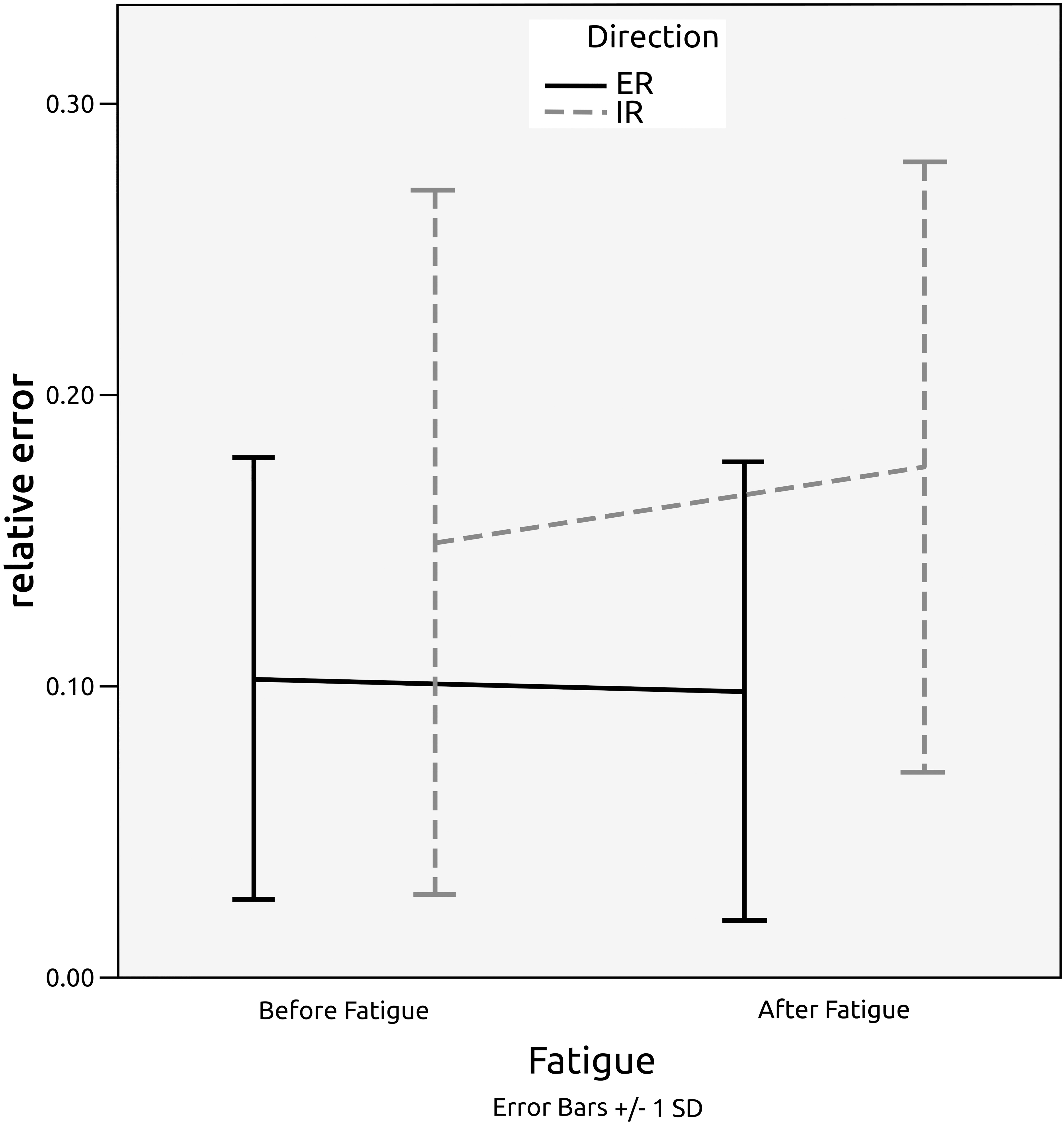
Force reproduction, graphic representation. Each curve represents the average moments of all subjects. Target (solid line) and AM (dotted line) are indicated.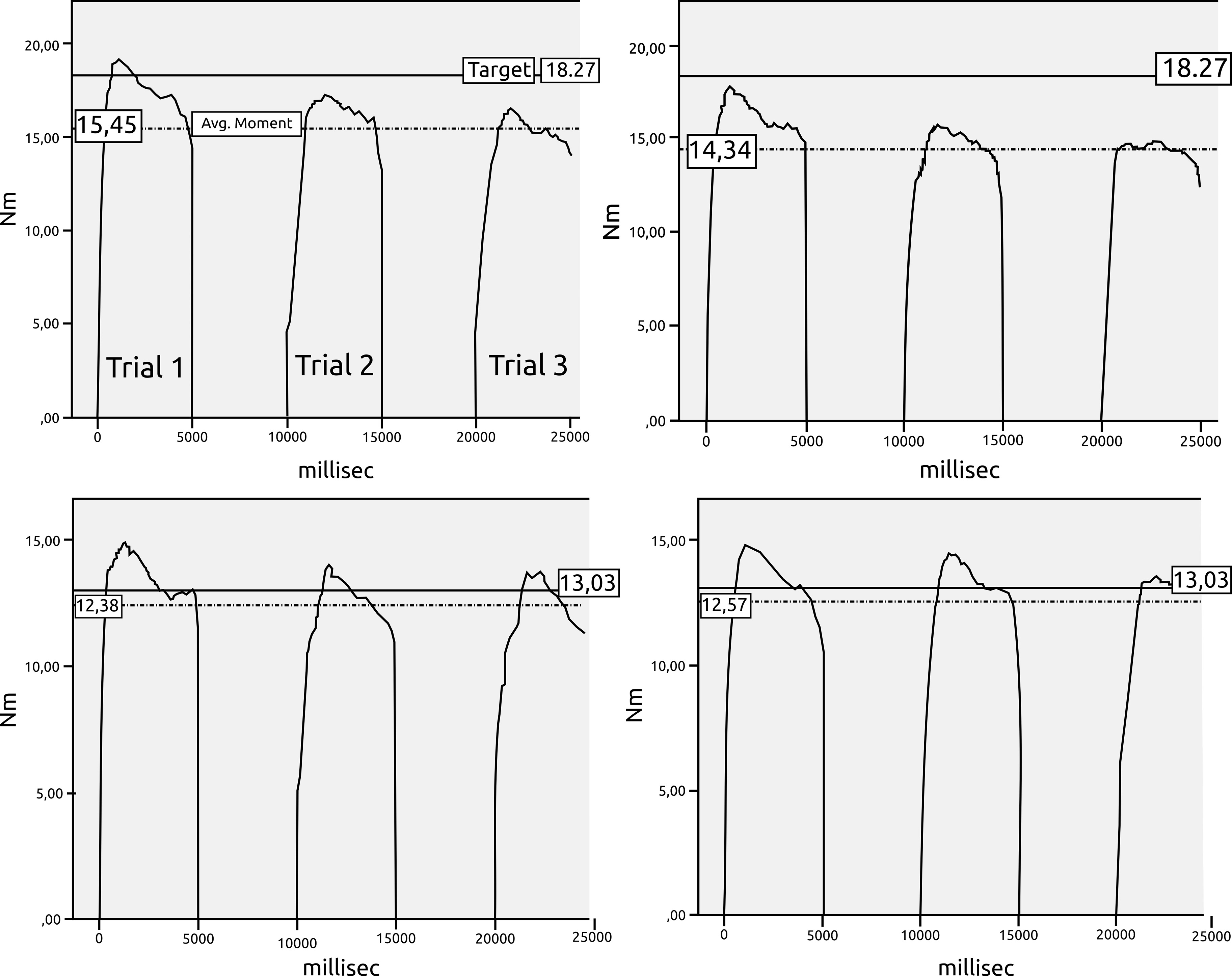
An effect of direction on FR was observed in the study. As described above, a significant error of the AM for internal rotation was made by the study subjects. This means that the subjects were remarkably better at reproducing external rotation forces than they were at reproducing internal rotation forces. A possible explanation for this difference can be found in the testing position. Subjects were tested in an end ROM position, meaning a full external rotation and 90
After completing the testing procedure, some interpretation was necessary. The velocity used in the fatigue protocol could have had an influence on the results. Fatigue was only applied at 120
The subjects in the current study always scored below the target value. During the trial period preceding the actual test, the target value was indicated on the computer screen with a horizontal line. Most of the subjects tried to reach this line as fast as possible, showing a kind of overshoot, but stayed under the drawn line during the rest of the trial (Fig. 3). Subject positioning was the same as in a previous research [5, 32]. In future research different starting positions should be tested. To eliminate the extra tension on the anterior capsule favoring external rotation FR, a more neutral position might be preferable. A neutral position (30
The criteria used to determine fatigue were the same as those in the study by Myers et al. [15]. The decrease in the strength of the external rotator muscles until they were below 50% of MVC was examined. Immediately after the fatiguing exercise, subjects were asked to reproduce 50% of the original MVC. As the subject was fatigued at this point, it could mean that they were being asked to produce a force very close to maximal force at that moment. If the target were lowered, the difference between the actual maximum force and the target would be bigger, and reproduction would probably be more difficult. There is a need for further studies to investigate whether or not fatigue significantly influences FR using reproduction of other MVC-related relative values (e.g. 10%, 20% and 30%).
The main limitation of this study is related to it being a single gender one. We believe that FR should be also investigated in women, using the same protocol, in order to find out whether gender affects this skill.
The results of this study reveal no influence of fatigue on FR and thus only the direction of movement is likely to be of importance. However, applying a specific test position subjects seemed to reproduce force better towards external rotation. A more detailed research on the effect of angular velocity during the fatigue protocol, other percentages of MVC for reproduction, different test-positions and the influence of a learning will possibly enhance our understanding of the impact of fatigue on force reproduction.
Footnotes
Conflict of interest
The authors declare no potential conflicts of interest with respect to the research, authorship, and/or publication of this article.