
Abstract
BACKGROUND:
Eccentric strength training changes muscle architecture but it is also an important factor for the prevention of injuries and their rehabilitation.
OBJECTIVE:
To determine the architectural adaptations of the semitendinosus (ST) after an eccentric strength training protocol with Nordic hamstring exercise (NHE), followed by a subsequent detraining period.
METHODS:
Twenty-three male individuals, aged 25.5
RESULTS:
The post-test scores indicated that eccentric strength training resulted in a significant increase in the fascicle length (FL) (t
CONCLUSION:
The NHE seems to generate adaptations in ST architecture, which, in addition, are also reversible after a detraining period. These results may have practical implications for injury prevention and rehabilitation programmes.
Introduction
The architectural characteristics of muscles have an impact on the ability to produce tension, as well as on the risk of muscle injury. Thus, understanding their nature and possible applications in rehabilitation and prevention is very important [1, 2, 3, 4, 5]. The pennation angle (PA), muscle thickness (MT) and fascicular length (FL) are architectural variables which change when a muscle is subject to mechanical stimuli [2, 3, 6, 7, 8].
Sport practice has a clear impact on hamstring injuries [9], accounting for between 12% and 29% of all injuries in event categories which involve high-intensity running [10, 11, 12, 13, 14], in addition to a high injury-related recurrence rate, ranging between 22% and 34% [15, 16].
Previous studies suggest two main mechanisms in hamstring injuries: length [17, 18] and speed [13, 15, 19, 20, 21, 22, 23]. The hamstring muscles reach their maximum length during the terminal stance phase of the gait cycle, when they are required to eccentrically decelerate the knee extension and hip flexion before weight-bearing [24]. When this occurs at high speed the load on the muscle increases greatly, contributing to high injury rates [16, 25].
Most studies focused on the femoral biceps muscles, whereas the semitendinosus muscle (ST) has been less widely researched. However, the latter plays a strong role in hip and knee functionality during activity [26] and in the rehabilitation of anterior cruciate ligament injuries [27, 28].
Muscle injuries seem to have an influence on its architecture, reducing FT and increasing PA, with respect to the uninjured contralateral muscles [5, 29, 30]. These changes alter the muscle function, since a greater FL is associated with a higher maximal velocity of contraction [4, 31] and a lower incidence rate of injury [5, 29, 30]. Hence, eccentric strength training became an effective method of prevention [13, 14, 32, 33, 34, 35] and rehabilitation [36, 37], as it demonstrated its ability to increase muscle FL values and to reduce the PA [7, 38]. The architectural changes are reversible after a detraining period without eccentric mechanical stimuli [38].
The changes in the muscle architecture have been studied by applying eccentric loading assessed through isokinetic dynamometry [7, 38, 39, 40]. Nevertheless, the use of isokinetic systems presents numerous drawbacks, such as high costs, complex handling and the fact that they are difficult to use in large groups of athletes [41]. In addition, they can also be considered “non-functional” since the use of the sitting position bears little resemblance to actual sporting activities [7, 38, 39, 40]. Therefore, there is a need to evaluate alternative eccentric hamstring training systems that have the potential to become widespread in the daily practice routine of athletes.
Numerous studies show improvements generated by the eccentric ‘Nordic hamstring exercises’ (NHE). Mjølsnes et al. [42] pointed out an improvement in muscle strength and dynamic control after 10 weeks of training, the Nordic Hamstring Strength Training group developed by Salci et al. [43] increased their eccentric hamstring strength after the training program, albeit without changes in the dynamic control ratio, Clark et al. [44] referred to a decrease of the maximum peak torque, whereas Brooks et al. [45], Gabbe et al. [33], Arnason et al. [34] and Van der Horst et al. [46] emphasized a lower incidence rate of hamstring injuries in athletes and soccer players. In addition, this exercise showed a significant relationship between the ‘break-point-angle’ and maximum peak moment of the hamstring measured with isokinetic dynamometry [41] and has induced architectural changes in said musculature [28]. Hence, the above-mentioned authors referred to its effectiveness as a basis for a functional eccentric training for athletes. NHE is a simple exercise, not requiring any additional material, which has been proven to highly activate hamstrings, especially the ST muscle [26, 28].
Due to the high injury rate in this region and the difficulties encountered in using the isokinetic equipment, an intriguing question arises concerning the efficiency of easily applicable eccentric exercise, such as NHE, to generate architectural changes in the ST muscle.
Methods
Participants
Twenty-three male individuals (aged 25.5
Eccentric training evolution with NHE
Eccentric training evolution with NHE
Once the informed consent form was signed, the protocol and previous training was carried out in the facilities of the Faculty of Education and Sport Sciences, University of Vigo, Spain.
The study lasted 13 weeks. The first week, participants undertook an initial assessment of the ST muscle architecture (pretest), and subsequently two sessions of familiarisation with NHE in order to ensure proper technical execution and maximise its effectiveness. Next, an eight-week period of eccentric training with NHE was performed. In week 9 of this training period, the second assessment of the muscle architecture (posttest) was performed. Finally, the participants underwent four weeks of detraining, avoiding any kind of mechanical stimulus of eccentric nature. After this period, the third assessment of the muscle architecture (retest) was carried out in week 13. All measurements were made at the same time of the day and under the same conditions for all participants.
The protocol was designed with the aim of promoting a progressive assimilation of the eccentric mechanical stimuli and reducing delayed onset muscle soreness (DOMS) in participants. Each training session lasted between 10 and 20 minutes, depending on the training volume of sets and repetitions according to the evolution of the programme (Table 1). All sessions were separated by at least 48 hours and preceded by twelve-minute standardised warm-up self-loading exercises. All participants were provided with constant visual and verbal feedback, to contribute to their motivation and correct technical execution.
Two-dimensional ultrasound image of the semitendinosus taken along the longitudinal axis of the posterior thigh. From these images, the superficial and deep aponeuroses could be determined, as well as the MT and fascicle angle with respect to the aponeurosis. The estimations of the FL can be performed through a trigonometric calculation, using the MT and the PA.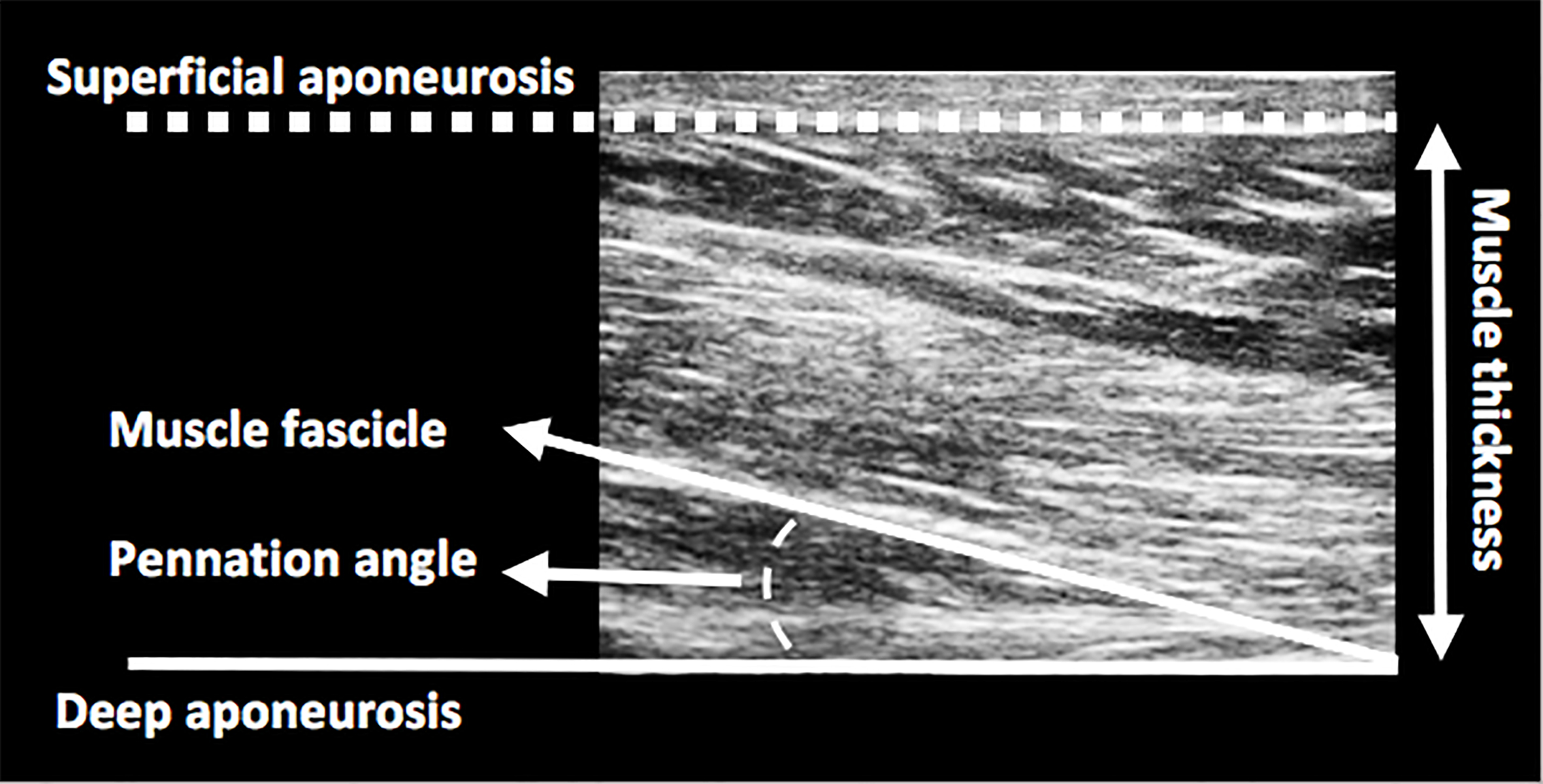
The use of ultrasound has proven to be a highly reliable method for assessing muscle architectural characteristics [5], and specifically those of the ST [47]. MT, PA and FL estimations were determined from ultrasound images obtained along the longitudinal axis of the muscle belly using B-mode ultrasound (12 Mhz frequency, 8 cm depth; 14
To obtain the images, the ultrasound probe was placed and aligned longitudinally and perpendicular to the posterior thigh in the scanning site on the participant’s skin, which was previously covered with a conductive gel. In order not to alter the accuracy of the measurements, the probe was handled carefully, ensuring minimal pressure [47, 48].
After the scan, an analysis was carried out by means of image processing software, MicroDicom, version 0.7.8 (Bulgaria). Following the procedure conducted by Blazevich et al. [49], six points were digitised for each of the images. The MT was defined as the distance between the superficial and intermediate aponeuroses of the ST. The PA was delimited between the intermediate aponeurosis and the direction of a muscle fascicle previously identified in the image (Fig. 1). The aponeurosis angle (AA) was determined as the angle between the line marked by the aponeurosis and a horizontal line drawn along the captured image [47, 49]. Finally, the FL was defined as the muscle fascicle length existing between both aponeuroses. Given that the total of the above-mentioned length cannot be observed in the field of view of the probe, an estimate was made using an equation validated by Blazevich et al. [49] and Kellis et al. [47].
The FL was expressed in absolute terms (cm) and also relative to muscle thickness (RFL). The images were collected and analysed by the same evaluator (MFJ) with previous experience in this protocol, who was blinded at all times to the participants’ identifiers during the analysis. The researcher participated previously in an intra-rater study to evaluate the reliability of the measurements. Twelve healthy subjects took part in the study in which 5 2D-ultrasound images of the ST were performed in the same anatomical region in different sessions, obtaining reliable measurements of the different architectural variables (intraclass correlations
Eccentric “Nordic hamstring” exercises
NHE, also known as ‘Nordic curl’, are eccentric exercises designed to strengthen the hamstrings (Fig. 2) [23, 25].
Nordic hamstring exercise: (a) start, (b) midpoint indicating the Nordic break-point angle, and (c) end.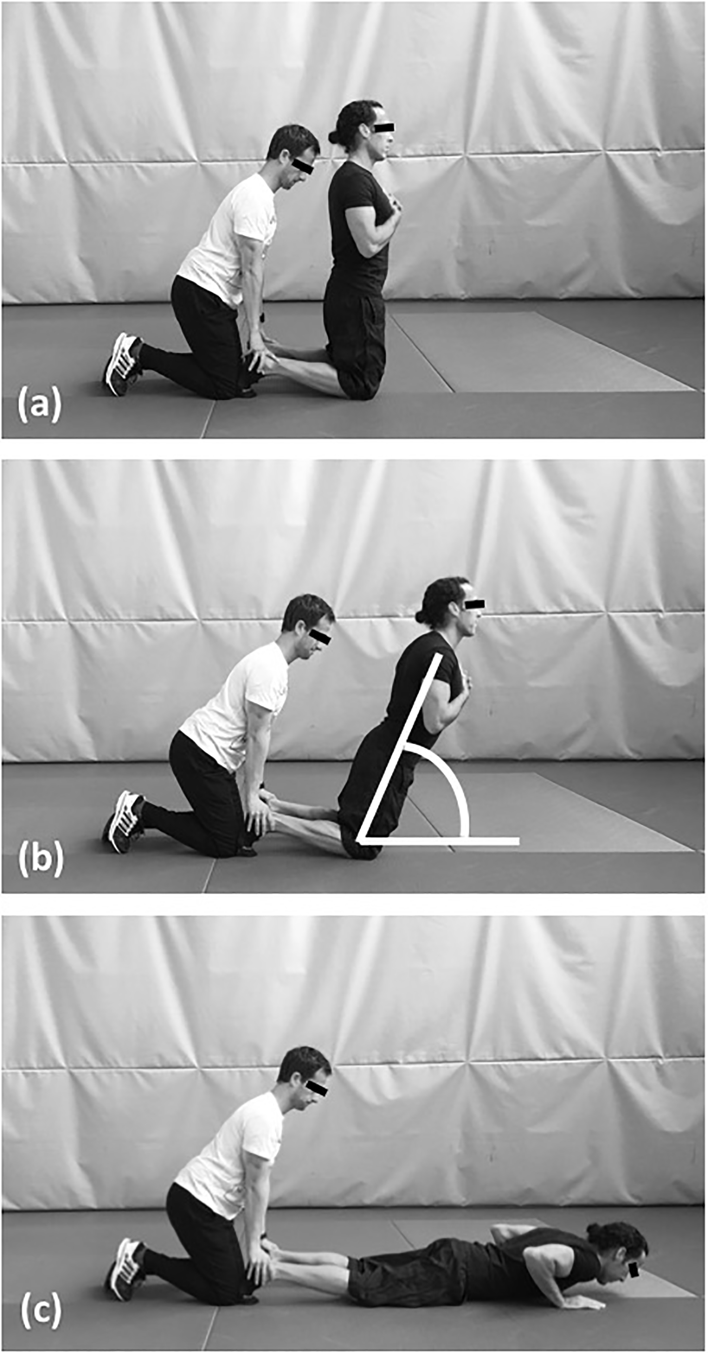
The data were reviewed to ensure the normality assumption by means of the Shapiro-Wilk test and homoscedasticity through Levene’s test. The Gree- nhouse-Geisser correction was used when the test of sphericity was violated (
The data analysis was performed using the IBM SPSS Statistics 24.0 statistical software (IBM Corporation, Chicago, IL, USA).
Changes in muscle architecture of the ST before (pretest) and after (posttest) the intervention and after the period of detraining (retest) (mean
standard error of the mean)
Changes in muscle architecture of the ST before (pretest) and after (posttest) the intervention and after the period of detraining (retest) (mean
The results indicate that the eccentric training protocol with functional NHE resulted in changes in FL, RFL, MT and PA variables. The obtained results are detailed below according to the different measured variables of the ST muscle architecture.
Fascicle length
Muscle FL was affected by the eccentric training protocol with functional NHE, F (2–44)
Muscle thickness
MT was affected by the eccentric training protocol with functional NHE, F (2–44)
Pennation angle
PA was affected by the eccentric training protocol with functional NHE, F (2–44)
Fascicle length relative to muscle thickness
Fascicle length relative to muscle thickness (RFL) was affected by the training protocol, F (2–44)
Table 2 outlines the changes in FL, RFL, MT and PA during the training and detraining period.
Discussion
The main contribution of this research study is to the field of practical application as NHE seems to induce architectural changes comparable to those obtained through isokinetic dynamometry.
The architectural study of the ST muscle has hardly been examined in the scientific literature [47], despite its importance in the hip and knee functionality, its synergy with the rest of the hamstrings [26] and its rehabilitation rate in terms of ACL injuries [27, 28].
Muscle architecture and NHE
The increase in FL of the ST at rest, observed in the present study, is similar to the results indicated by Potier et al. [7] who, after an 8 w eccentric strength training programme with curl leg, obtained a significant increase of 34% in FL of the Biceps Femoris long head (BFlh). On the other hand, Timmins et al. [38], using a 6 w eccentric training protocol with isokinetic dynamometry, obtained a significant increase of 16% in FL of the BFlh. Likewise, Bourne et al. [28] achieved a 16% increase in the same muscle using a 10 w eccentric training protocol with NHE. Similarly, our study reported a significant increase of 7.44% in FL at rest of the ST. The differences in the duration of the intervention (from 6 to 10 weeks), in the training techniques (curl leg, isokinetic dynamometry and NHE) and in the involved muscle (BFlh and ST) may explain the variations between the studies, although all are in agreement on the increase of FL after an eccentric strength training that could be due to the increase of sarcomeres in series [51].
The PA results are not as conclusive, since significant decreases were observed, in line with the findings of Timmins et al. [38], but at variance with those obtained by Potier et al. [7] with a decrease of 3.1%, despite the fact the authors pointed to this result as “counter-intuitive”. Since there are no previous studies on the architectural changes undergone by ST after an eccentric training protocol, further studies on this variable are needed.
The significant increase observed in MT of the ST is consistent with the results obtained after an eccentric training period by Baroni et al. [39] and Malas et al. [40] in the rectus femoris and vastus lateralis. However, the fact that these results match those obtained by Bourne et al. [28] is even more relevant due to the similarity of the protocol, although in the latter the imaging technique had been the MRI. After a 10 w eccentric training with NHE, the authors observed a significant increase in the ST muscle volume. This finding is of interest since 1–6 y from an ACL injury, a decrease in the ST muscle volume is observed [27]. Hence, NHE could be a valuable element in the rehabilitation of this type of injury.
Muscle architecture and detraining
The results also show that the changes in muscle architecture induced by NHE were reversed after a four-week detraining period. There was a significant decrease of FL and MT, whereas PA increased. In spite of focusing on other muscle groups and other type of eccentric training, these results are comparable to those found by Narici and Carretelli [52], Seynnes et al. [6] and Timmins et al. [38]. This has made us realise the need to maintain this type of mechanical stimuli in order to preserve the derived architectural changes although further studies will be needed to establish where the limit of the muscular “memory” is and what is the optimal way to maintain it.
Practical application for prevention and rehabilitation
Shorter muscle fascicles are more prone to muscle damage than longer fascicles [5, 53, 54]. Muscle injuries change its architecture, decreasing FL, increasing PA and making it more sensitive to relapses [5, 29]. Moreover, as already mentioned, ACL injuries cause a decrease in the ST muscle volume [27, 28]. These reasons seem sufficient to introduce eccentric strength content in the training plan of athletes at risk of suffering hamstring injuries and those recovering from an ACL injury; this content has proven to be an element of protection and rehabilitation of these types of injuries [36, 37]. There was already evidence of the potential benefits of eccentric training, but these were hardly transposed to the daily routine of athletes, as they had been obtained with specialised equipment and under conditions beyond their reach [7, 38, 39, 40].
Limitations
There are certain limitations to our study. On the one hand, the use of 2D ultrasound for the architecture assessment should be mentioned; it requires to a certain degree an estimation because FL is not entirely visible in the ultrasound image.
Although the estimation equation used in this study was validated [47], there is still a potential error that should be reduced in future works based on 2D ultrasound. On the other hand, only the ST architecture was evaluated, and considering that each hamstring has unique architectural characteristics, it would not be appropriate to generalise the results to all knee flexors.
Finally, establishing the application conditions of this type of training will be necessary, so that lasting architectural effects could be obtained in different populations of athletes.
Conclusion
The results obtained in the current research are similar to those obtained in previous studies that used isokinetic dynamometry in their design. In practical terms, NHE has proven to be relevant in bringing upon structural changes in this muscle that may be implicated in the prevention and rehabilitation of hamstring and anterior cruciate ligament (ACL) injuries.
Footnotes
Acknowledgments
The authors would like to thank Juan Martínez Fernández (Football Club Rubin Kazan and Spanish Society of Ultrasound in Physiotherapy) for their helpful contributions to the production of this manuscript.
Conflict of interest
The authors declare no conflicts of interest.