
Abstract
The early diagnosis of skin cancer is of paramount importance for effective patient treatment. Dermoscopy, a non-surgical technique, utilizes precise equipment to examine the skin and plays a crucial role in identifying specific features and patterns that may indicate the presence of skin carcinoma. In recent times, machine learning (ML) methods have been developed to recognize and classify dermoscopic images as either malignant or benign. Deep learning (DL) systems, including Convolutional Neural Networks (CNNs), as well as various ML models like Random Forest (RF) classifiers and Support Vector Machine (SVM), are employed to extract relevant features from these images. This study introduces the Crow Search Algorithm with Deep Transfer Learning Driven Skin Lesion Detection on Dermoscopic Images (CSADTL-SLD) technique. The CSADTL-SLD method starts with the application of a median filter (MF) to remove noise from the images and utilizes the GoogleNet model for feature extraction. GoogleNet is well-regarded for its capacity to capture intricate and meaningful patterns within the data, which are essential for accurate lesion characterization. Furthermore, the CSADTL-SLD technique applies the Crow Search Algorithm (CSA) for parameter tuning of the GoogleNet model. After feature selection, the system employs the MLP classification model for precise lesion categorization. The comprehensive results of this research demonstrate the superiority of the CSADTL-SLD algorithm, showing significant enhancements in skin lesion detection accuracy and robustness when compared to existing methods. This approach holds promise as an effective solution for automating the detection and classification of skin lesions in dermoscopic images.
Introduction
Melanoma remains a highly aggressive form of skin lesion and is currently on the rise [1]. It undergoes ongoing examination regarding the computerized analysis of suspicious skin cancers for malignancy, utilizing images captured by digital cameras [2]. Analysing such images often proves challenging due to the presence of disruptive factors such as light reflections and variations in skin surface illumination. The most critical step in diagnosing melanoma involves the segmentation of cancerous regions from healthy skin [3, 4]. Conducting a visual examination of a suspicious skin surface by a dermatologist often constitutes the initial step in the detection of a potentially cancerous lesion. Precision in identification is of utmost importance, as certain types of cancers may bear similarities. Moreover, the accuracy of Computer-Aided Diagnosis (CAD) approaches that of a skilled dermatologist’s identification [5]. In the absence of technology-assisted tools, dermatologists currently analyse melanoma with an accuracy rate ranging from 65 to 80%. In cases where suspicion arises, dermatoscopic images are obtained using an exceptionally high-resolution camera for the purpose of conducting visual inspections [6, 7]. Lighting conditions are currently quantified, and filters are employed during the recording process to reduce skin reflections, enabling a more in-depth visualization of skin layers. This professional support has yielded a 49% improvement in the diagnosis of skin cancer. Presently, the classification of skin cancer has become a focal point within the machine learning community [8]. Automated categorization of cancers is being increasingly utilized in medical analysis to assist healthcare professionals, providing swift access to life-saving detections. In non-hospital settings, smartphone applications are being deployed for this purpose. Manual diagnoses of skin cancer are time-consuming and require significant human resources, making it a challenging process [9]. Various techniques for the automatic identification of skin cancers have recently emerged, benefiting from technological advancements. Both conventional and deep learning methods play a pivotal role in the medical sector [10].
In this study, we are developing a technique called Crow Search Algorithm with Deep Transfer Learning Driven Skin Lesion Detection on Dermoscopic Images (CSADTL-SLD). The CSADTL-SLD approach incorporates the use of a median filter (MF) for noise removal and employs the GoogleNet model for the feature extraction phase. GoogleNet is well-known for its ability to capture intricate and relevant patterns in the data, which is essential for precise lesion characterization. Subsequently, the CSADTL-SLD technique utilizes the Crow Search Algorithm (CSA) to fine-tune the parameters of the GoogleNet model. After feature selection, the CSADTL-SLD approach utilizes the MLP classification model for accurate lesion classification. This research demonstrates the superiority of this proposed method, showcasing significant enhancements in skin lesion detection accuracy and robustness when compared to existing methodologies.
Literature review
In [11], the authors introduced an interpretable approach that relied on a stacked ensemble of CNNs for the early detection of melanoma skin cancers. Within this analysis, transfer learning (TL) techniques were incorporated into the stacked ensemble architecture, while multiple CNN submodules designed to address the same classification tasks were also assembled. To generate the final predictive results, a novel technique known as the meta-learner was employed to make predictions for each sub-model.
Bhimavarapu and Battineni [12] proposed the integration of deep learning (DL) methods to automate the classification of melanoma through dermoscopic images. They introduced a Fuzzy-based GrabCut-stacked CNN (GC-SCNN) method, which was validated using pre-trained images. Additionally, the authors [13] developed an automatic approach for categorizing dermoscopic images, distinguishing between malignant and benign skin cancers. Consequently, this research introduced an enhanced DL-based performance using CNN. To mitigate the risk of overfitting, techniques such as dropout, regularization, and data augmentation were employed, which were common practices in CNN approaches.
In [14], the authors presented introduced a deep learning (DL) approach using YOLO (You Only Look Once) that relied on the utilization of DCNNs. This algorithm was applied to the task of diagnosing melanoma, using both dermoscopic and digital images. Their method yielded faster and more accurate results compared to conventional CNNs. It’s worth noting that this approach combined specific resourceful techniques in a two-step segmentation process to achieve its outcomes.
In [15], the authors devised a hybrid methodology for the classification of melanoma skin cancer, which was utilized for the examination of potentially problematic tumors. This novel approach relied on the integration of three distinct diagnostic methods, namely CNN and two traditional machine learning (ML) algorithms. These methods were trained using specific sets of features that encompassed descriptors related to the color, border, and texture of the skin cancer.
The proposed model
This research has introduced an automated system for detecting skin lesions in dermoscopic images, referred to as the CSADTL-SLD technique. In the initial phase, a noise removal process based on MF is employed. The combination of GoogleNet, CSA, and MLP classification presents a potent and effective approach for skin lesion detection. Figure 1 illustrates the comprehensive workflow of the CSADTL-SLD system.
Overall flow of CSADTL-SLD system.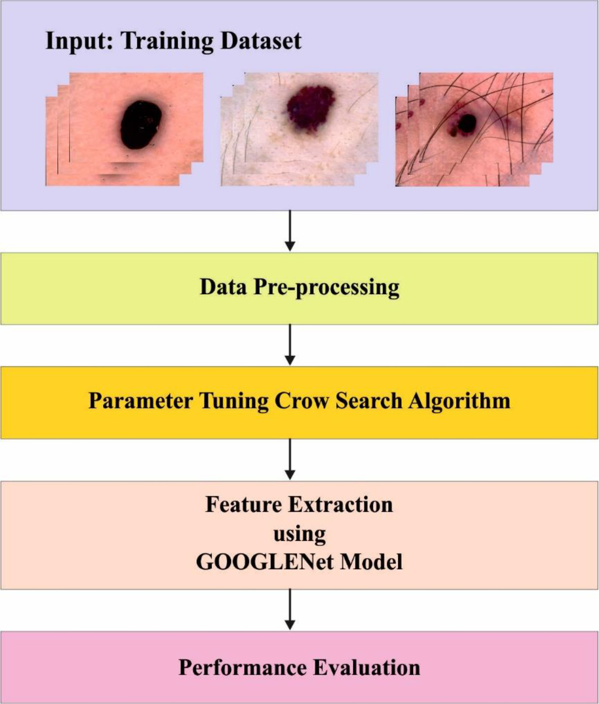
In the initial phase, the noise removal processes are initiated using a median filter (MF) [16]. Data pre-processing is employed to enhance the image quality through the application of the MF system. This involves a non-linear signal processing method tailored to the characteristics of the available data. In this method, the noisy digital image is transformed using a mask, which comprises the median value of the surrounding pixels. The pixel values within the mask are arranged based on their grey values, and the median value of this group is used to replace the noisy pixel value, thus refining the overall image quality. The result obtained from the MF operation is expressed as
Where
The GoogleNet model, also known as Inception v1, is a pioneering convolutional neural network architecture that revolutionized image classification tasks. It is characterized by its innovative use of inception modules, which consist of parallel convolutional layers with different filter sizes, enabling the network to capture features at various scales efficiently. These modules are stacked to form a deep neural network while maintaining computational efficiency. GoogleNet also introduced the concept of global average pooling, eliminating the need for fully connected layers and reducing the model’s parameters. This model’s inception modules and design principles have inspired subsequent iterations and architectures in the field of deep learning, making it a significant milestone in the advancement of computer vision. The construction of GoogLeNet consists primarily of three convolution layers with filter sizes of 7
Parameter tuning using CSA
In this study, CSA is employed for the optimal selection of GoogleNet parameters. CSA is a method belonging to the SI (Swarm Intelligence) model, which simulates the natural foraging behavior of crows in the atmosphere [17]. In essence, the CSA’s functionality relies on the behaviors of the crows, which encompass living within the swarm, storing the locations of food in memory, tracking other crows to pilfer their food, and ultimately thwarting other crows from discovering its own food locations. The CSA method employment to solve an optimizing issue can be depicted as
Here, every row in the matrix crow depicts one probable solution.
Later, the crows start travelling in the searching space and try in discovering and memorizing the outcome with optimum fitness attained priory in the matrix
In normal CSA, the process of search is illustrated via most 2 cases. In the initial case, the owner crow
Where
Secondly, the crow
else
In this context,
The Artificial Neural Network (ANN) mentioned here is a widely employed architecture within the realm of supervised learning, particularly suited for tasks involving issue classification [18]. It stands as one of the most straightforward and commonly utilized neural network structures. The core of this architecture is the Multi-Layer Perceptron (MLP) classifier, which comprises numerous layers of nodes (neurons) interconnected in a feed-forward manner. The initial layer serves as the input layer, while the final layer acts as the output layer, responsible for receiving the input data, which consists of features, and producing the ultimate classification decision.
In between these input and output layers, one or more hidden layers may exist, each housing a set of neurons. These hidden layers play a crucial role in processing and transforming the input data, allowing the network to capture intricate patterns and relationships within the data. The interconnected neurons within these layers work collaboratively to extract valuable features and make sense of complex information. Overall, the MLP architecture is a fundamental and effective tool in the domain of supervised learning, especially in scenarios where classification tasks are of paramount importance.
Performance validation
In this section, we systematically assess the effectiveness of the CSADTL-SLD methodology in detecting skin lesions. This evaluation is conducted on a dataset comprising 2000 instances, as meticulously documented in Table 1. To provide visual context, a selection of sample images from this dataset is thoughtfully illustrated in Fig. 2. This rigorous testing allows us to gauge the methodology’s performance in the context of real-world skin lesion detection scenarios, providing valuable insights into its practical utility.
Description on database
Description on database
Sample images.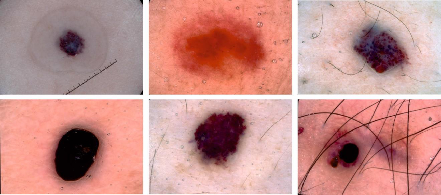
Table 2 and Fig. 3 present the results of skin lesion recognition achieved using the CSADTL-SLD method. The findings indicate that the CSADTL-SLD system effectively distinguishes between three classes. When utilizing 70% of the training set (TR set), the CSADTL-SLD technique yields an average accuracy, sensitivity, specificity, F-score, and Matthews correlation coefficient (MCC) of 94.24%, 84.70%, 93.54%, 86.20%, and 80.43%, respectively. Furthermore, when applying 30% of the test set (TS set), the CSADTL-SLD approach demonstrates similar performance with average accuracy, sensitivity, specificity, F-score, and MCC values of 94.22%, 84.10%, 93.33%, 86.09%, and 80.57%, respectively. These results underscore the effectiveness and consistency of the CSADTL-SLD methodology in accurately classifying skin lesions.
Skin lesion recognition outcomes of the CSADTL-SLD methodology on a 70:30 split of the training set (TR set) and the test set (TS set)
Comparative outcome of CSADTL-SLD approach with recent methods
Average outcomes of the CSADTL-SLD algorithm on a 70:30 split of the training set (TR set) and the test set (TS set).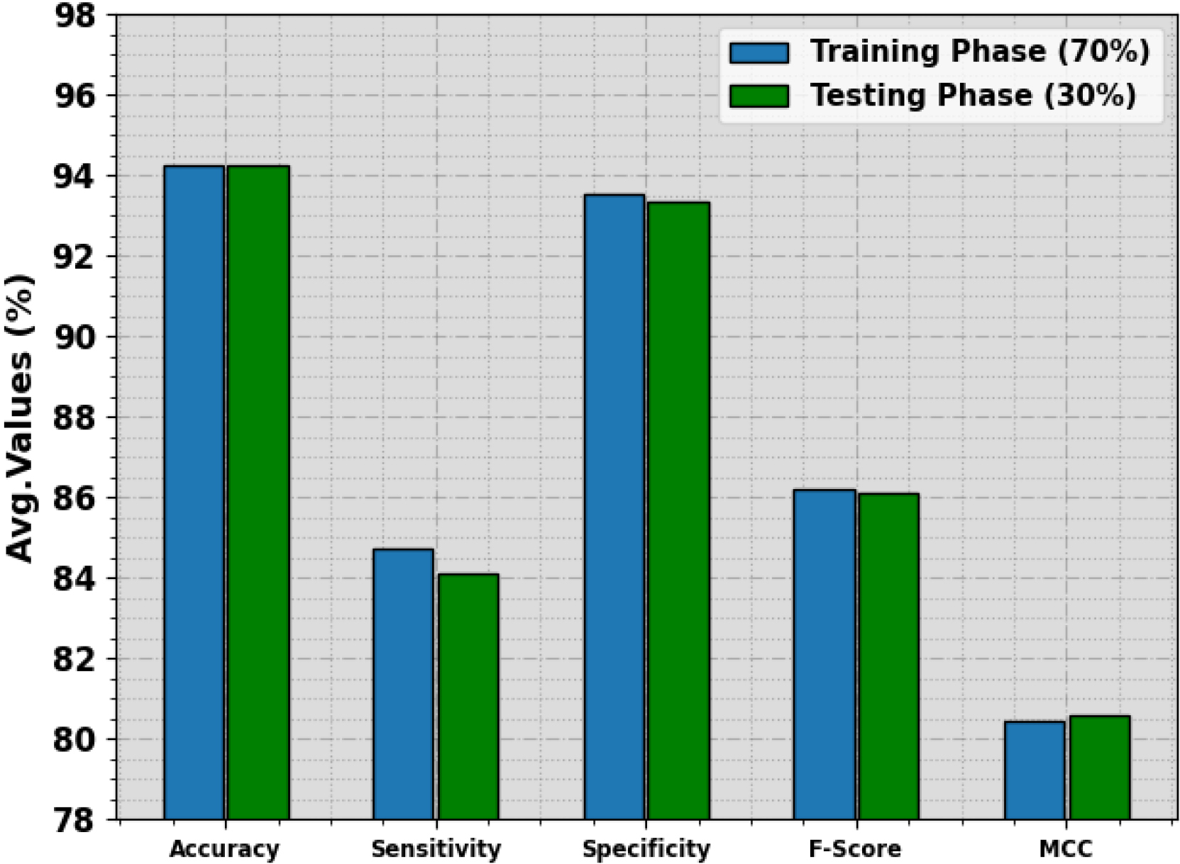
PR curve of the CSADTL-SLD approach.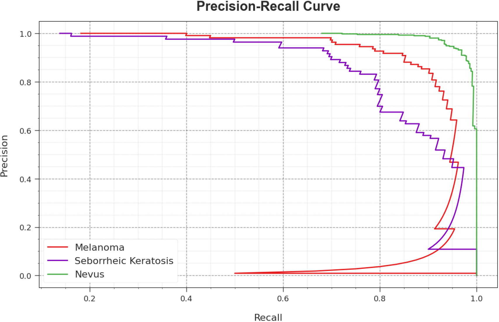
Figure 4 showcases a comprehensive investigation into the precision-recall (PR) performance of the CSADTL-SLD algorithm when applied to the test database. The findings clearly demonstrate a substantial enhancement in PR outcomes as a result of using the CSADTL-SLD approach. Notably, it becomes apparent that this system consistently achieves higher PR values across all three classes, signifying its improved precision and recall capabilities. This observation suggests that the CSADTL-SLD algorithm excels in the accurate identification and classification of skin lesions.
ROC of the CSADTL-SLD approach.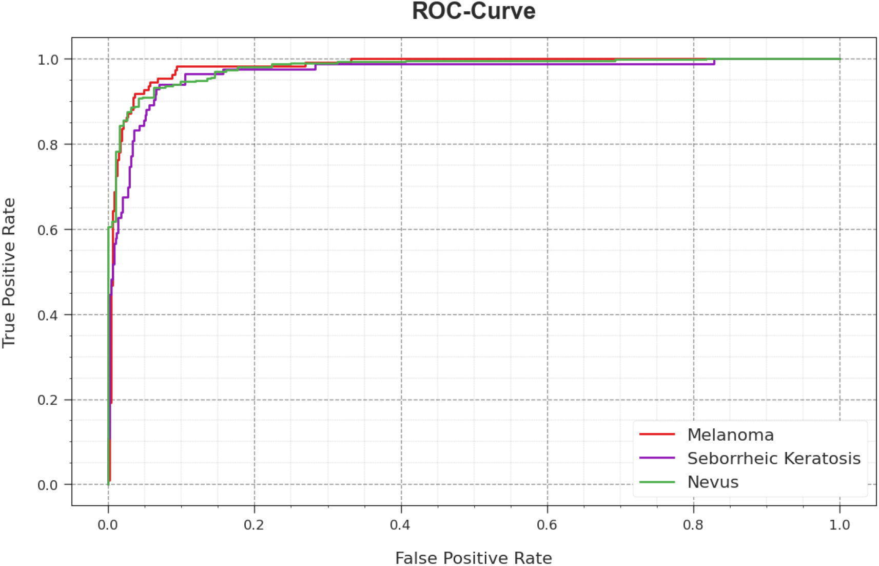
Figure 5 displays a Receiver Operating Characteristic (ROC) analysis of the CSADTL-SLD approach applied to the test database. The results of this analysis reveal that the CSADTL-SLD system consistently achieves higher ROC values, indicating its superior performance in distinguishing skin lesion classes. It is evident that the CSADTL-SLD methodology excels in terms of ROC outcomes across all three classes. These findings underscore the model’s ability to effectively discriminate between different types of skin lesions, thus establishing its strength in enhancing diagnostic accuracy and classification in comparison to alternative methods.
The performance of the CSADTL-SLD technique is assessed alongside existing systems, as detailed in Table 3 and illustrated in Fig. 6. The data presented in the table underscore the superior performance of the CSADTL-SLD approach in the realm of skin lesion detection. Specifically, when considering accuracy, the CSADTL-SLD technique outperforms its counterparts, achieving a higher accuracy rate of 94.24%. In contrast, other models, such as MAFCNN-SCD, NB, KELM, MSVM, MobileNet, and DenseNet-169, demonstrate lower accuracy values, ranging from 85.03% to 92.22%. Furthermore, in terms of sensitivity, the CSADTL-SLD method excels with a notably higher sensitivity rate of 84.70%. In contrast, the alternative models, including MAFCNN-SCD, NB, KELM, MSVM, MobileNet, and DenseNet-169, exhibit lower sensitivity values, ranging from 74.17% to 77.07%. Lastly, with respect to specificity, the CSADTL-SLD algorithm again outperforms its counterparts, offering a significantly higher specificity rate of 93.54%, while other models show lower specificity values, ranging from 83.19% to 88.67%.These results emphasize the consistent and superior performance of the CSADTL-SLD technique in skin lesion classification when compared to existing systems, demonstrating its potential to significantly enhance diagnostic accuracy and classification in the field.
Comparative outcome of CSADTL-SLD approach with recent systems.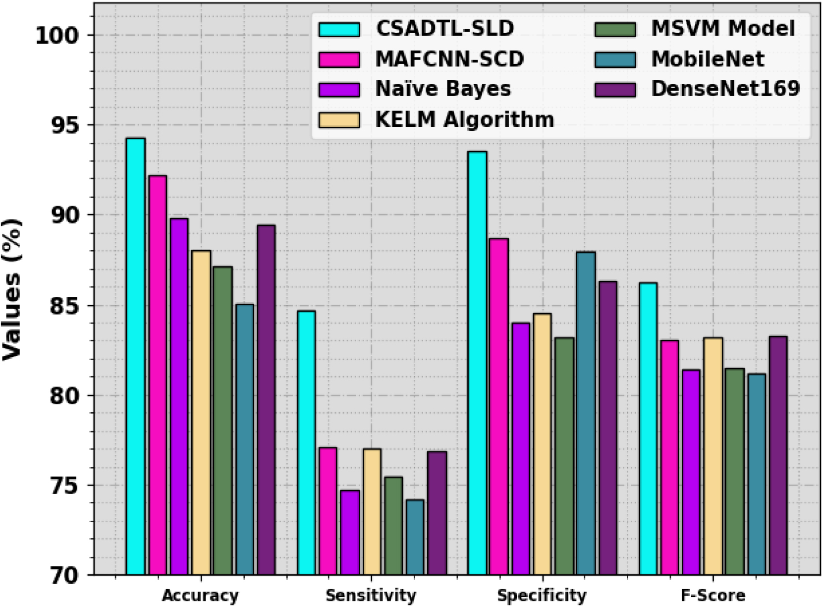
This research introduces an automated approach, the CSADTL-SLD technique, designed for skin lesion detection in dermoscopic Images. The initial phase involves noise reduction through median filtering. To extract meaningful features crucial for precise lesion characterization, the CSADTL-SLD technique leverages the GoogleNet model, known for its proficiency in capturing intricate data patterns. Subsequently, parameter tuning for the GoogleNet model is executed using CSA (possibly a parameter optimization technique). After feature selection, the system employs the MLP classification model for the accurate categorization of skin lesions. The comprehensive results demonstrate the superiority of this system, showcasing substantial enhancements in the accuracy and robustness of skin lesion detection when compared to existing methods. This approach stands out as an effective and efficient solution for automating the detection and characterization of skin lesions in dermoscopic images.