
Abstract
There is growing evidence that COVID-19 not only affects the lungs but beyond that the endothelial system. Recent studies showed that this can lead to microcirculatory impairments and in consequence to functional disorders of all inner organs. The combination of endothelial dysfunction with a generalized inflammatory state and complement elements may together contribute to the overall pro-coagulative state described in COVID-19 patients leading to venular as well as to arteriolar occlusions.
In December 2019, an outbreak of pneumonia due to a novel corona virus – the severe acute respiratory syndrome corona virus 2 (SARS-CoV-2) – occurred in Wuhan, Hubei province, China. The virus causing the corona virus disease 2019 (COVID-19) has since spread to many countries resulting in a pandemic [1]. Symptoms most commonly reported include fever, cough, shortness of breath and loss of sense of taste and smell. Older patients with co-morbidities are more likely to develop respiratory failure due to severe alveolar damage [2]. In more severe cases, the disease can also show a rapid progression to organ failure, with complications, such as shock, acute respiratory distress syndrome (ARDS), acute cardiac injury, acute kidney injury, disseminated intravascular coagulopathy (DIC), which may ultimately prove fatal [3]. Recent observations suggest that respiratory failure in COVID-19 is not driven by the development of ARDS alone [4], but that macro-vascular as well as micro-vascular thrombotic processes may play a role [5, 6]. It is becoming apparent that severe cases of COVID-19 are characterized by hyper inflammation and a thrombotic phenomenon. Such major adverse clinical events seem to suggest that in advanced stages of this disease one target is the endothelium, one of the largest organs in the human body. Whether vascular derangements in COVID-19 are due to endothelial cell dysfunction is currently unknown.
The main transmission route is through virus containing droplet or aerosol but also smear transmission is possible. The virus replicates in the upper respiratory tract decent to lower respiratory tract. The spread of the virus in the host is thought to occur also via the vascular system and transgression into mucosal tissues of nose, throat and especially of the lungs, where the endothelium comes into contact with SARS CoV-2 at an early stage. The virus enters endothelial cells by endocytosis via the binding of its spike glycoprotein to a cellular receptor which facilitates viral attachment to the surface of target cells [7–9]. Angiotensin-converting enzyme 2 (ACE2) was identified as the main receptor for severe acute respiratory syndrome corona virus (SARS-CoV 2) [10], which is abundantly expressed in the lungs, the respiratory epithelium and alveolar monocytes [11] and may explain the many cases of rapidly occurring lung failure. However, ACE2 is also expressed by endothelial cells [12–14] in the heart and the macro vascular system, gut, kidneys, liver, central nervous system, and adipose tissue [15–17]. As the density of ACE2 differs in the various tissues – very high in the lungs – the receptor density may correlate with the severity of the disease in those tissue [18–20]. In addition, there are further receptors on the surface of human cells which can mediate the entry of SARS-CoV-2, including transmembrane serine protease 2 (TMPRSS2 [19]), sialic acid receptors [21], and extracellular matrix metalloproteinase inducer (CD147 [22]). These four receptors are known to be expressed by endothelial cells [23–26].
Whereas unperturbed endothelial cells provide very potent anti-coagulant properties [27], exposure to inflammatory stimuli can rapidly lead to a procoagulant behavior. Very recently, Varga et al. found evidence of direct SARS-CoV-2 infection of endothelial cells in several organs and diffuse endothelial inflammation associated with apoptosis [28]. They described this state as endotheliitis with viral elements within endothelial cells and accumulation of inflammatory cells, with evidence of endothelial and inflammatory cell death. Endothelial cell injury can strongly activate the coagulation system via exposure of tissue factor and other pathways. Endothelial dysfunction refers to a systemic condition in which the endothelium loses its physiological properties, including the tendency to promote vasodilation, fibrinolysis, and anti-aggregation [27]. Narrowing of organ supplying arteries as well as microcirculatory disturbances in liver, spleen and kidneys in patients with severe COVID-19 were already described recently [29].
COVID-19-endotheliitis could explain the impaired microcirculation in different vascular beds [30] and their clinical sequelae in patients with COVID-19. During inflammatory activation or apoptosis, endothelial cells become pro-coagulant [31] and release microvesicles (MV) which can affect the function of target cells through surface interaction and receptor activation, cellular fusion and the delivery of intra-vesicular cargo [31]. Among others, endothelial MV have been described to affect hemostasis [27]. Figure 1 shows a picture with apoptotic human umbilical venous endothelial cells with massive signs of blebbing and detached microvesicles.
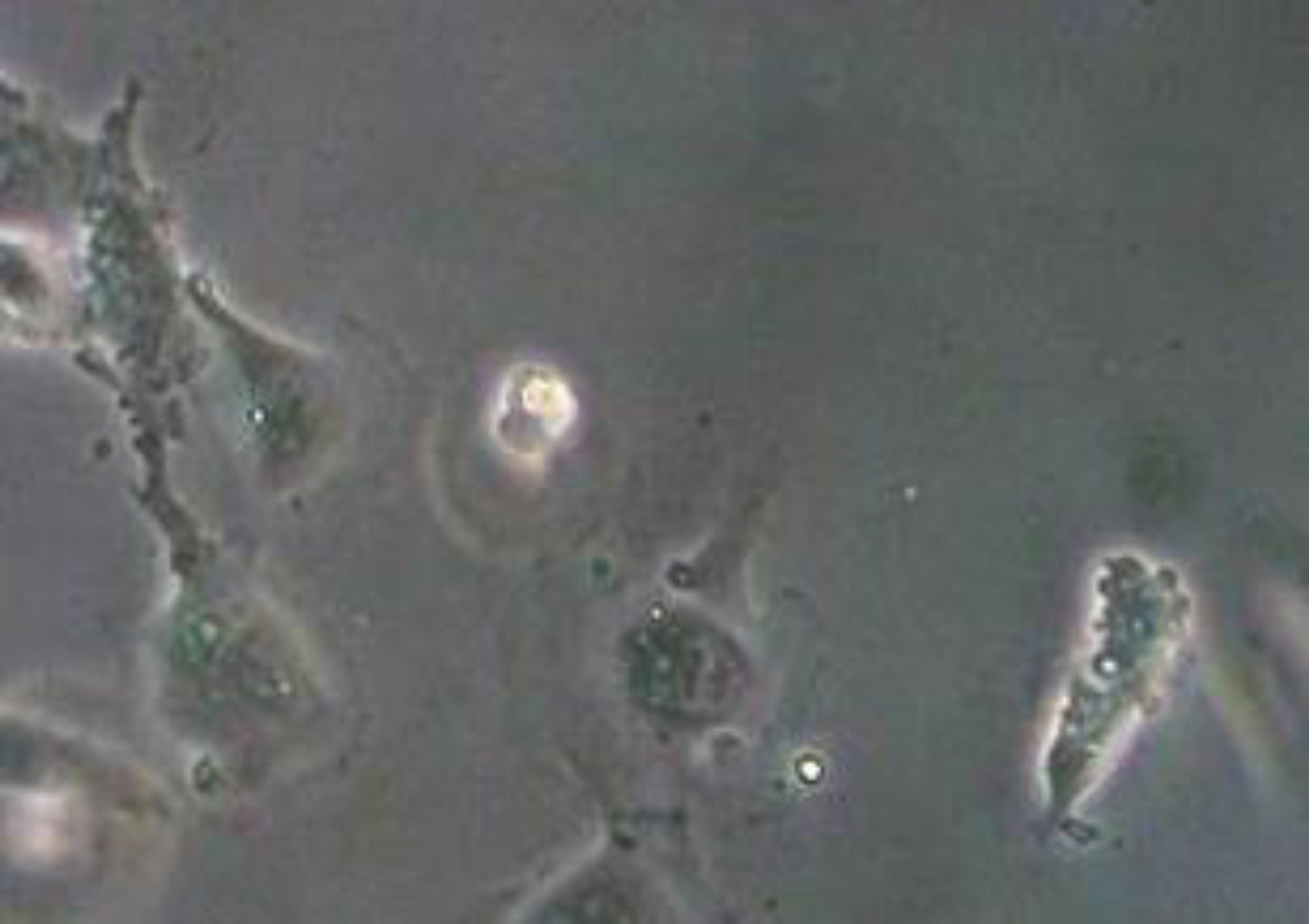
Apoptotic endothelial cells with signs of blebbing and detached microvesicles (cLSM, objective ×40).
Pathological investigations and also imaging studies confirmed the COVID-19 disease as a thrombo-inflammatory process that initially affects the lungs and in consequence the perfusion, which consecutively can affect all organs of the body. It is well known that a variety of viruses can affect the coagulation system including HIV, Dengue virus, and Ebola virus [32, 33]. Very recently, Spiezia et al. reported that SARS-CoV-2 may predispose patients to thrombotic disease, both in the venous and arterial circulations [34]. Excessive inflammation, platelet activation, endothelial dysfunction, and stasis related to the infection were described [35], which can result in severe hypercoagulability and predispose to thrombosis. That has been related to deaths in critically ill COVID-19 cases [36]. A single-center retrospective cohort study of 183 patients with confirmed COVID-19 evaluated coagulation abnormalities that mimic disseminated intravascular coagulation (DIC) [37]. According to the International Society on Thrombosis and Hemostasis definition of DIC, 15 of 21 non-survivors (71%) were classified as having overt-DIC (≥5 points) at any time during follow-up, whereas only 1 of 162 survivors (0.6%) met these criteria (P < 0.001). Likewise, Tang et al. reported that 71.4% of non-survivors and 0.6% of survivors of COVID-19 showed evidence of overt DIC [38]. This is in line with another study, in which a clear correlation between D-dimer levels, disease progression and chest CT features suggesting venous thrombosis were reported [39]. Also, pulmonary embolism is with 30% significantly more frequent in COVID-19 patients [40] than usually occurring in critically ill patients without COVID-19 infection (1.3%, [41]) or in emergency department patients (3 to 10% [42]). Tee et al. were able show that in the lung a vascular areas occur which most likely represent 3–5 mm micro infarcts [43] which confirms the micro vascular involvement in the course of the disease.
In conclusion, it seems that COVID-19 is a disease affecting the lungs and, beyond that, the endothelial system. Recent studies show that this can lead to microcirculatory impairments, and in consequence to functional disorders of all inner organs. The combination of endothelial dysfunction with a generalized inflammatory state and complement elements may together contribute to the overall pro-coagulative state described in COVID-19 patients.