
Abstract
BACKGROUND:
Patients with advanced pancreatic cancer (APC) and liver metastases have much poorer prognoses than patients with other metastatic patterns.
OBJECTIVE:
This study aimed to develop and validate a radiomics model to discriminate patients with pancreatic cancer and liver metastases from those with other metastatic patterns.
METHODS:
We evaluated 77 patients who had APC and performed texture analysis on the region of interest. 58 patients and 19 patients were allocated randomly into the training and validation cohorts with almost the same proportion of liver metastases. An independentsamples
RESULTS:
The constructed nomogram demonstrated good discrimination in the training (AUC
CONCLUSIONS:
This study presents a radiomics nomogram incorporating RS and CA19-9 to discriminate patients who have APC with liver metastases from patients with other metastatic patterns.
Introduction
Pancreatic cancer is one of the most highly lethal diseases with almost as many deaths as cases worldwide [1, 2]. Among all types of distant metastases in APC, liver metastases is the most common, and it occurs in more than half of patients with APC [3, 4]. In our previous study, patients with APC and liver metastases showed much poorer prognoses compared with patients who had other patterns of metastases (6.1 months vs. 10.9 months,
Computed tomography (CT) is a commonly used examination for the initial diagnosis and staging of pancreatic cancer. However, the information acquired from CT in the clinical practice is limited due to inadequate elucidation of CT images [6]. For example, the size-based and shape-based features derived from CT images (e.g., longest tumor diameter) are sometimes associated with marked variability in outcomes and response, such as pseudoprogression in patients receiving immunotherapy [7]. Recently, the rapidly evolving field of radiomics has made it possible to translate medical images into high-throughput quantitative data [8, 9], and TA provides an opportunity for more precise diagnosis and treatment [10, 11, 12]. In addition, much evidence has shown that TA may be used to characterize and identify specific subsets of cancers. For example, previous studies have shown that a CT-based radiomics signature (RS) could help identify patients with lung adenocarcinoma patients who have high risks of distant metastases and specific histologic subtypes [13, 14].
In pancreatic cancer, previous studies have shown that radiomics features have both diagnostic and prognostic value. For example, Guo et al. showed that CT imaging features and texture parameters could help differentiate pancreatic neuroendocrine carcinoma from pancreatic ductal adenocarcinoma [15]. Sandrasegaran et al. showed that CT texture features, especially kurtosis and the mean value of positive pixels, were significantly associated with overall survival (OS) in pancreatic cancer [16]. In addition, Attiyeh et al. found that CT texture features were associated with SMAD4 status and stromal content in pancreatic cancer [17]. However, few studies have used radiomics features to characterize specific subtypes of pancreatic cancer.
Therefore, the aim of this study was to develop and validate a radiomics nomogram that incorporated both RS and clinicopathologic variables to discriminate patients who have APC with liver metastases from patients with other metastatic patterns.
Patients and methods
Patients
The primary cohort of this study consisted of 77 patients with APC enrolled in Changzhou No. 2 People’s Hospital. The following inclusion criteria were applied: (1) newly diagnosed and pathologically confirmed pancreatic adenocarcinoma; (2) no concurrent cancer at another organ site; and (3) complete records of baseline clinicopathologic features available. Of all the patients, 40 had liver metastases, and 37 had other metastatic patterns. The patients were randomly allocated into two stratified cohorts that included almost the same proportion of liver metastases: a training cohort (
CT image acquisition
Contrast-enhanced CT examinations were performed on a 128-row dual-source CT scanner (SOMATOM Definition Flash, Siemens, Germany) with 120 kV, tube current modulation and a 1 mm reconstructed section thickness. All patients were instructed to fast for at least 8 hours before administration of 70 ml of intravenous contrast (Iohexol 300 mg/mL) at a rate of 3 mL/s. After injection of the contrast agent, patients underwent double helical scanning at the arterial phase and portal venous phase while holding their breaths. The portal venous phase was selected as a region of interest (ROI), because pancreatic cancer boundaries have been most consistently seen in this phase [17, 18].
Image processing
ROIs were drawn on each slice with primary pancreatic cancer and metastatic tumors on the portal venous phase using the Semantic Segmentation Editor (Hitachi Automotive and Industry Laboratory). Then all ROIs were imported into a freeware software (Local Image Features Extraction, or LIFEx, version 5.10,
Workflow of image processing and model construction. Abbreviations: RFC, Random Forest Classifier; RS, radiomics signature.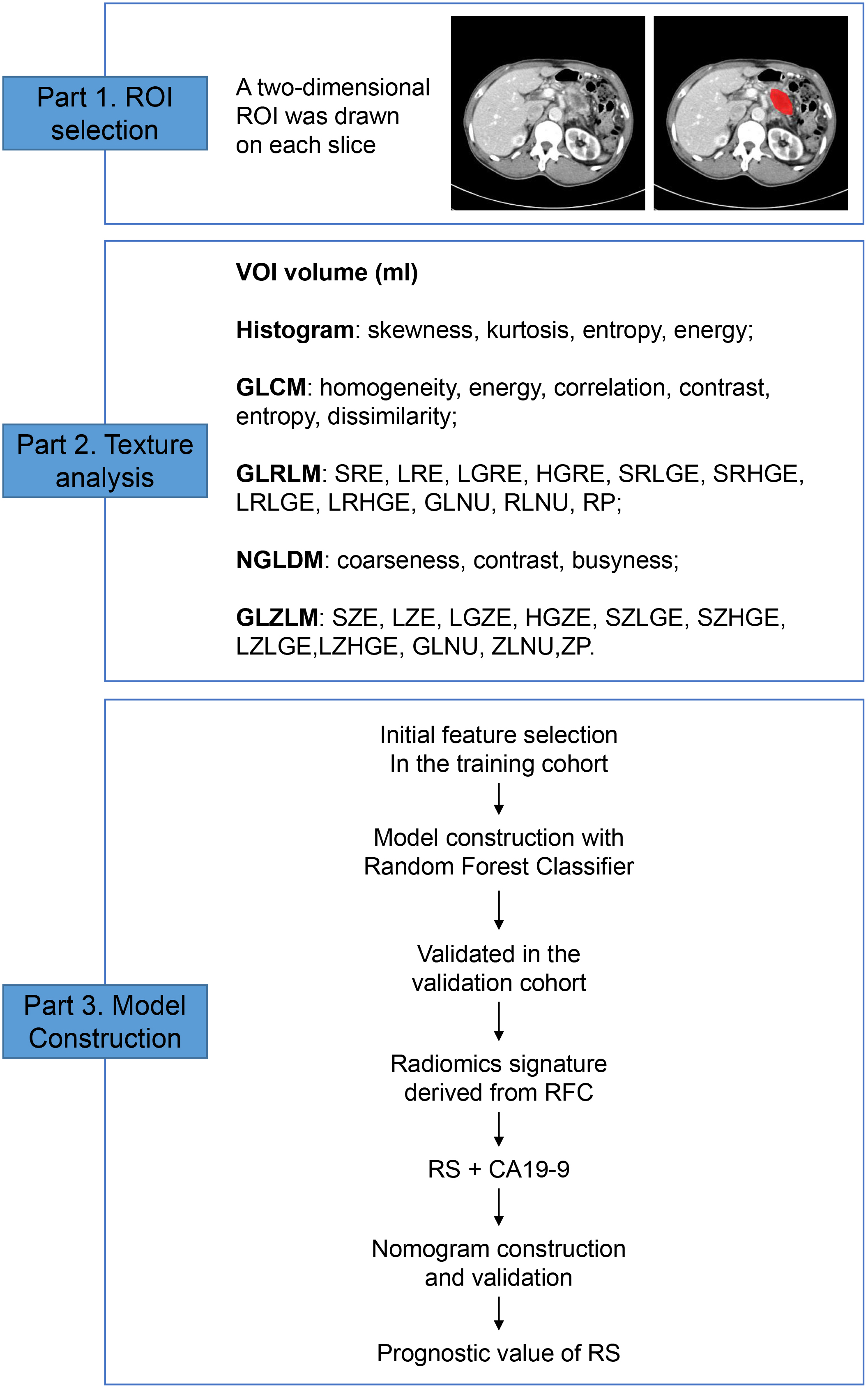
Baseline clinicopathologic characteristics of patients with APC
Comparison between baseline clinicopathologic characteristics of patients with APC according to liver metastases
Statistical analysis was conducted with R software (version 3.6.1, Institute for Statistics and Mathematics, Vienna, Austria) and Python (version 3.6.8). The univariable correlations between clinicopathologic characteristics and liver metastases were assessed with the Pearson Chi-Square test or Fisher’s exact test for categoric variables and with an independent-samples
Results
Patient characteristics
Table 1 demonstrated the baseline clinicopathologic characteristics of patients with APC in both the training cohort and the validation cohort. All the variables, including age, gender, body mass index (BMI), hypertension, diabetes mellitus, smoking, drinking, liver metastases, primary tumor location and CA19-9 were comparable between the two groups. In the training cohort, 30 patients (51%) had liver metastases, and 28 patients (48.3%) had other metastatic patterns. In the validation cohort, 10 patients (52.6%) had liver metastases and 9 patients (47.4%) had other metastatic patterns.
Correlations between clinicopathologic characteristics and liver metastases
In the training cohort, patients with liver metastases had a higher level of CA19-9 compared with patients without liver metastases (
Principal parameters calculated by texture analysis in the training cohort
Principal parameters calculated by texture analysis in the training cohort
The correlations between texture parameters.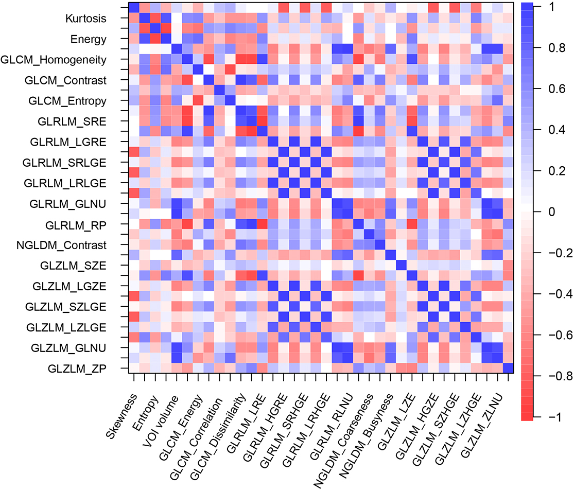
In the training cohort, no significant differences in VOI and NGLDM parameters were observed between the two groups with independentsamples
Pearson’s correlation coefficient was used to investigate the correlations between texture parameters in the training cohort (Fig. 2). The results demonstrated significant correlations between some pairs of these parameters. For example, skewness showed significant negative correlations with GLRLM_HGRE (
The ROC curve and AUC of Random Forest Classifier models in the training cohort.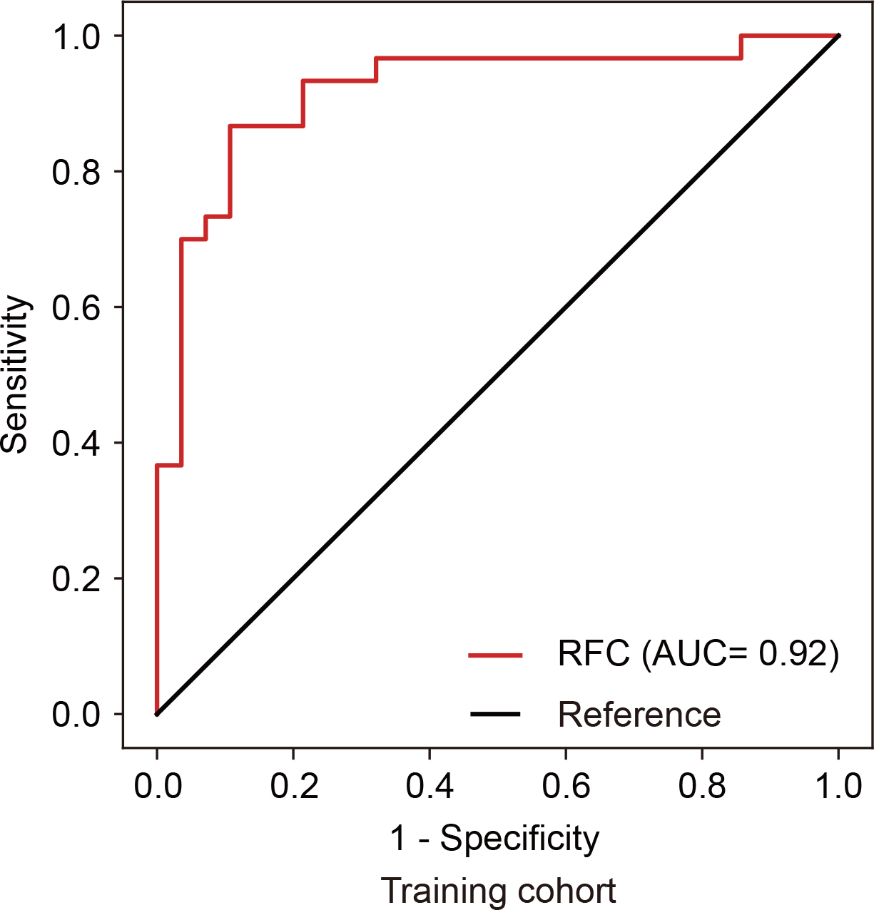
Developed radiomics nomogram to discriminate advanced pancreatic cancer patients with liver metastases or other metastatic patterns.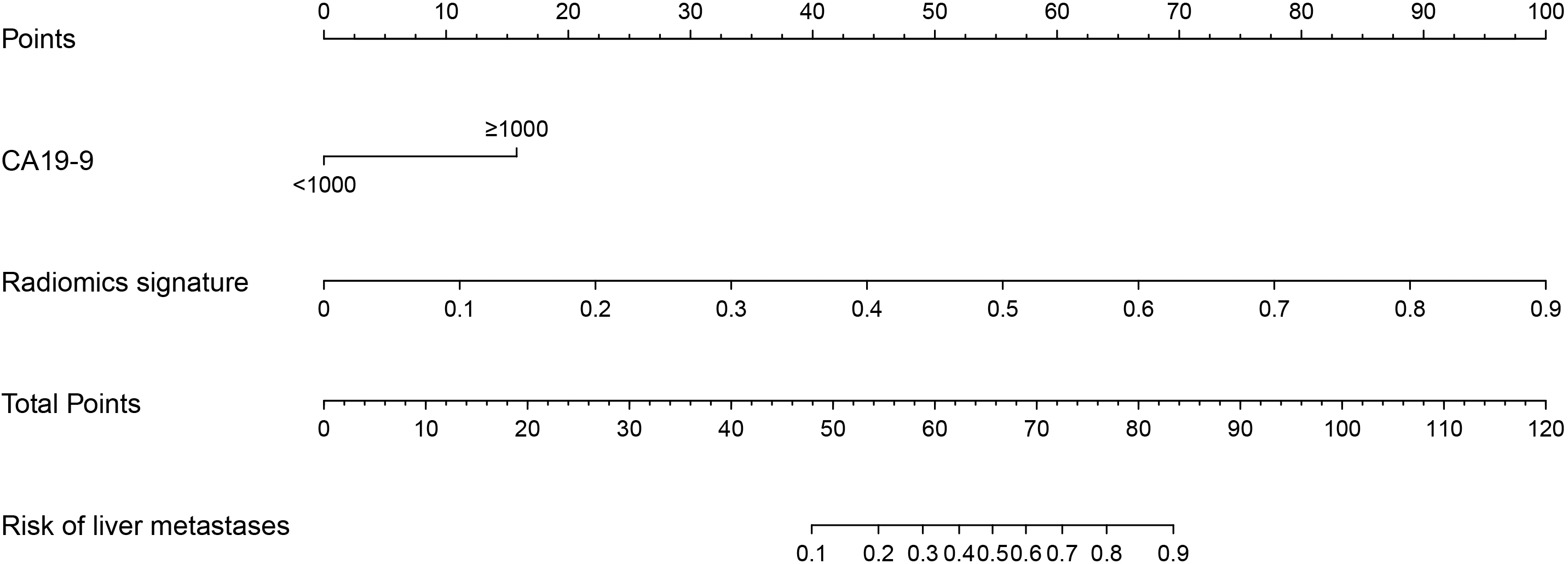
Calibration plot and decision curve of the radiomics nomogram.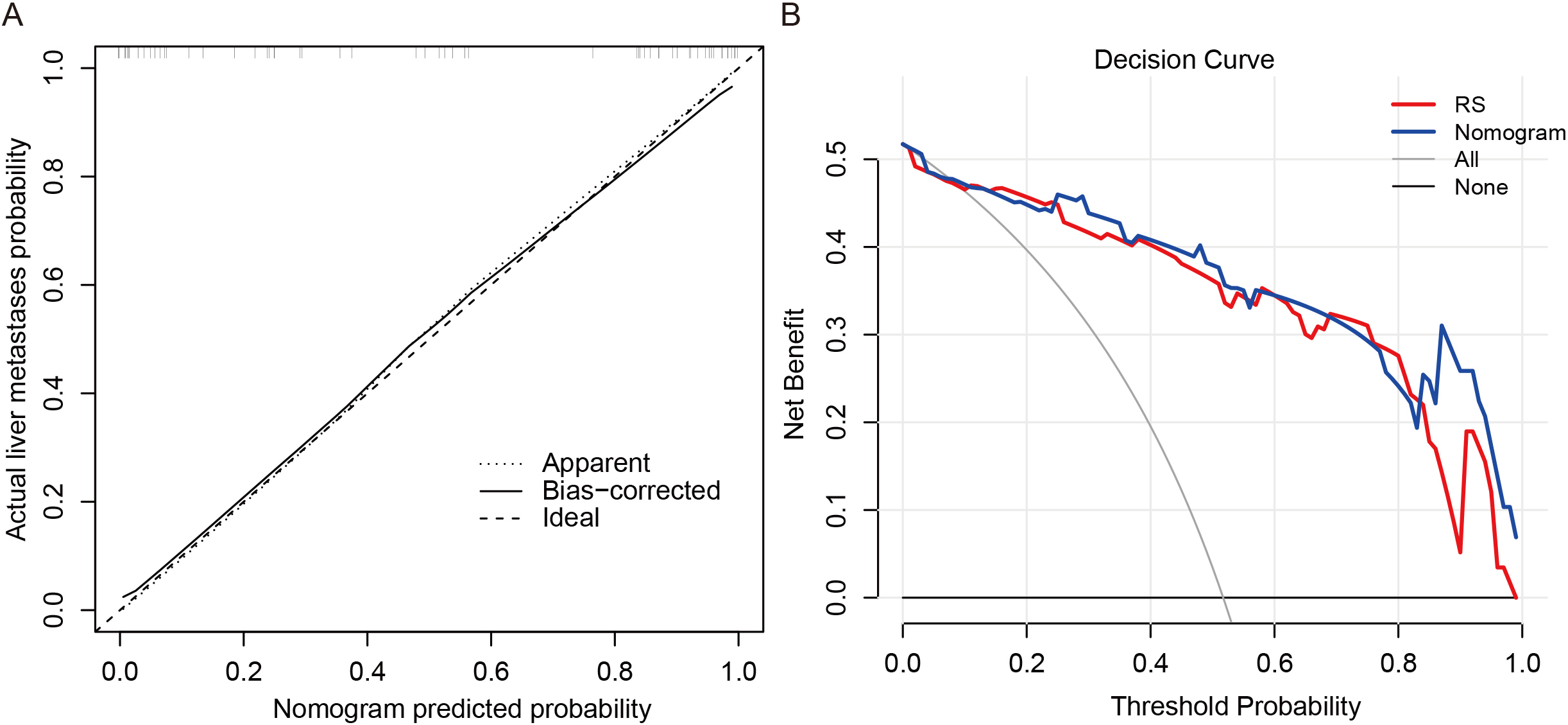
Kaplan-Meier estimates OS according to radiomics signature in the training cohort (A) and validation cohort (B).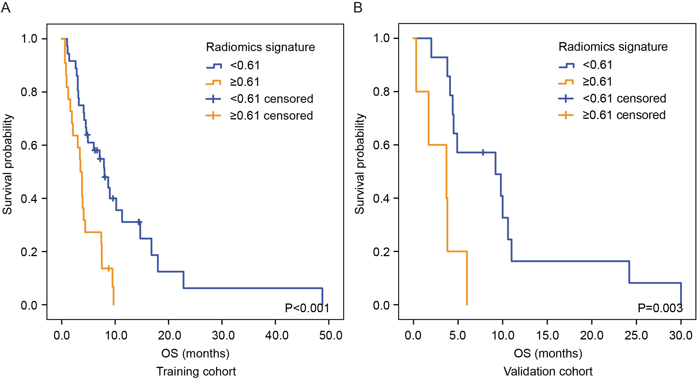
Eighteen texture parameters (shown in Table 3) with significance values
Given the significant difference in the CA19-9 level between patients with or without liver metastases, a nomogram containing both the RS and CA19-9 was constructed to predict liver metastases (Fig. 4). The nomogram showed good discrimination with an AUC of 0.93 in the training cohort and an AUC of 0.81 in the validation cohort. Internal validation of the nomogram was performed with a calibration plot (Fig. 5A). The decision curve analysis of the RS and the nomogram is presented in Fig. 5B. The decision curve showed that, if the threshold probability was
Prognostic value of the RS
We also investigated the prognostic value of the RS with the Kaplan-Meier methods and the log-rank test. In the training cohort, patients with an RS
Discussion
In this study, we showed a strong discrimination ability between patients who had APC with liver metastases and patients with other metastatic patterns by developing an RS. A nomogram was constructed using both the RS and CA19-9, and it showed good performance in the training cohort (AUC
Previous studies have shown that a variety of texture features correlates with metastasis in cancer. For example, kurtosis and GLCM-Energy have been associated with progression-free survival and tumor metastasis-dependent factors, such as yes-associated protein [23, 24]. Beckers et al. [25] found that uniformity was an independent and significant predictor of liver metastases in patients with colorectal cancer. Sheen et al. [21] demonstrated that GLZLM_SZLGE was an independent predictor of metastasis in osteosarcoma. Becker et al. [26] also found that texture features from the histogram and the gray-level matrices could be used to predict metastases in mice. In this study, we found that 18 texture features correlated with liver metastases; among them, GLZLM_LZHGE showed the smallest
Conventional multiparametric analysis has been widely used in previous studies for parameter selection and model construction [12]. Recently, machine-learning algorithms have shown superiority in this aspect, especially in medical imaging analysis [27, 28, 29, 30, 31]. To construct radiomics models in this study, 18 potential predictors were selected from 36 candidate radiomics features. This study used RFC to acquire an RS, which showed an acceptable discrimination ability (AUC
The CA19-9 level did not demonstrate enough discrimination ability alone on the basis of univariable association with liver metastases (
This study had several limitations. First, the retrospective design and relatively small sample size can result in unavoidable bias and reduce. Second, the texture features of the liver were not considered. Recently, Lee [4] found that hepatocytes can direct the formation of a pro-metastatic niche in the liver by coordinating myeloid cell accumulation and fibrosis, which suggests that liver characteristics may affect the metastasis of pancreatic cancer. Previous studies have shown that a TA of a whole-liver CT may predict which patients with colorectal cancer are at risk of developing early (
In conclusion, this study presents a radiomics nomogram incorporating both the RS and CA19-9. This nomogram can be used to discriminate patients who have APC with liver metastases from patients with APC and other metastatic patterns.
Authors’ contributions
Conception: T.Z., J.H. and L.W.
Interpretation or analysis of data: T.Z., X.D., Y.Z., J.H. and L.W.
Preparation of the manuscript: T.Z. and J.H.
Revision for important intellectual content: X.D. and M.L.
Supervision: J.H. and L.W.
Funding
This research was funded by grants from the National Natural Science Foundation of China (81902955), Natural Science Foundation of Jiangsu Province (BK201901 61), Project of Jiangsu Shuangchuang Doctor (QT2019 04), Foundation of Changzhou Sci & Tech Program (CJ20190096), the Youth Science and Technology Project of Changzhou Health and Family Planning Commission (QN201817).
Supplementary data
The supplementary files are available to download from http://dx.doi.org/10.3233/CBM-210190.
Footnotes
Acknowledgments
None.
Conflict of interest
The authors declare no conflict of interest.