
Abstract
BACKGROUND:
Neuronatin (NNAT) determined by immunohistochemistry is a negative prognostic biomarker for breast cancer, independent of the major clinicopathological markers.
OBJECTIVE:
Here, we investigated whether NNAT is also a predictive biomarker for pathological remission after neoadjuvant chemotherapy.
METHODS
: One hundred and four breast cancer patients, treated with systemic neoadjuvant chemotherapy were included in this retrospective study. NNAT was detected in formaldehyde fixed, paraffin embedded primary cancer tissue by immunohistochemistry and an immuno-reactive score (IRS) determined. Pathological remission was scored according to Sinn and by evaluation of cytopathic effects. NNAT-IRS was correlated with clinicopathological parameters as well as relapse free and overall survival and for pathological remission after neoadjuvant therapy.
RESULTS:
NNAT IRS was an independent prognostic marker for relapse free and overall survival and the time from diagnosis to the “tumor-free” state. NNAT IRS was associated with Luminal-A tumors and correlated slightly negative with age and lymph-node metastasis. There was no significant correlation of NNAT-IRS with Sinn’s remission score, but with cytopathic effects of chemotherapy.
CONCLUSIONS:
We confirmed the prognostic impact of NNAT-IRS in an independent cohort of neoadjuvantly treated patients. Additionally, a correlation with a score for pathological remission under systemic neoadjuvant chemotherapy for breast cancer was found.
Introduction
Breast cancer is the most frequent neoplasm in women worldwide and accounted for 2,088,849 new cases and 626,679 deaths worldwide in 2018 [1]. It is a heterogeneous disease and therapy is mostly planned according to TNM stage and immuno-histochemical analysis, determining hormone receptor status and mitosis count (Ki-67 proliferation index) [2]. Based on gene expression data, breast cancer is classified in the luminal-A and -B subtype, HER2 overexpressing and basal subtypes [3]. The immuno-histochemical determination of hormone receptor status and Ki-67 proliferation index is widely used as an estimate for these molecular subtypes in a clinical setting [4]. Whereas the estrogen receptor positive subtypes are treated with targeted therapies blocking the estrogen receptor or inhibiting aromatase [5, 6, 7], HER2/Neu overexpressing cases receive an antibody therapy against this epidermal growth factor receptor [8]. For the basal, triple negative subtypes, no established targeted therapy is in routine clinical use, but chemotherapy is applied regularly [9]. In the last decade a shift from the traditional therapy scheme, starting with surgery and then continuing with an appropriate adjuvant chemotherapy frequently combined with radiotherapy, towards systemic neoadjuvant chemotherapy has occurred [10]. Several studies showed that this is in several aspects superior over adjuvant treatment, however an increased rate of local reoccurrence might occur [11]. Nevertheless, neoadjuvant chemotherapy has become especially important for locally advanced breast cancer [12, 13].
Neoadjuvant therapy is considered effective, when pathologic complete remission (PCR) of the tumor occurs. A complete remission is accomplished when no remaining cancer cells are found after therapy in the surgically removed tumor residuals. As complete remission is frequently not achieved, remission is scored i.e. according to Sinn [14]. In this scoring system, tumor residuals are classified in four groups. Sinn score 0 indicates no effect of therapy, Sinn 1 shows significant tumor sclerosis, inflammation and cytopathic effects, Sinn 2 is characterized by extensive tumor sclerosis with only a few remaining tumor cells resulting in a residual tumor of less than 0.5 cm. In Sinn 3, no invasive tumor is seen and in Sinn 4, no tumor at all is found. Nevertheless, alternative scoring systems have also been developed [15].
We have recently showed that neuronatin (NNAT) determined by immuno-histochemistry is an independent unfavorable prognostic marker for breast cancer [16]. Neuronatin is a proteolipid occurring in two forms encoded by different splice products [17]. Such proteolipids function in the regulation of ion channels and NNAT is proposed to be particularly involved in Ca
We here report on a follow up study of our retrospective evaluation of the prognostic value of NNAT for breast cancer [16]. We additionally investigated whether NNAT, determined by immuno-histochemistry, is also associated with pathological remission in neoadjuvantly treated breast cancer patients.
Systemic treatment of patients stratified for hormone receptor expression. Significance was tested between treatments for receptor positive and negative cases using Fisher’s exact test. A significance value
0.05 is indicated in italic. Monoclonal antibody (Ab), Tyrosine kinase inhibitor (TKI)
Systemic treatment of patients stratified for hormone receptor expression. Significance was tested between treatments for receptor positive and negative cases using Fisher’s exact test. A significance value
Experimental subjects
Female patients (104) with primary breast cancer undergoing neoadjuvant systemic chemotherapy from the department of gynecology of the Otto von Guericke University (Magdeburg) were included into this retrospective study from 2010 to 2016. A positive ethical vote was obtained from the ethics committee of the Otto von Guericke University of Magdeburg at the medical faculty (AKZ 114/13) according to the statement of the central German ethics commission on the reuse of patient material for research [31]. Clinical and pathological data were collected from the patients’ files in the pathology and gynecology department of the Otto von Guericke University. A mean follow up of 217 weeks (min
Antibodies and Immuno-histochemistry
In contrast to our earlier study, an automated method for NNAT staining was applied. For evaluation of this procedure, some slides of the former patient cohort were stained and the results compared with the results of the manual procedure. After establishing the improved staining method, specimen from the new cohort were stained as follows: In short, 2
Analysis of gene expression databases
We accessed the METABRIC gene expression data for breast cancer via cBioportal [32] and plotted the co-expression data on this website. The 20 most significant co-expressed genes were selected and used for an overrepresentation analysis on the enrich-database website [33].
Statistical analysis
All statistical calculation were done with SPSS vers. 26 (IBM). For evaluation of a correlation of categorical parameters, cross tabulation with Fisher’s exact p was applied. For correlation of ordinal parameters, Spearman correlation factor was calculated. Survival probability distribution was analysed using the Kaplan Meier method. Equality of the survival curves was tested using the log rank and the Breslow method. Univariate and multivariate cox proportional hazard regression was used to determine hazard ratios and significance of prognostic factors. Generally, a
Microphotographs demonstrating NNAT-immuno-histochemistry results. All tumors depicted here, are classified as “no special type” (NST). A–C and G–I show examples for NNAT staining intensity at low magnification (A, B weak intensity 1; C, medium intensity 2; G, H, I strong intensity 3). Higher magnifications corresponding to the specimens depicted in A–C and G–I are shown in D–F and J–L. Scale bars represent 250 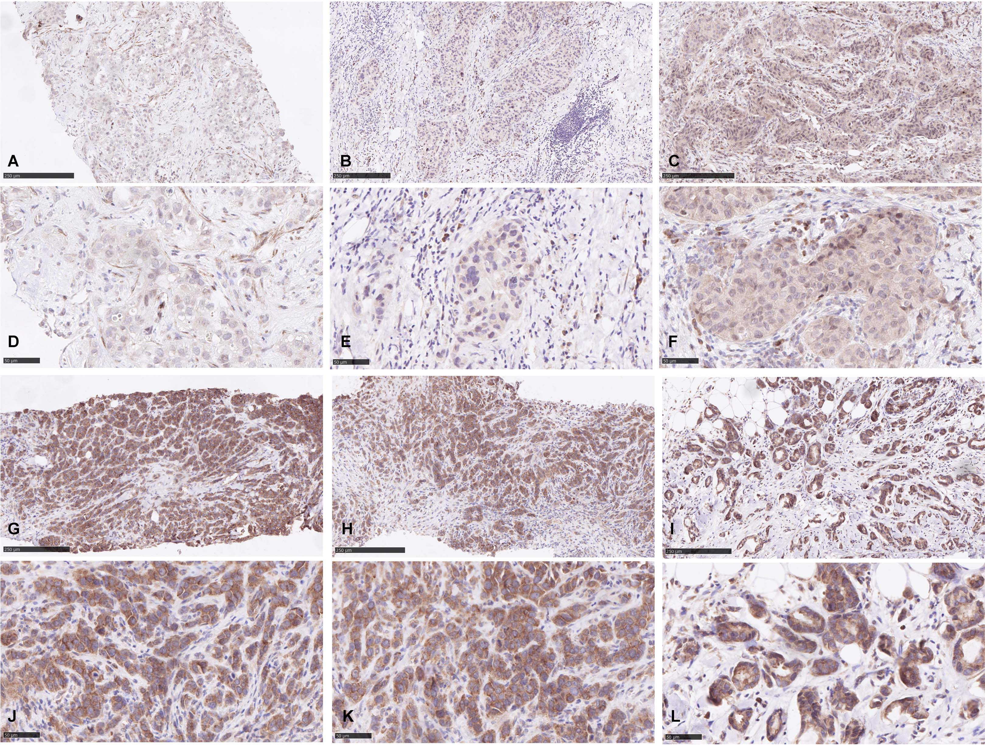
Correlation with clinicopathological parameters
NNAT staining intensity and percent positive tumor cells were determined and an immuno-reactive score (IRS) calculated (Fig. 1). Then we investigated a possible association of this NNAT-IRS with standard parameters (Table 2) such as age, hormone receptor – and lymph node status. This was done in cross-tabulation tests and by determining the Spearman correlation factor. For cross tabulation, the cut off for NNAT low versus high was set to 17, which was the optimal cutoff in survival analysis (see below). In this cross tabulation tests, NNAT turned out to be correlated only to the Luminal-B subtype. Additionally, in Spearman correlation analysis, a weak negative correlation with age and lymph node status and a positive correlation with Ki-67 proliferation index was seen (Table 3).
Study cohort with respect to the distribution of clinico-pathological parameters and NNAT expression. NNAT was considered as high when IRS was higher than 17. Fisher’s exact p or Spearman correlation
is given
Study cohort with respect to the distribution of clinico-pathological parameters and NNAT expression. NNAT was considered as high when IRS was higher than 17. Fisher’s exact p or Spearman correlation
Spearman correlation analysis of NNAT IRS with clinicopathological parameters. ER, PR and HER2 are represented as immunoreactive score. Ki-67 as % positive tumor cells. Positive lymphnodes were classified according to TNM stage as shown in Table 2
Survival analysis was done using the Kaplan-Meier algorithm. We first investigated the time from diagnosis until the patients were declared as “tumor-free” and underwent surgery. Interestingly, the presence of high NNAT was associated with a longer median time from diagnosis until being tumor free (28.4 and 31.3 weeks Breslow estimator (generalized Wilcoxon)
Kaplan-Meier survival curves, demonstrating the impact of NNAT-IRS on prognosis. Time from diagnosis to tumor-free state (2A) was analyzed and significance determined using the Breslow estimator. Relapse free survival was analyzed for the time from diagnosis to relapse (2B) and overall survival was defined as time from the tumor free state until death (2C).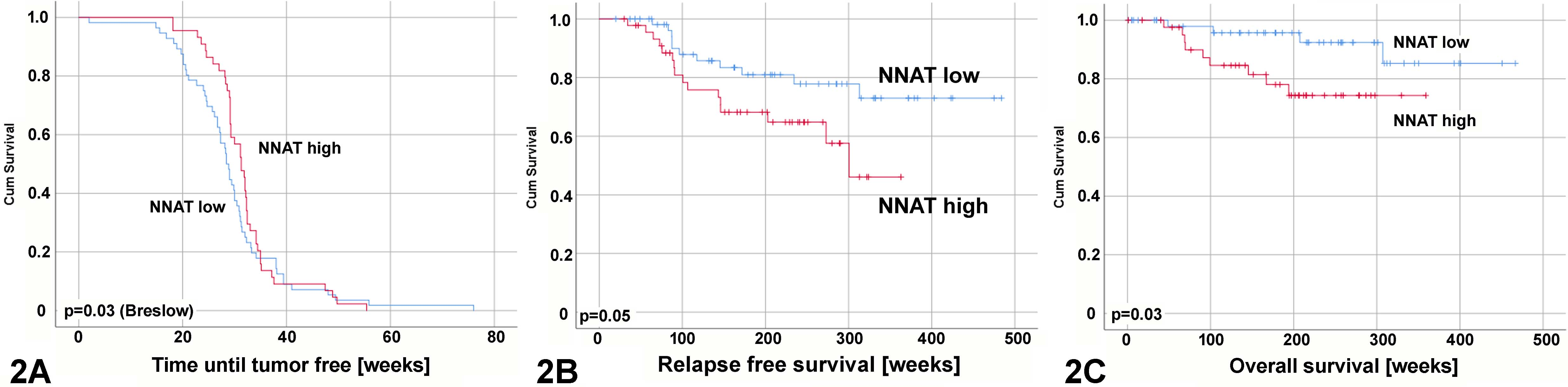
Kaplan Meier survival analysis of the time from diagnosis to the tumor free state. Survival curves are stratified for lymph node-, Ki-67-, ER-, PR-, HER2-status, luminal-A versus -B and triple negative breast cancers. Every tumor subtype is then further analyzed for the impact of NNAT expression. Number of cases and significance (Breslow estimator) is given.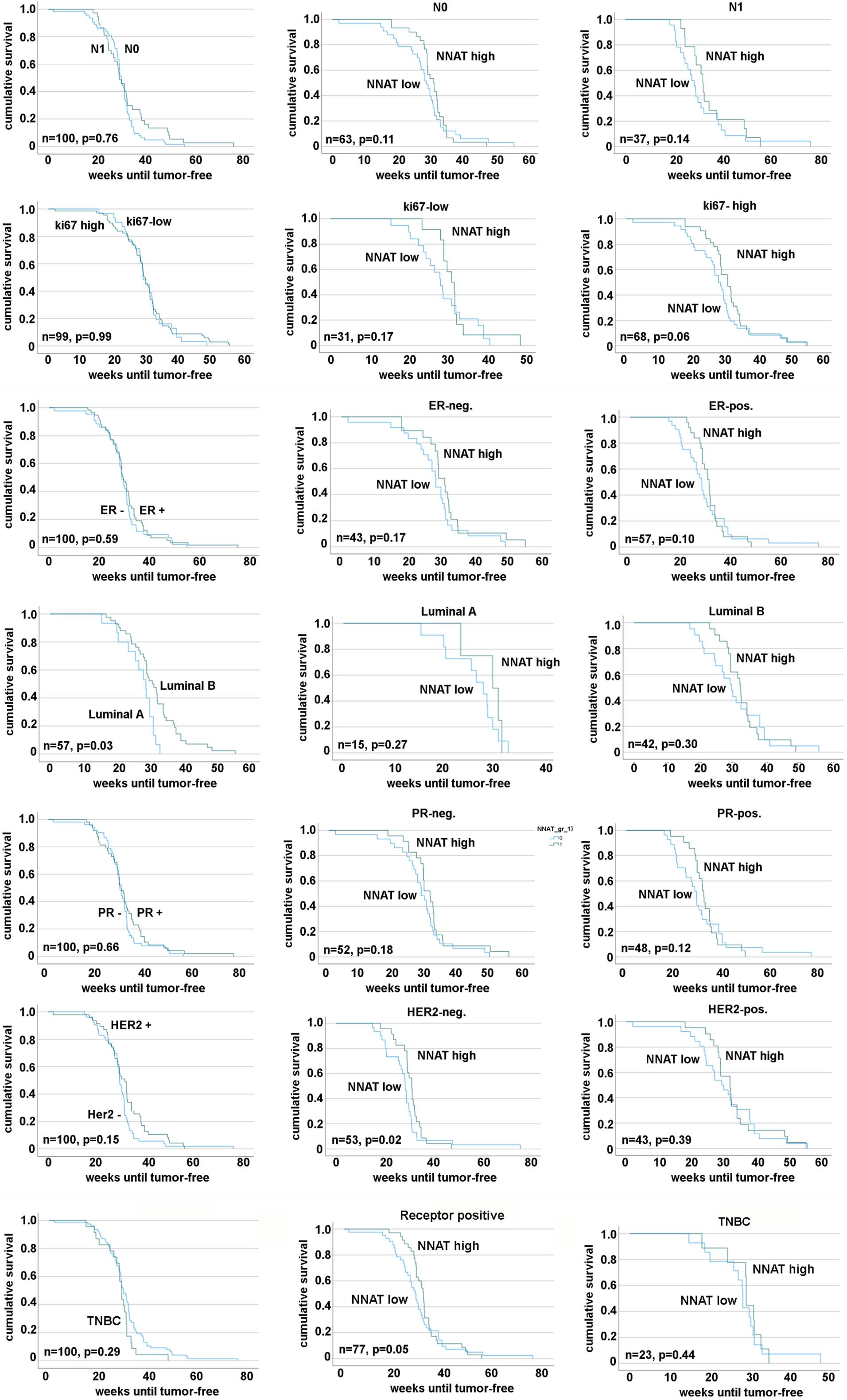
In terms of relapse free survival (RFS), we analyzed both, the time from diagnosis and from the tumor-free state until occurrence of a local relapse. Here, the time from diagnosis to relapse depended significantly on NNAT-IRS (log rank
When analyzing tumor subtypes separately, lymph node status was the only significant prognostic factor for RFS (Fig. 4). When the subtypes were further stratified for NNAT expression, NNAT became significant for lymph node positive (N1) and tumors that express any of ER, PR or HER2. However, there were clear trends (
Kaplan Meier survival analysis of relapse free survival from time of diagnosis to relapse. Survival curves are stratified for lymph node-, Ki-67-, ER-, PR-, HER2-status, luminal-A versus -B and triple negative breast cancers. Every tumor subtype is then further analyzed for the impact of NNAT expression. Number of cases and significance (log-rank p) is given.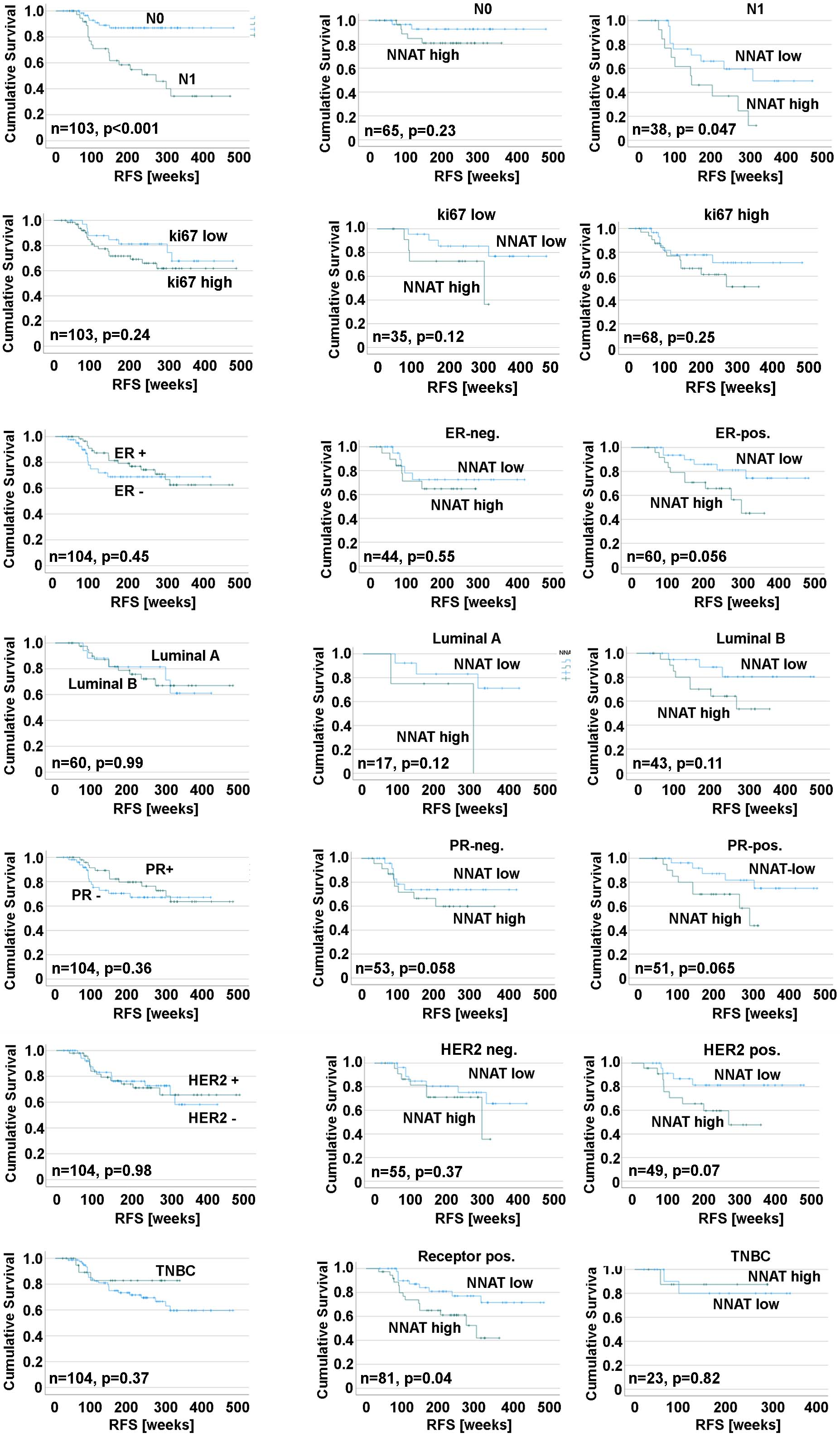
KaplanMeier analysis of overall survival determioned from tumor free state until death. Survival curves are stratified for lymph node-, Ki-67-, ER-, PR-, HER2-status, luminal-A versus -B and triple negative breast cancers. Every tumor subtype is then further analyzed for the impact of NNAT expression. Number of cases and significance (log-rank p) is given.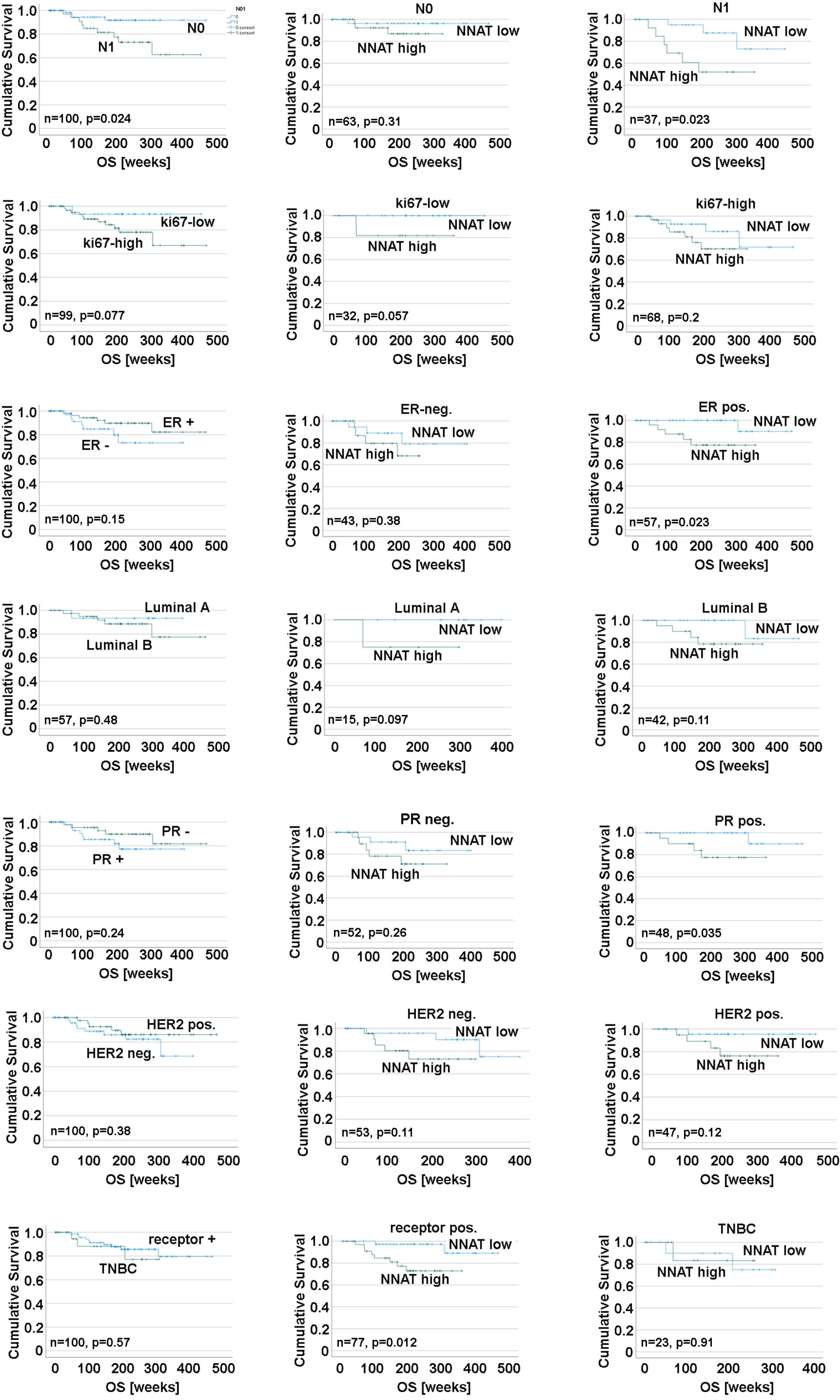
Cox regression analysis. RFS was calculated from date of diagnosis to event. OS was calculated from date being “tumor-free” to death. For multivariate cox regression analysis, NNAT HR was adjusted to ER, PR, HER2 and N status. Only significant factors represented in the final cox regression model are shown
Kaplan-Meier survival analysis for relapse free survival depending on NNAT and PPAR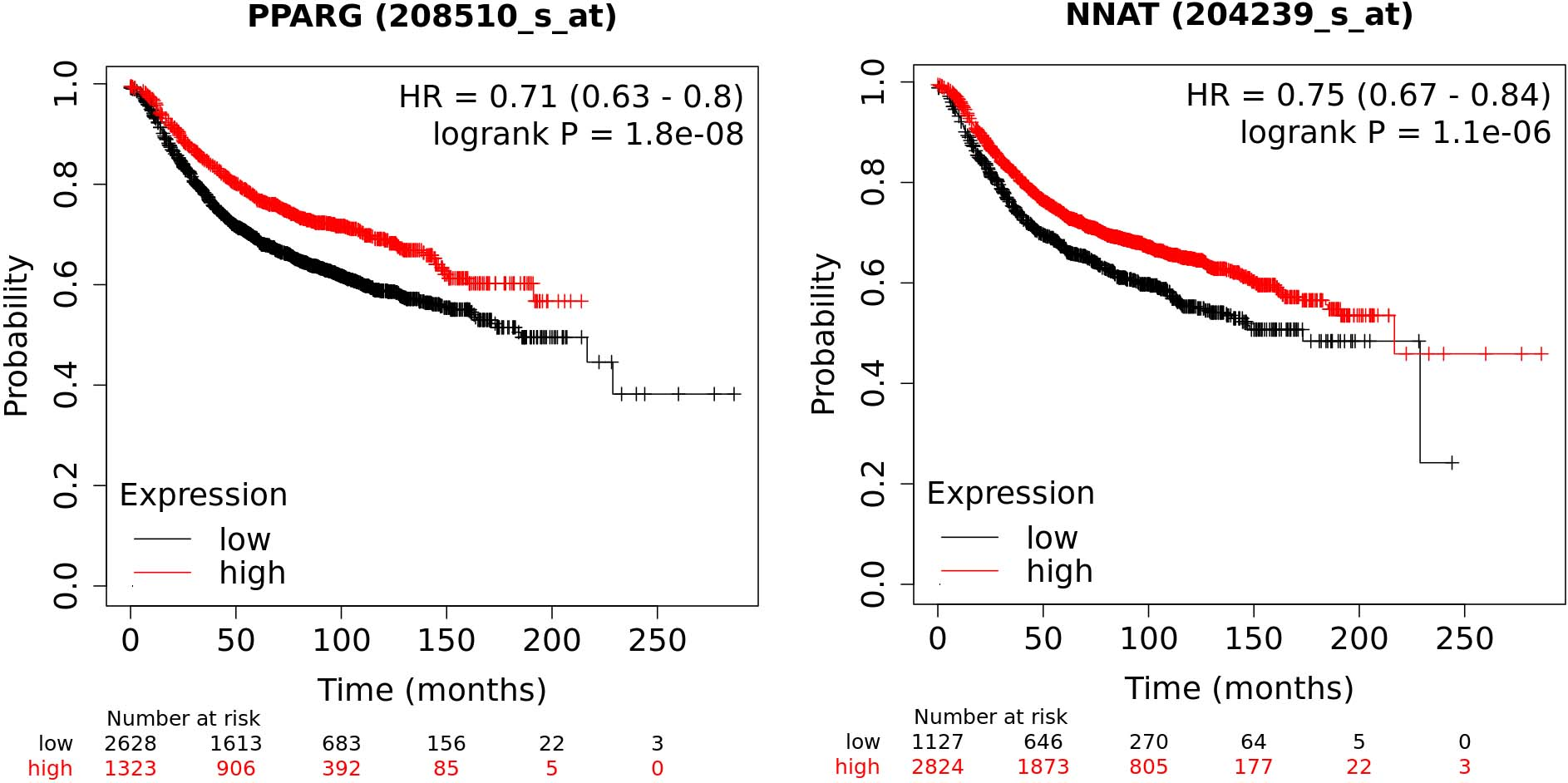
In cox regression analysis (Table 4) NNAT turned out to be a significant prognostic factor for OS, but not RFS although a statistical trend could be observed. For RFS, only the presence of infiltrated lymph nodes (N
We then correlated NNAT with scores for remission after neoadjuvant chemotherapy. First, we investigated the regression grade according to Sinn [14]. This rather crude score classified most tumors (61 %) in grade 1 characterized by tumor sclerosis, inflammation and substantial cytopathic effect. Here we saw no correlation in either cross tabulation nor in Spearman correlation (Tables 2 and 3) with NNAT-IRS. We then prepared a more refined scoring for the cytopathic effect, ranging from 0 (no effect) to 3 (severe effect). Applying this score, resulted in a significant correlation with NNAT-IRS (Table 3). Thus, NNAT-IRS was clearly related with this indicator for pathological remission.
Database analysis
In order to develop further ideas on the molecular function of NNAT, we identified co-expressed genes in the METABRIC database of breast cancer [34]. The 20 most significantly correlated genes (Table 5) were used for an overrepresentation study performed on the enrich database website [33]. Here, the most significant hits were associated with lipid metabolism, especially regulated by PPAR
Co expression analysis for NNAT using the METABRIC [34] gene expression data
Co expression analysis for NNAT using the METABRIC [34] gene expression data
Overrepresentation analysis of NNAT co-expressed genes using the enrichr-database [33]. Only significant terms (adjusted
In this study, we were interested in the predictive potential of NNAT on pathological remission upon systemic neoadjuvant chemotherapy. Furthermore, we aimed at reproducing our earlier data on NNAT and relapse free and overall survival in a novel cohort of breast cancer patients. We also established an automated staining procedure for NNAT, which makes this biomarker more appropriate for routine diagnostics. In contrast to our earlier study, only patients treated by systemic neoadjuvant chemotherapy were included in this study and the treatment schemes have changed over the years. As a result, HER2-positive tumors were regularly targeted with specific antibodies such as trastuzumab. Additionally, due to the selection for neoadjuvant chemotherapy, the study cohort contained tumors that are more aggressive. Nevertheless, the impact of NNAT on survival data and the correlation to clinico-pathological features are similar to our earlier study. However, there are some minor differences to the results from the previous study, as i.e. we now observed a more pronounced effect in ER-positive cases than in negative cases for overall survival (Fig. 5).
The second result concerned the predictive potential of NNAT-IRS for the neoadjuvant treatment. Surprisingly, NNAT-IRS significantly correlated with the length of the interval from diagnosis to the “tumor-free state” (Fig. 2A). This interval reflects the speed of shrinking of the tumor and the possible occurrence of a progress under therapy. The dependency of this interval upon NNAT also influences the survival analysis. We therefore saw a difference in statistical significance, when starting the analysis from the date of diagnosis or the time of being “tumor-free”.
We then investigated the correlation of NNAT-IRS with the remission score according to Sinn or determined by the cytopathic effects caused by the therapy. The majority of cases was within the group of ”Sinn 1” defined by “increased tumor sclerosis and/or resorption with clear cytopathic effect” [14]. Using this scoring system, no statistical significance could be found. We therefore decided to replace it with a more detailed score based on the morphology of the cytopathic effects caused by the therapy. Indeed, for this score a highly significant correlation with NNAT-IRS was found. Unfortunately, we could not analyze the impact of NNAT on the type of therapy in more detail, as therapy regimes in this cohort were too divers. Nevertheless, we conclude, based on the dependence of the time until “tumor free” and the correlation with the cytopathic score that NNAT might be predictive for the efficacy of neoadjuvant chemotherapy. Nevertheless, this should be investigated in a larger cohort with more clearly defined therapy regimes.
As the molecular function of NNAT for cancer biology is still not completely understood, more efforts should also be taken for a detailed functional analysis of this protein and the associated pathways. We have pointed out that his proteolipid modulates cellular Ca
Unexpectedly, the NNAT co-expressed genes in breast cancer were significantly associated with “PPAR
In conclusion, we here present evidence that NNAT could become a more important prognostic biomarker for breast cancer in the future. This was also evident for the prediction of the remission upon neoadjuvant chemotherapy, but only when a remission score based on the cytopathic effect was used. Nevertheless, the molecular function of NNAT should be investigated in more detail to identify potential therapeutic targets related to neuronatin.
Footnotes
Acknowledgments
The authors wish to thank the members of the histochemistry laboratory of the Institute of Pathology, Magdeburg for their great support and excellent work. This investigation has been funded by institutional funds only.
Conflict of interest
The authors declare that no conflict of interest exists.
Author contributions
Conception: NN, JH, TK, AI
Interpretation or analysis of data: WP, NN, PC
Preparation of the manuscript: WP, NN, PC
Revision for important intellectual content: JH, NN
Supervision: NN, PC