To explore the pathogenesis of oral submucosal fibrosis (OSF) by analyzing the impact of Platelet Derived Growth Factor (PDGF)-BB on oral mucosal fibroblasts (FB) and PDGFR-/Phosphoinositide 3-kinase (PI3K)/serine/threonine protein kinase (AKT) signaling pathway.
METHODS:
The isolated and purified oral mucosal fibroblasts were divided into four groups: the control group (CON, 10% FBS DMEM), the PDGF-BB group (40 ng/ml PDGF-BB), the PDGF-BB+IMA group (40 ng/ml PDGF-BB and 60 mol/L IMA), and the PDGF-BB+LY294002 group (40 ng/ml PDGF-BB and 48 mol/L LY294002). Primary human FB cells were isolated and cultured for detecting the effects of PDGF-BB on -smooth muscle actin (-SMA) by indirect immunofluorescence. 3-(4,5-dimethyl-2-thiazolyl)-2,5-diphenyl-2-H-tetrazolium bromide, Thiazolyl Blue Tetrazolium Bromide (MTT) method and scratch test were used to detect the proliferation and migration of FB. Western blots were used to detect the synthesis of type I collagen (Col I) and the expression of PDGFR-/PI3K/AKT signaling pathway-related proteins. The effects of PDGFR- inhibitor and PI3K inhibitor were observed.
RESULTS:
Compared with group CON, group IMA, and group LY294002, -SMA was upregulated in group PDGF-BB ( 0.05), with higher OD490 nm value ( 0.05), narrower average scratch width, and higher relative cell migration rate ( 0.05). The expression levels of Col I, p-PDGFR-, p-PI3K, and p-AKT were higher in group PDGF-BB ( 0.05).
CONCLUSIONS:
PDGF-BB induces FB to transform into myofibroblasts (MFB) through the PDGFR-/PI3K/AKT signaling pathway, and promotes the proliferation, migration, and collagen synthesis.
OSF is precancerous condition that can lead to a special type of carcinoma of oral mucosal squamous cells with a high degree of malignancy and a 5-year survival rate of less than 50% [1, 2]. The transformation and proliferation of fibroblasts (FB) to myofibroblasts (MFB) will cause the deposition of extracellular matrix (ECM), such as collagen [3], eventually leading to various fibrotic diseases, including OSF [4]. PDGF is a strong class of fibroblast stimulating factors [5, 6] and has five types (PDGF-AA, PDGF-AB, PDGF-BB, PDGF-CC, and PDGF-DD) [7]. PDGF-BB can induce a variety of cells to transform into MFBs [8], but its role in the occurrence and development of OSF remains to be explored.
Previous studies have found that the PI3K/AKT signaling pathway plays an important role in cell proliferation, migration, differentiation, and angiogenesis [9]. The activation of this signaling pathway can promote the proliferation of a variety of fibroblasts and collagen synthesis [10]. In addition, some studies have shown that PDGFR- is upregulated in the vascular endothelial cell membrane, fibroblast membrane, or epithelial cell membrane of OSF patients’ tissues [11]. Based on this, we speculate that PDGF-BB may promote the conversion of oral mucosal FB to MFB through the PDGFR-/PI3K/AKT signal pathway and affect its biological behaviors, such as proliferation, migration, and collagen synthesis, thereby promoting the occurrence and development of OSF.
Materials and methods
Specimen collection
The specimens were the gum tissues obtained from the extraction of molar teeth in a healthy 20-year-old Chinese female periodontal patient on March 27, 2018. The patient was in good health without systemic disease and periodontal inflammation. This study was conducted in accordance with the declaration of Helsinki. This study was conducted with approval from the Ethics Committee of Xiangya Hospital of Central South University. Written informed consent was obtained from all participants.
Culture and identification of oral mucosal FB
The gum flap tissues were cultured at 37C and 5% CO for 15 days, followed by digestion with 0.25% trypsin (which was terminated by DMEM high glucose medium containing 15% FBS), centrifugation, resuspension, and re-culture at 37C and 5% CO to obtain the first-generation oral mucosal FB. After successful isolation and continuous culture for 2 generations, the purified oral mucosal fibroblasts were obtained and identified by detecting the expression of vimentin using the indirect immunofluorescence method.
Grouping
The isolated and purified oral mucosal fibroblasts were divided into four groups: the control group (CON, 10% FBS DMEM), the PDGF-BB group (40 ng/ml PDGF-BB), the PDGF-BB+IMA group (40 ng/ml PDGF-BB and 60 mol/L IMA), and the PDGF-BB+LY294002 group (40 ng/ml PDGF-BB and 48 mol/L LY294002).
Indirect immunofluorescence
Indirect immunofluorescence was used to detect the expression of vimentin protein in FB and effects of PDGF-BB on -smooth muscle actin (-SMA). For detecting the expression of vimentin protein in FB, rabbit anti human vimentin antibody (CST, Darmstadt, Germany) was added after the second generation of oral mucosa fibroblasts were fixed and transmembrane. The cell nucleus was stained with Hoechst, and goat anti rabbit IgG Cy2 (Beijing comwin Biotechnology Co., Ltd., Beijing, China) was used as the second antibody. For detecting the effects of PDGF-BB on -SMA, each group of cells was inoculated with 10 cells/well on glass coverslips in a 24-well plate for 6 days, then the cells were fixed and transmembrane, mouse anti human -SMA antibody (CST, Darmstadt, Germany) was added. The cell nucleus was stained with Hoechst, and goat anti mouse IgG Cy3 (Beijing comwin Biotechnology Co., Ltd., Beijing, China) was used as the second antibody. The results were observed under fluorescence microscope.
MTT
MTT method was used to detect the effects of PDGF-BB on FB proliferation. The oral mucosa fibroblasts in logarithmic growth period were collected and cultured in conditioned medium for 0, 24, 48, 72 and 96 hours respectively. MTT solution was added and incubated for 4 hours. 150 l DMSO solution was added. The absorbance at 490 nm of each well was measured by enzyme marker.
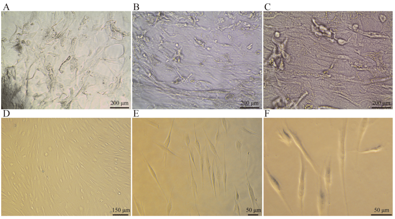
Morphological observation of primary cells under inverted microscope. A–C: the cells migrate out of the tissue blocks and amplify into the surrounding area. D–F: the growth state of cells in medium.
Cell scratch test
Cell scratch test was used to detect the effects of PDGF-BB on FB migration. Draw three parallel lines on the back of the six-well plate with a ruler and a mark pen, inoculate each group of cells with 10 cells/well in the six-well plate, and culture them in a 5% CO 37C constant temperature incubator for 4 days; after 4 days, the confluency of the cells is about 90%. The tip of a 200 l micropipette was used to draw horizontal lines perpendicular to the back and make scratches at the bottom of the well plate, which should have uniform width and form cross with the parallel lines on the back. The plate was then washed twice with PBS to wash off the shed cells. Serum-free medium was then added for 6 h, 12 h, 18 h, and 24 h continuous culture, respectively, before result observation. Observe under the inverted microscope, measure the scratch area and length of each group of cells with Image Pro Plus software, According to the formula that the value of scratch width scratch area/scratch length, the average width of cell scratches in each group was calculated, and the migration rate (initial scratch width value scratch width of corresponding point) value/initial scratch width 100%, migration rate is calculated.
Western blot
Western blot was used to detect the effects of PDGF-BB on FB collagen synthesis and expressions of PDGFR-/PI3K/AKT signaling pathway related protein. Each group of cells was inoculated with 10 cells/well in a 6-well plate for 4 days, and 80 l RIPA cell lysate was added. The OD value of the protein to be measured was measured with a microplate reader at the wavelength of 562 nm, and the concentration of the sample to be measured was calculated according to the standard curve. The protein sample was added into the electrophoresis tank, and the separation gel was immersed in precooled buffer of membrane transfer after electrophoresis for 30 min at 80 V, and the membrane was transferred for 2 h and sealed for 1 h; the first antibody was incubated in refrigerator at 4C overnight, and the second antibody was incubated at 37C for 1 h. Image J software was used to analyze the gray values of the target protein and the internal reference bands.
Statistical analysis
SPSS18.0 software was used to statistically analyze and compare the data among groups, and the results were expressed as mean standard deviation (). The comparison among groups used the group-designed test, with 0.05 as the test standard and 0.05 being considered as statistical significance.
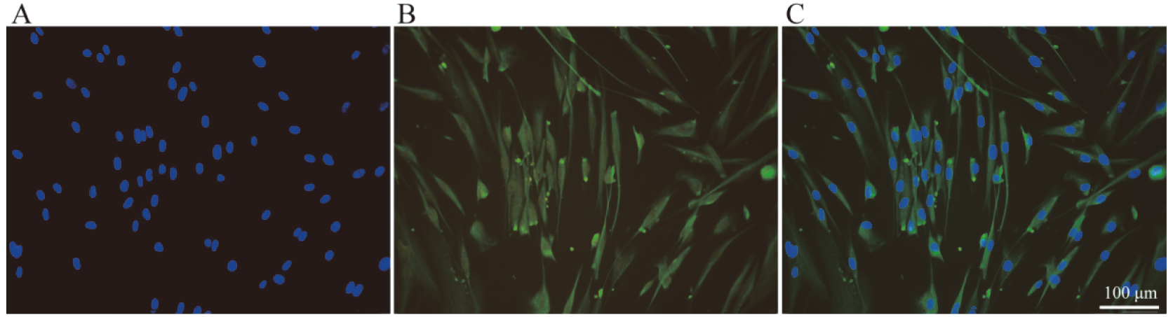
Expression of vimentin in the primary cells under a fluorescent microscope. The cells express vimentin positively, and the rate is more than 99% (immunofluorescence staining, 200).
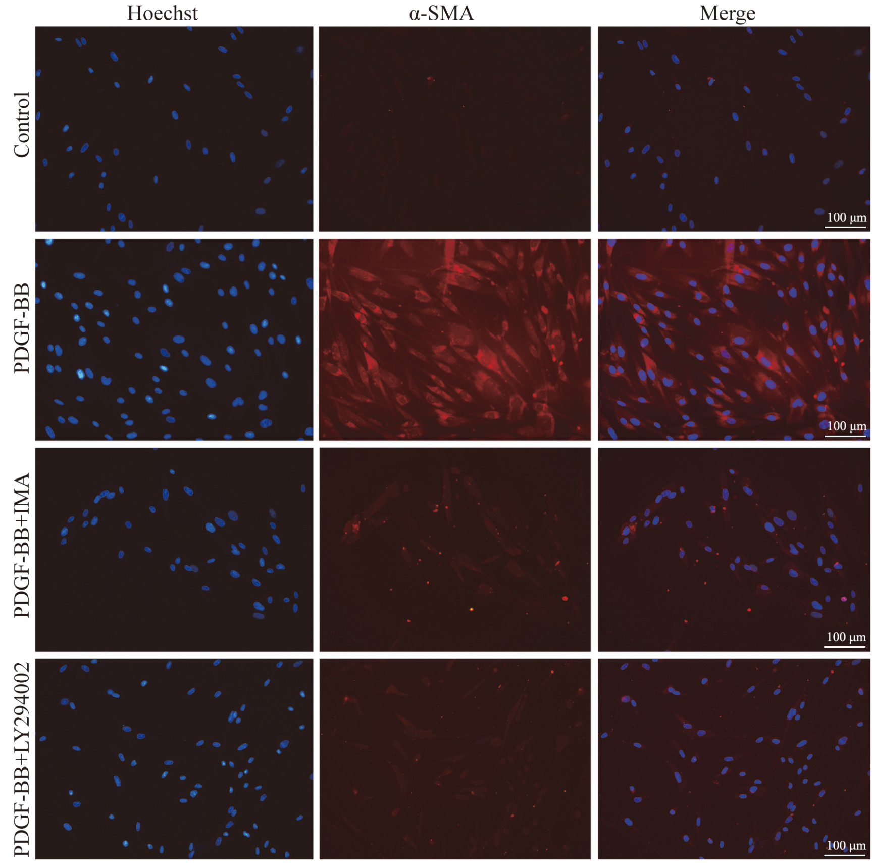
Expression of -SMA in oral mucosal fibroblasts under indirect immunofluorescence assay ( 200). Almost no -SMA positive cells can be observed in group CON and almost all of the cells (100%) are positive for -SMA in group PDGF-BB. However, less than 10% of the cells are positive for -SMA in group PDGF-BB+IMA and group PDGF-BB+LY294002.
Results
Isolation and culture of oral mucosal fibroblasts
Morphological observation of primary cells (Fig. 1): The newly digested and separated cells are bright spherical, with clear outline and single or aggregate cell clusters. After 30 minutes of inoculation, a large number of cells adhered to the wall, some of them began to stretch, and most of the adherent cells were spindle shaped after 2 hours. The fibroblasts of oral mucosa were cultured for about 4 days, the cells proliferated obviously, and the fusion degree was up to 90%. The cells of the second to fourth generations were uniform in shape, long fusiform, with clear boundaries between cells.
Detection of vimentin protein expression: The results showed that almost all of the cells ( 99%) expressed vimentin protein in the cytoplasm (Fig. 2), indicating that fibroblasts were successfully isolated and cultured.
PDGF-BB promoted conversion of oral FB to MFB
The oral mucosal fibroblasts in group CON did not express -SMA, but the cytoplasm of each cell in group PDGF-BB showed bright red fluorescence, and the positive expression rate of -SMA was 100%. Group PDGF-BB+IMA (PDGFR- inhibitors) and group PDGF-BB+LY294002 (PI3K inhibitors) had significantly weaker fluorescence intensity than group PDGF-BB, and the positive expression rate of -SMA was less than 10% (Fig. 3).
Almost no -SMA positive cells can be observed in group CON and almost all of the cells (100%) are positive for -SMA in group PDGF-BB. However, less than 10% of the cells are positive for -SMA in group PDGF-BB+IMA and group PDGF-BB+LY294002.
PDGF-BB promoted proliferation
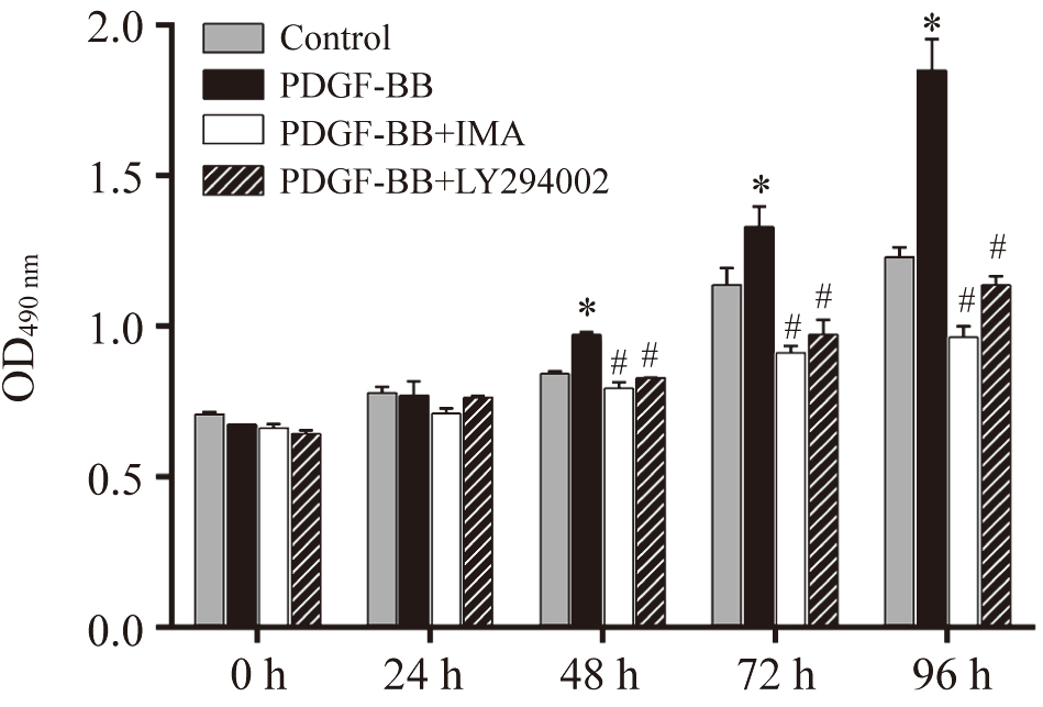
After 48 h culture, the cells’ OD490 nm value in group PDGF-BB was higher than group CON ( 0.05), and the difference in OD490 nm value between the two groups became more significant as the culture time prolonged ( 0.01). The OD490 nm values in group PDGF-BB+IMA and group PDGF-BB+LY294002 were significantly lower than group PDGF-BB at 48 h, 72 h, and 96 h, respectively (Fig. 4).
Effects of PDGF-BB, IMA and LY294002 on the proliferation of oral mucosa fibroblasts. 0.05 vs group CON; 0.05 vs group PDGF-BB.
PDGF-BB promoted migration
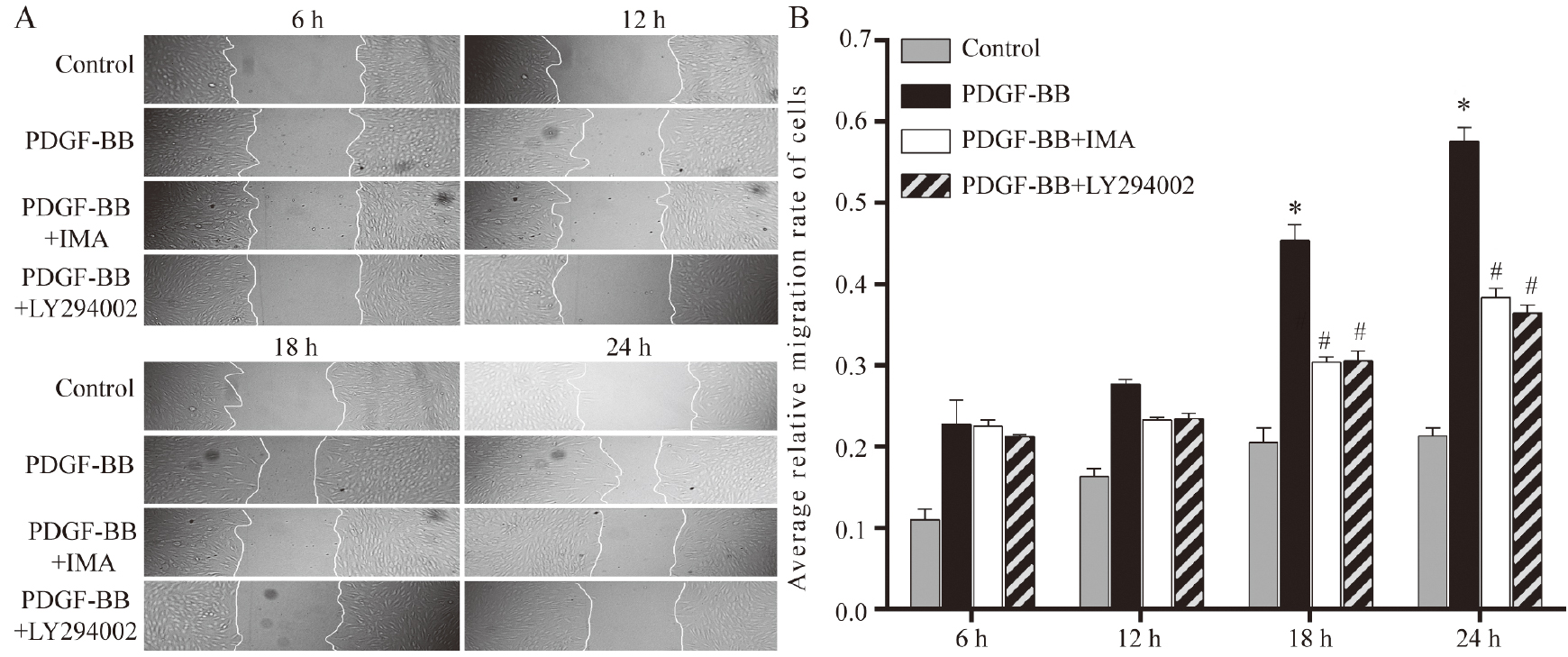
With the increase of culture time, the cells in group PDGF-BB gradually grew and migrated to the scratch area. After 18 to 24 h culture in serum-free medium, the average scratch width of the cells in group PDGF-BB was significantly reduced compared to group CON. In group PDGF-BB+IMA and group PDGF-BB+LY294002, the average scratch widths gradually decreased with the increase of culture time. However, compared with group PDGF-BB, after 18 to 24 h culture in serum-free medium, the average scratch widths in group PDGF-BB+IMA and group PDGF-BB+LY294002 were significantly smaller than group PDGF-BB (Fig. 5A). The average relative mobility of cells in group PDGF-BB was significantly larger than those in group CON, group PDGF-BB+IMA, and group PDGF-BB+LY294002 (all 0.05) (Fig. 5B).
Migration of oral mucosal fibroblasts detected by cell scratch assay. A: Real-time photographs show the relative migration widths in group CON, group PDGF-BB, group PDGF-BB+IMA and group PDGF-BB+LY294002 ( 50); B: Comparison of mean relative mobility among group CON group PDGF-BB, group PDGF-BB+IMA and group PDGF-BB+LY294002 at 6 h,12 h,18 h, and 24 h. 0.05 vs group CON; 0.05 vs group PDGF-BB.
PDGF-BB promoted synthesis of COI I protein
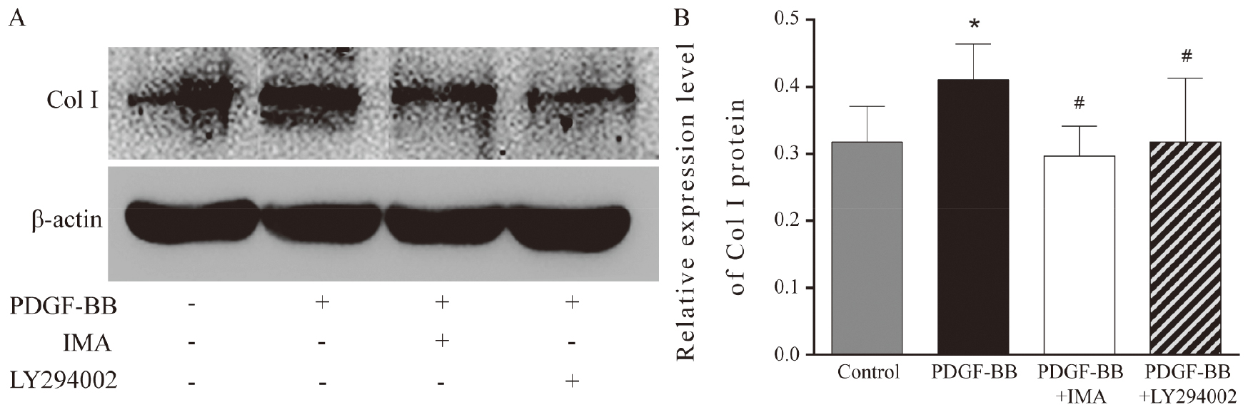
The relative expression of COI I protein in group PDGF-BB was significantly higher than those in group CON, group PDGF-BB+IMA, and group PDGF-BB+LY294002 (Fig. 6).
Synthesis of COI I protein in oral mucosal fibroblasts detected by western blot. 0.05 vs group CON; 0.05 vs group PDGF-BB.
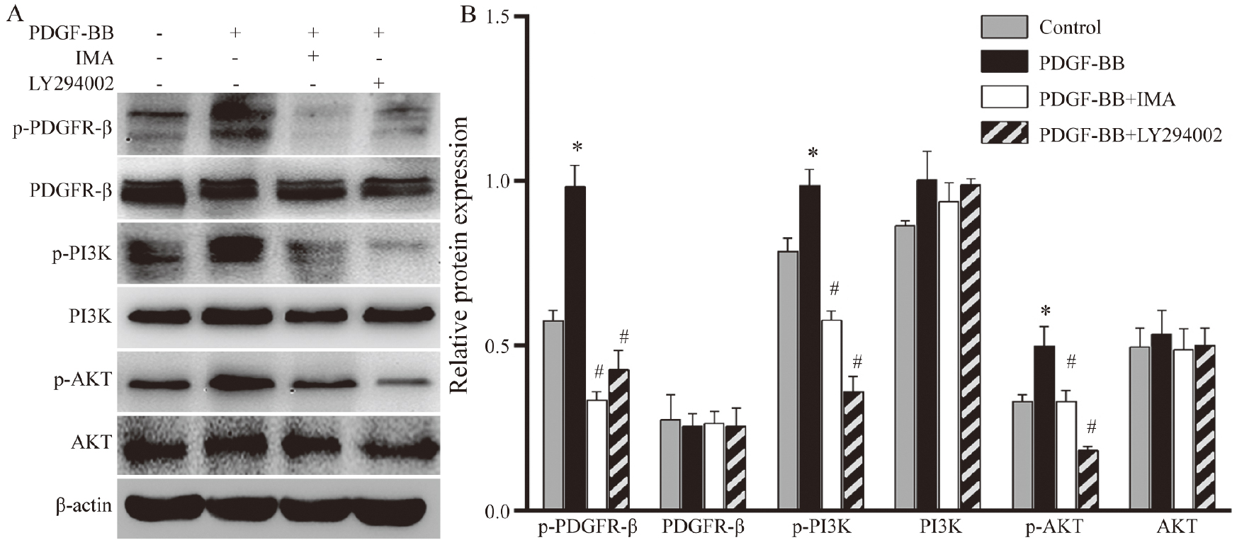
Compared with group CON, the levels of p-PDGFR-, p-PI3K, and p-AKT in group PDGF-BB increased significantly ( 0.05); however, in group PDGF-BB+IMA and group PDGF-BB+LY294002, the contents of p-PDGFR-, p-PI3K, and p-AKT were significantly lower than group PDGF-BB ( 0.05). There were no differences in the expressions of PDGFR-, PI3K and AKT protein among the groups ( 0.05) (Fig. 7).
Expression changes of related proteins in PDGFR-/PI3K/AKT signaling pathway by western blot. 0.05 vs group CON; 0.05 vs group PDGF-BB.
Discussion
Oral squamous cell carcinoma (OSCC) is the most common solid tumor in the oral cavity [12]. Accumulating evidence has revealed a role for an anti-tumor immune response via the T cell-mediated mechanisms [13] since peripheral blood mononuclear cells (PBMCs) from OSCC patients have presented cytotoxic CD8 T cell response [14]. Oral submucosal fibrosis (OSF) is a chronic inflammatory disease and has been classified as a precancerous state by WHO. The presence of autoantibodies in sera of OSF patients is the most characteristic and direct evidence of OSF being an autoimmune disease [15, 16]. Currently, different institutes have obtained inconsistent results about the roles of autophagy in the occurrence and development of tumors, as well as its therapeutic effects due to their different research directions [17]. Fibroblasts and myofibroblasts are important cells associated with OSF. Studies have confirmed that the abnormality and persistence of myofibroblasts are important signs of fibrotic diseases [18, 19, 20]. When stimulated, myofibroblasts can secrete a large number of cytokines and chemokines to promote the synthesis of collagen, glycosaminoglycans, or fiber connexins while inhibit and reduce the degradation of ECM, which causes a large accumulation of collagens in ECM and causes fibrosis of tissues and organs [21, 22]. This study shows that PDGF-BB induces oral mucosal fibroblasts to convert into myofibroblasts through the PDGFR-/PI3K/AKT signaling pathway, and promotes their proliferation, migration, and collagen synthesis.
-SMA is a signature protein expressed in MFBs while not expressed in other cells, such as fibroblasts. Therefore, we can detect the effects of PDGF-BB on the conversion of oral mucosal fibroblasts by detecting the expression of -SMA [23]. Our results show that after 7-day culture of oral mucosal fibroblasts in conditioned medium containing PDGF-BB, almost all of the cells expressed -SMA, indicating that PDGF-BB can promote the conversion of oral mucosal fibroblasts to MFBs. In the occurrence and development of fibrotic diseases, the proliferation and migration of fibroblasts and MFBs play an important role [24]. PDGF-BB is an important mitogenic factor, and its expression is elevated in the tissues of OSF patients. Our study results show that PDGF-BB promotes the proliferation of oral mucosal fibroblasts in time- and dose-dependent maners; at the same time, after PDGF-BB stimulation, the migration ability of cells significantly increased, which may be related to PDGF-BB’s promotion of oral mucosal fibroblasts into MFBs. One of the main features of oral submucosal fibrosis is the deposition of collagens, of which type I collagen accounts for 60% of the extracellular matrix. Western blot results show that PDGF-BB can promote the synthesis of Col I in oral mucosal fibroblasts. The abnormality and persistence of MFBs are important signs of fibrotic diseases, such as OSF. They can deposit extracellular matrix by secreting a large amount of collagens (such as Col I), which eventually leads to fibrosis [25]. Our results indicate that PDGF-BB can promote the conversion, proliferation, migration, and collagen synthesis of oral mucosal fibroblasts, suggesting that the high expression of PDGF-BB in OSF tissues may promote the occurrence and development of OSF.
At present, it is considered that arecoline in betel nuts is the main factor that induces OSF [26, 27]. Therefore, whether arecoline can promote the change of PDGF-BB expression in oral mucosal tissue, thereby promoting the conversion, proliferation, migration, and collagen synthesis of oral mucosal fibroblasts still needs further studies.
In this study, western blot was used to detect the expression of PDGFR-/PI3K/AKT signaling pathway-related proteins, namely PDGFR-, p-PDGFR-, PI3K, p-PI3K, AKT, and p-AKT. The results showed that the expression of p-PDGFR-, p-PI3K and p-AKT in oral mucosal fibroblasts significantly increased after PDGF-BB stimulation, and such effects can be inhibited by PDGFR phosphorylase inhibitor (IMA) and PI3K/AKT signaling pathway inhibitor (LY294002). IMA and LY294002 can significantly inhibit PDGF-BB-induced conversion of oral mucosal fibroblasts to MFBs, and reduce their proliferation, migration and collagen synthesis. The results suggest that PDGF-BB promotes the conversion, proliferation, migration, and collagen synthesis of oral mucosal fibroblasts through the PDGFR-/PI3K/AKT signaling pathway. The PDGF/PDGFR signaling has been shown to be associated with a variety of cancers, inflammation, fibrosis, restenosis, or atherosclerosis. Borkham-Kamphorst [28] introduced PDGFR- soluble receptor expression vector (sPDGFRb), through the tail vein, into the animal model of liver fibrosis caused by bile duct ligation and found that the degree of liver fibrosis was reduced and the expression of Col I and -SMA decreased significantly, suggesting that PDGFR- is involved in the formation of liver fibrosis. The downstream pathways of PDGF- are complex, and the PI3K/AKT signaling pathway plays an important role in fibrotic diseases. In animal models of pulmonary fibrosis and liver fibrosis, the PDGFR-/PI3K/AKT signaling pathway is involved in proliferation, transformation, and collagen synthesis in fibroblasts [29, 30]. PDGF, CTGF, and IGF-1 can promote the anti-apoptotic ability of lung fibroblasts through the PI3K/AKT signaling pathway [31], and it has been proven that in rat models of diabetic cardiomyopathy, IGF-1 can activate the expression of Bcl-2 protein through the PI3K/AKT signaling pathway and play an anti-apoptotic role in the myocardium, which in turn allows the persistence of myocardial fibroblasts and MFBs [10].
Although we have confirmed via in vitro experiments that PDGF-BB promotes the conversion, proliferation, migration, and collagen synthesis of oral mucosal fibroblasts through the PDGFR-/PI3K/AKT signaling pathway, there are still many places in this research that need further investigation. For example, we can detect the expressions of PI3K/AKT signaling-related mRNAs at the transcription level to further verify its role in conversing oral mucosal fibroblasts. In addition, we can further clarify the effects and mechanisms of PDGF-BB on the conversion, proliferation, migration, and collagen synthesis of oral mucosal fibroblasts by silencing the PDGFR- receptor gene. The occurrence and development of OSF are affected by complex in vivo environments instead of a certain pathway in cells. Future studies will need to explore other pathways, and construct OSF animal models for in vivo experiments to further clarify the effects and mechanism of PDGF-BB on oral mucosal fibroblasts so as to provide experimental evidence for elucidating the mechanism of OSF development.
Footnotes
Acknowledgments
Funded by: National Natural Science Foundation of China, No. 81271154.
Conflict of interest
The authors declare no conflict of interest.
References
1.
ArakeriG.RaiK.K.HunasgiS.MerkxM.A.W.GaoS. and BrennanP.A., Oral submucous fibrosis: an update on current theories of pathogenesis, J Oral Pathol Med46 (2017), 406–412.
2.
BrennanP.A. and ArakeriG., Oral submucous fibrosis-an increasing global healthcare problem, J Oral Pathol Med46 (2017), 405.
3.
SharmaA.KumarR.JoharN. and SabirH., Oral submucous fibrosis: an etiological dilemma, J Exp Ther Oncol12 (2017), 163–166.
4.
JimenezS.A.HitrayaE. and VargaJ., Pathogenesis of scleroderma. Collagen, Rheum Dis Clin North Am22 (1996), 647–674.
5.
HeldinC.H.ErikssonU. and OstmanA., New members of the platelet-derived growth factor family of mitogens, Arch Biochem Biophys398 (2002), 284–290.
6.
FredrikssonL.LiH. and ErikssonU., The PDGF family: four gene products form five dimeric isoforms, Cytokine Growth Factor Rev15 (2004), 197–204.
7.
BartoschekM. and PietrasK., PDGF family function and prognostic value in tumor biology, Biochem Biophys Res Commun503 (2018), 984–990.
8.
CorsinoviD.GiannettiK.CericolaA.NaefV. and OriM., PDGF-B: the missing piece in the mosaic of PDGF family role in craniofacial development, Dev Dyn248 (2019), 603–612.
9.
ZhangH.BajraszewskiN.WuE.WangH.MosemanA.P.DaboraS.L.GriffinJ.D. and KwiatkowskiD.J., PDGFRs are critical for PI3K/Akt activation and negatively regulated by mTOR, J Clin Invest117 (2007), 730–738.
10.
VittalR.HorowitzJ.C.MooreB.B.ZhangH.MartinezF.J.ToewsG.B.StandifordT.J. and ThannickalV.J., Modulation of prosurvival signaling in fibroblasts by a protein kinase inhibitor protects against fibrotic tissue injury, Am J Pathol166 (2005), 367–375.
11.
ZhouS.ZhuY.HeZ.ZhangD.GuoF.JianX. and ZhangC., Long non-coding RNA expression profile associated with malignant progression of oral submucous fibrosis, J Oncol2019 (2019), 6835176.
12.
JiangC.YuanF.WangJ. and WuL., Oral squamous cell carcinoma suppressed antitumor immunity through induction of PD-L1 expression on tumor-associated macrophages, Immunobiology222 (2016), 651–657.
13.
WangJ.SunF.LinX.LiZ.MaoX. and JiangC., Cytotoxic T cell responses to streptococcus are associated with improved prognosis of oral squamous cell carcinoma, Exp Cell Res362 (2018), 203–208.
14.
WangJ.YangL.MaoX.LiZ.LinX. and JiangC., Streptococcus salivarius-mediated CD8+T cell stimulation required antigen presentation by macrophages in oral squamous cell carcinoma, Exp Cell Res366 (2018), 121–126.
15.
WangJ.YouJ.WangL.WangH.TianT.WangW.JiaL. and JiangC., PTMA, a new identified autoantigen for oral submucous fibrosis, regulates oral submucous fibroblast proliferation and extracellular matrix, Oncotarget8 (2017), 74806–74819.
16.
JiangC.YangD. and WangJ., Correlations of abnormally upregulated CC chemokine ligand 18 (CCL18) with clinical stage and cervical lymph node metastasis status in serum and tumor tissue of patients with oral squamous cell carcinoma, Int J Clin Exp Pathol9 (2016), 6317–6325.
17.
JiangC.JinS.JiangZ. and WangJ., Inhibitory effects of silibinin on proliferation and lung metastasis of human high metastasis cell line of salivary gland adenoid cystic carcinoma via autophagy induction, Onco Targets Ther9 (2016), 6609–6618.
18.
RajalalithaP. and ValiS., Molecular pathogenesis of oral submucous fibrosis-a collagen metabolic disorder, J Oral Pathol Med34 (2005), 321–328.
19.
DaiJ.P.ChenJ.BeiY.F.HanB.X. and WangS., Influence of borneol on primary mice oral fibro-blasts: a penetration enhancer may be used in oral submucous fibrosis, J Oral Pathol Med38 (2009), 276–281.
20.
MoutasimK.A.JeneiV.SapienzaK.MarshD.WeinrebP.H.VioletteS.M.LewisM.P.MarshallJ.F.FortuneF.TilakaratneW.M.HartI.R. and ThomasG.J., Betel-derived alkaloid up-regulates keratinocyte alphavbeta6 integrin expression and promotes oral submucous fibrosis, J Pathol223 (2011), 366–377.
21.
KisselevaT., The origin of fibrogenic myofibroblasts in fibrotic liver, Hepatology65 (2017), 1039–1043.
22.
Gyftaki-VenieriD.A.AbrahamD.J. and PonticosM., Insights into myofibroblasts and their activation in scleroderma: opportunities for therapy? Curr Opin Rheumatol30 (2018), 581–587.
23.
KatjaB.VanR.C.IrisS.WickertL.FloegeJ. and GressnerA.M., Expression patterns of PDGF-A, -B, -C and -D and the PDGF-receptors alpha and beta in activated rat hepatic stellate cells (HSC), Cytokine31 (2005), 349–357.
24.
Borkham-KamphorstE.van RoeyenC.R.OstendorfT.FloegeJ.GressnerA.M. and WeiskirchenR., Pro-fibrogenic potential of PDGF-D in liver fibrosis, J Hepatol46 (2007), 1064–1074.
25.
SandulacheV.C.ParekhA.Li-KorotkyH.DoharJ.E. and HebdaP.A., Prostaglandin E2 inhibition of keloid fibroblast migration, contraction and transforming growth factor1 (TGF-β1) induced collagen synthesi, Wound Repair Regen15 (2007), 122–133.
26.
ChungC.H.YangY.H.WangT.Y.ShiehT.Y. and WarnakulasuriyaS., Oral precancerous disorders associated with areca quid chewing, smoking, and alcohol drinking in southern Taiwan, J Oral Pathol Med34 (2005), 460–466.
27.
MurtiP.R.BhonsleR.B.GuptaP.C.DaftaryD.K.PindborgJ.J. and MehtaF.S., Etiology of oral submucous fibrosis with special reference to the role of areca nut chewing, J Oral Pathol Med24 (1995), 145–152.
28.
Borkham-KamphorstE.HerrmannJ.StollD.TreptauJ.GressnerA.M. and WeiskirchenR., Dominant-negative soluble PDGF-beta receptor inhibits hepatic stellate cell activation and attenuates liver fibrosis, Lab Invest84 (2004), 766–777.
29.
CzochraP.KlopcicB.MeyerE.HerkelJ.Garcia-LazaroJ.F.ThieringerF.SchirmacherP.BiesterfeldS.GalleP.R.LohseA.W. and AffiliationsS.K., Liver fibrosis induced by hepatic overexpression of PDGF-B in transgenic mice, J Hepatol45 (2006), 419–428.
30.
ZhangL.LiY.LiangC. and YangW., CCN5 overexpression inhibits profibrotic phenotypes via the PI3K/Akt signaling pathway in lung fibroblasts isolated from patients with idiopathic pulmonary fibrosis and in an in vivo model of lung fibrosis, Int J Mol Med33 (2014), 478–486.
31.
WuS.H.WuX.H.LuC.DongL. and ChenZ.Q., Lipoxin A(4) inhibits proliferation of human lung fibroblasts induced by connective tissue growth factor, Am J Respir Cell Mol Biol34 (2006), 65–72.