
Abstract
BACKGROUND:
Increasing evidence shows that long non-coding RNAs (lncRNAs) play a key role in the development of various cancers. Zinc finger antisense 1 (ZFAS1) is a novel lncRNA with previously demonstrated associations with several types of cancer. Here we examined the expression and potential function of the ZFAS1 in nasopharyngeal carcinoma (NPC).
METHODS:
We detected ZFAS1 expression in GSE12452, a human microarray dataset, and NPC cell lines. Small interfering RNA against ZFAS1 was used to elucidate the cellular functions of ZFAS1 using MTT, colony formation, cell cycle, cell apoptosis, transwell invasion and migration and western blot assays. An activator of the PI3K/AKT signaling pathway (740Y-P) was used to determine the contribution of PI3K/AKT. RESULTS: ZFAS1 was significantly upregulated in NPC tissues and cell lines. Silencing ZFAS1 significantly inhibited cell proliferation and invasion, arrested cell cycle progression and promoted cell apoptosis, as well as reduced epithelial–mesenchymal transition. Moreover, 740Y-P could rescue the effects of ZFAS1 knockdown on proliferation, apoptosis and invasion in 5-8F cells.
CONCLUSIONS:
ZFAS1 might play an oncogenic role in NPC and facilitate cell proliferation and invasion via the PI3K/AKT signaling pathway in NPC cells.
Introduction
Nasopharyngeal carcinoma (NPC) is one of the most common malignant tumors in the head and neck, with a high incidence in southern China and Southeast Asia [1, 2]. With the use of intensity-modulated radiation therapy (IMRT) and concurrent chemoradiotherapy for NPC treatment, the overall survival rate for NPC patients has been significantly improved (70%) [3]. Local/regional recurrence and distant metastasis are the most common causes of failure in NPC treatment [4]. Different pathological types of NPC have differences in biological behaviors as well as prognosis, and the undifferentiated form of NPC is associated with the worst prognosis [5]. The development and progression of NPC are influenced by genetic susceptibility, environmental factors, dietary factors and Epstein-Barr virus infection [6]. Detection of Epstein-Barr virus DNA in plasma is of great value in diagnosing NPC progression after IMRT [7]. With the development of gene diagnosis and gene therapy, the identification of specific molecular markers for NPC will be critical in improving the diagnosis and prognosis for NPC patients as well as further clarifying of the pathogenesis of nasopharyngeal carcinoma.
Long noncoding RNA (lncRNA) represents a class of RNA molecules over 200 nt in length without an open reading frame 3[8, 9]. While early studies concluded that lncRNAs could not encode proteins and had no biological functions [10, 11], research has now demonstrated that lncRNAs play important cellular roles in various processes. In addition, recent studies have demonstrated potential functions for lncRNAs in carcinogenesis and tumor suppressor pathways. LncRNAs have been shown to regulate cell growth, metastasis and apoptosis in most tumor types via transcriptional, post-transcriptional and epigenetic mechanisms [12, 13]. Several supporting evidences have shown that a number of lncRNAs, including LINC00460 [14], XIST [15], AFAP1-AS1 [16], MALAT1 [17], play a signifcant role in nasopharyngeal carcinoma.
Zinc finger antisense 1 (ZFAS1) is a newly identified lncRNA that is located at chromosome 20q13 and possibly shares bidirectional promoters with ZNFX1 [18, 19]. ZFAS1 is the “host gene” of three CD/D box small nucleolar RNAs (snoRNAs): snord l2c, snord l2b and snord l2 [20]. Askarian et al. found that ZFAS1 was downregulated in breast cancer, while other reports indicated that ZFAS1 was upregulated in gastric cancer [21], colorectal cancer [22], lung cancer [23]and glioma [24] as a carcinogenesis. However, the expression and the function of ZFAS1 in NPC remain unknown.
Here we explored the significance and pathogenic mechanism of ZFAS1 in the development of NPC, and we hope it can help us to explore the molecular mechanism of NPC and to identify potential molecular markers related to early diagnosis and prognosis, which is of great significance for clinical diagnosis and treatment.
Materials and methods
Microarray data analysis
GSE12452 is a human microarray dataset that was obtained from a public GEO database (
Cell culture
NPC cell lines (5-8F, CNE1, CNE2, C666) and a normal nasopharyngeal epithelium cell line (NP69) were purchased from ATCC Company. NP69 cells were cultured in RPMI 1640 (Corning, USA) with 10% fetal bovine serum (FBS) (Gibco, USA); CNE1, CNE2, 5-8F and C666 cells were cultured in DMEM medium (Hyclone, USA) with 10% FBS (Gibco). All cell lines were cultured at 37
Cell transfection
The ZFAS1-specific small interfering RNA (siZFAS1) and a scrambled negative control (siNC) were purchased from GenePharma Company (China). The sequences were as follows: siZFAS1: 5
Quantitative real-time PCR (qRT-PCR)
Cells were harvested by trypsinization (YIKE, China) and RNA was obtained using Trizol (Sangon, China). RNA (1
Cell proliferation assays
Cell proliferation was evaluated using a Cell Proliferation Reagent Kit I (MTT) (Roche Applied Science, Germany). CNE2 and 5-8F cells were plated in 96-well plates (1
Colony formation assay
NPC cells transfected with siNC or siZFAS1 were plated with 200 cells per well in a six-well plate and then cultured for 1–2 weeks at 37
Wound healing assay
CNE2 and 5-8F cells were plated in six-well plates (3
Transwell invasion assay
Transwell assays were performed using transwell chambers (Corning, USA). Matrigel (50
Cell apoptosis assay
Cell apoptosis was evaluated using APC-Annexin V/propidium iodide (PI) (Sigma, USA) double staining according to the manufacturer’s protocol. NPC cells transfected with siNC or siZFAS1 were harvested, washed with PBS and re-suspended in binding buffer with a cell density of 1
Cell cycle arrest
NPC cells transfected with siNC or siZFAS1 were harvested, resuspended in 75% ethanol and fixed over-night at 4
Western blot analysis
Treated cells were collected and lysed with RIPA (Radio-Immunoprecipitation Assay) buffer (Beyotime, China). Proteins were separated by 10% SDS-polya-crylamide gel electrophoresis (SDS-PAGE) and transferred to 0.22
Activator treatment
To investigate the PI3K/Akt signaling pathway, 5-8F cells transfected with siZFAS1 for 24 h were incubated with PI3K agonist 740Y-P (50
Statistical analysis
Statistical analyses were performed by unpaired, two-tailed Student’s t-test using GraphPad Software Version 6.0. (GraphPad Software Inc., San Diego, CA, USA).
Results
lncRNA ZFAS1 is overexpressed in NPC tissues and cell lines
To determine the expression profile of lncRNA ZFAS1 in NPC, we analyzed GEO data from the previous study by Sengupta et al. [25] (GSE12452), which included 31 NPC tissues and 10 normal nasopharyngeal tissues. R software Affy version 1.50.0 [26] was used to correct the background of the expression value of the data and the normalized preprocessing of the expression spectrum data. Data were annotated by the platform annotation file. We found that ZFAS1 was highly expressed in NPC tissues compared with normal nasopharyngeal tissues (
ZFAS1 expression is upregulated in NPC tissues and NPC cells. (A) Relative expression of ZFAS1 in nasopharyngeal carcinoma tissues compared with normal tissue was analyzed using GEO dataset GSE12452. (B) Expression of lncRNA ZFAS1 in four nasopharyngeal carcinoma cell lines and a normal nasopharyngeal line was determined by RT-PCR. (C) qRT-PCR of ZFAS1 in 5-8F and CNE2 cells transfected with ZFAS1 siRNA or negative control (siNC). (D) MTT assays in 5-8F and CNE2 cells transfected with ZFAS1 siRNA or negative control (siNC) and cultured for the indicated times. (E, F) Colony-forming growth assays showed reduced colony formation of 5-8F and CNE2 cells after silencing of ZFAS1. 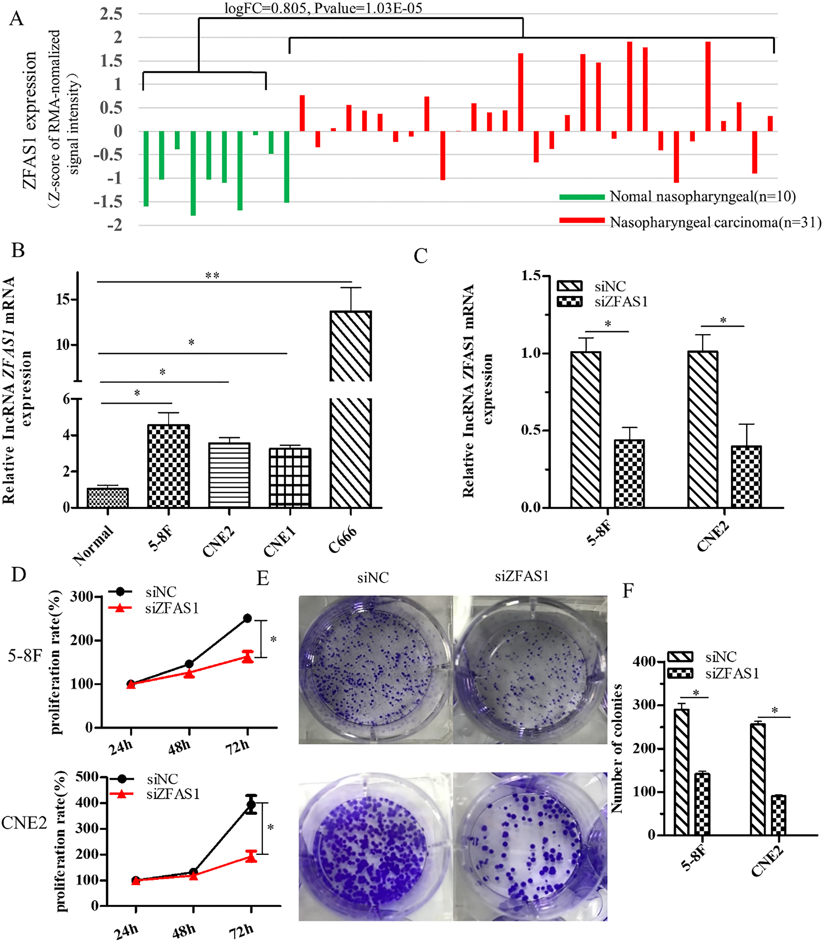
To examine whether ZFAS1 was involved in the progression of nasopharyngeal carcinoma, we used siRNA to knockdown ZFAS1 expression in 5-8F and CNE2 cells (Fig. 1C). MTT assay demonstrated that ZFAS1 silencing significantly inhibited proliferation in both 5-8F and CNE2 cells after 72 h (
To explore the potential role of ZFAS1 in cell proliferation, we investigated the effect of ZFAS1 knockdown on the cell cycle and apoptosis of 5-8F and CNE2 cells using flow cytometry. In cells with ZFAS1 knockdown, the proportion of cells in G0/G1 phase increased and S phase cells decreased to varying degrees (Fig. 2A and B). Cell apoptosis assay measured by Annexin V-APC/PI staining revealed that silencing of ZFAS1 enhanced the cell apoptosis rate of 5-8F and CNE2 cells compared with the siNC groups (Fig. 2C and D). Together these findings suggested that ZFAS1 might stimulate cell proliferation by promoting cell cycle progression and inhibiting NPC cell apoptosis.
ZFAS1 regulates cell cycle and inhibits cell apoptosis by reducing p53, p21 proteins and increasing Bcl-2 protein. (A, B) Effects of ZFAS1 knockdown on the cell cycle. Downregulating ZFAS1 expression caused the percentages of cells in G0/G1 phase to increase and cells in S phase cells to decrease. (C, D) ZFAS1 silencing dramatically induced apoptosis in 5-8F and CNE2 cells. (E, F) Western blot analysis of the indicated proteins in 5-8F and CNE2 cells transfected with ZFAS1 siRNA or negative control (siNC). 
To more closely examine the molecular basis underlying the role of ZFAS1 in regulating the cell cycle and cell apoptosis, we examined the levels of related proteins. Western blot analysis demonstrated that the expression levels of cell cycle-related proteins p21and p53 were increased, while anti-apoptotic protein Bcl-2 was decreased in 5-8F and CNE2 cells after ZFAS1 silencing (Fig. 2E and F). This indicates that ZFAS1 may inhibit cell apoptosis and regulate the cell cycle by inhibiting the expression of p53 and p21 and promoting the expression of Bcl-2.
Knockdown of lncRNA ZFAS1 suppresses migration and invasion of NPC cells and represses endothelial-mesenchymal transition (EMT) in NPC cells
To further investigate the effect of ZFAS1 in NPC cells, we performed wound-healing and Transwell assays to detect the role of ZFAS1 in cell migration and invasion. In the wound-healing assay, the migration rate of ZFAS1 siRNA-transfected cells was inhibited at 24 and 48 h compared with control cells (Fig. 3A and B). Similarly, the transwell assay result showed that invasion and migration were weakened in ZFAS1-silenced 5-8F and CNE2 cells compared with the siNC group (Fig. 3C). These findings suggested that ZFAS1 could enhance the migration and invasion abilities of NPC cells in vitro.
We next speculated whether ZFAS1 was involved in EMT, which plays an important role in cancer migration and invasion [27, 28]. We thus examined the levels of EMT-related proteins and found that the expression of the epithelial marker E-cadherin was upregulated, while the expressions of the mesenchymal markers N-cadherin and Vimentin were decreased after siZFAS1 transfection [29] (Fig. 3D and E). Together this suggests that ZFAS1 might promote the migration and invasion of NPC by enhancing EMT progression.
ZFAS1 regulates the activity of the PI3K/AKT-mediated signaling pathway in NPC cells
Considering that the PI3K/AKT pathway is abnormally activated in various types of tumors and is closely related to tumor proliferation as well as EMT progression [30], we speculated that the effects of ZFAS1 in regulating the proliferation and invasion of NPC cells may occur through the PI3K/AKT signaling pathway. We thus performed western blot analysis to examine the PI3K/AKT pathway in NPC cells with ZFAS1 knockdown. We found that ZFAS1 deficiency significantly reduced the level of phosphorylated (and activated) AKT (p-AKT), while the expression of total AKT (AKT) did not change significantly (Fig. 4A and B).
Downregulation of ZFAS1 inhibits the migration and invasion of 5-8F and CNE2 cells and inhibits EMT process. (A, B) Silencing ZFAS1 expression inhibited the migration of NPC cells in wound-healing assays. (C, E) Silencing ZFAS1 expression inhibited the invasion ability of NPC cells in Transwell assays. (D, F) Western blot analysis demonstrated that N-cad and Vim were downregulated while E-cad protein was upregulated in 5-8F and CNE2 cells after silencing ZFAS1. 
Effects of the PI3K/Akt signaling pathway agonist (740Y-P) on the role of ZFAS1 in NPC cells. (A, B) Western blot analysis demonstrated that p-AKT was downregulated while AKT was unchanged after silencing ZFAS1 in 5-8F, CNE2 cells. (C-K) An agonist of the PI3K/AKT signaling pathway (740Y-P) was used in 5-8F cells transfected with siZFAS1. (C, D, F) MTT and colony-forming assays demonstrated that 740Y-P reversed the effect of ZFAS1 inhibition of 5-8F cells proliferation. (E, G) 740Y-P reversed the effect of ZFAS1 inhibition on 5-8F cells invasion. (H, J) 740Y-P reversed the effect of ZFAS1 inhibition on 5-8F cells cell cycle progression. (I, K) 740Y-P reversed the effect of ZFAS1 inhibition on 5-8F cells cell apoptosis. 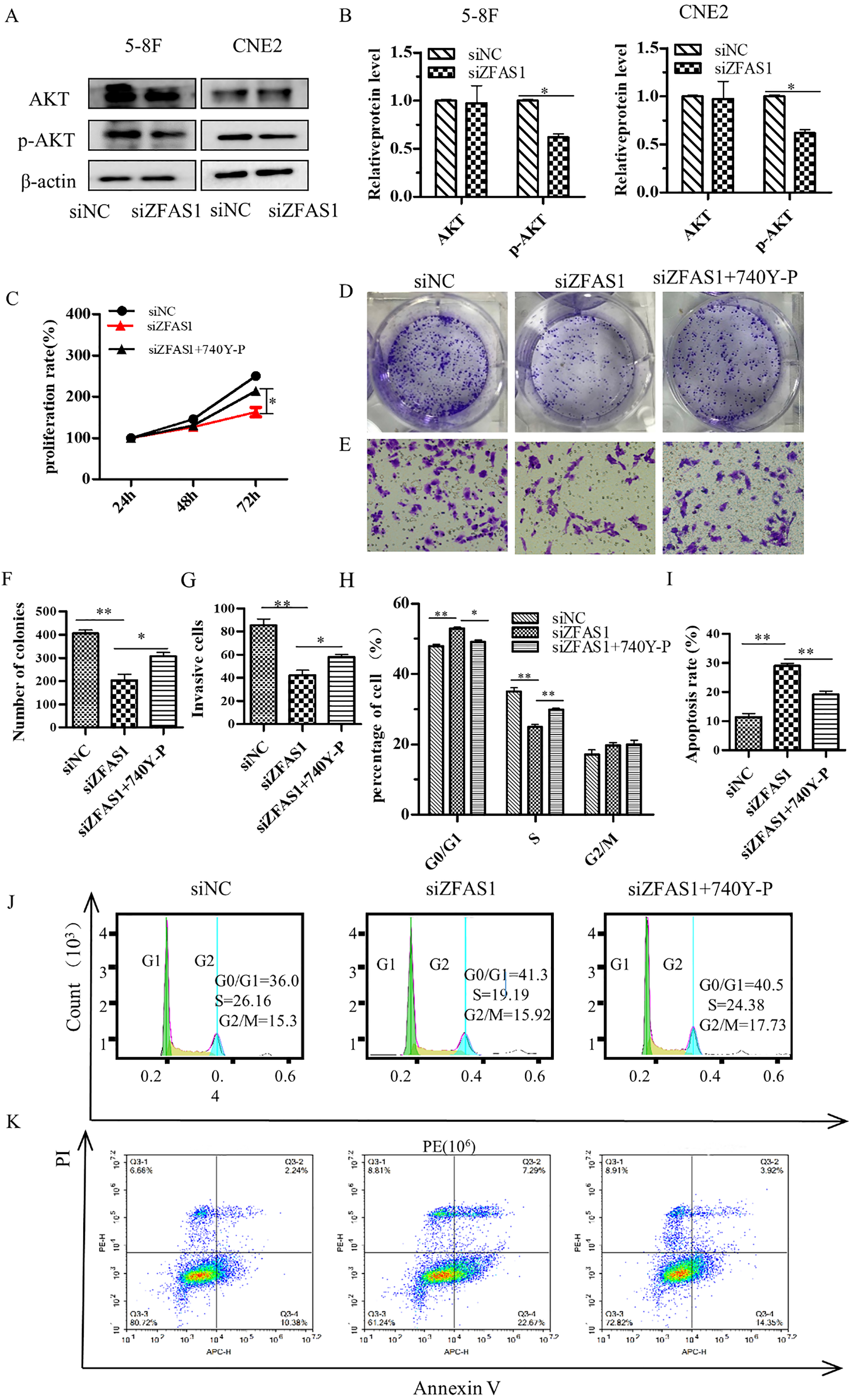
To confirm the involvement of the PI3K/AKT pathway, we conducted a series of rescue experiments using 740Y-P, an activator for the PI3K/AKT pathway. We found that treatment with 740Y-P in ZFAS1 knockdown NPC cells restored the level of the inhibition of cell proliferation and invasion ability caused by ZFAS1 siRNA in MTT, colony formation and transwell assays (Fig. 4C–E). Furthermore, 740Y-P could block the G0/G1 phase arrest and induction of cell apoptosis caused by ZFAS1 siRNA (Fig. 4F–I). Finally, treatment of 5-8F cells transfected with siZFAS1 with 740Y-P restored the levels of p-AKT, Bcl-2, p21, p53 as well as EMT-related proteins (Fig. 5A and B). Thus, we propose that the biological functions of ZFAS1 in NPC cells occurs in part through the PI3K/AKT signaling pathway.
The PI3K/AKT signaling pathway is involved in the effects of ZFAS1 in NPC cells. (A, B) The PI3K/AKT signaling pathway agonist (740Y-P) was used in 5-8F cells transfected with siZFAS1. Western blot analysis was performed for the indicated proteins. (C) ZFAS1 may promote cell proliferation and invasion by activating PI3K/AKT-mediated signaling. 
Recent research has demonstrated that some lncRNAs may either promote or inhibit cancer during the occurrence and development of tumors [31, 32], through mechanisms that involve genome imprinting processes, histone modification, chromatin remodeling and cell cycle regulation [12, 33] . Previous studies showed that the lncRNA ZFAS1 was highly expressed in colorectal cancer [34], hepatocellular carcinoma [35], gastric cancer [36], glioma [37]and lymphoma yet lowly expressed in breast cancer [20], with its functions as carcinogens or tumor suppressors. These findings indicate that ZFAS1 may exert oncogenic or tumor suppressor functions in different types of tumors. Our findings reported here indicate that ZFAS1 could serve as a potential marker for nasopharyngeal carcinoma.
In this study, we found that ZFAS1 was highly expressed in NPC through our analysis of the microarray data of GSE12452. These findings were also observed in NPC cell lines. We then performed a series of in vitro assays to clarify the functions of ZFAS1 in NPC. Our results showed that knockdown of ZFAS1 inhibited NPC cell proliferation and invasion, which is consistent with the findings in gastric cancer cells [18] and and colon cancer cells [22]. Knockdown of ZFAS1 in NPC cells induced cell apoptosis and arrested the cell cycle at G1 phase, which is consistent with the findings in colon cancer [34] and gastric cancer [21]. These results indicate that highly expressed ZFAS1 in NPC may exert oncogenic functions. However, further study is required to determine whether high level of ZFAS1 is associated with NPC grading, staging or prognosis.
Our results showed that silencing ZFAS1 significantly arrested the cell cycle and induced apoptosis in NPC cells. Further analysis showed that ZFAS1 could regulate the cell cycle by inhibiting the p53/p21 axis. Several studies have shown that p53 regulates the restriction point at G1-S phase and G2-M phase transitions [38], and inhibits DNA replication [39]. p21 is a transcription target of p53 and functions as an inhibitor of several cyclin/CDK complexes that regulate progression at G1-S [40]. In other words, knockdown of ZFAS1 could upregulated the expression of p53 and p21, which made more NPC cells arrest in G1-S phase, leading to the inhibition of cell proliferation. Besides, the Bcl-2 anti-apoptotic protein maintains mitochondrial membrane integrity, activates Ca
We also explored the molecular mechanism by which knockdown of ZFAS1 inhibited NPC cell growth and metastasis in vitro. EMT is the process of epithelial cells transforming into stromal cells, which occurs during invasion and metastasis of many kinds of tumors, indicating poor prognosis [27]. Here we found that after knocking down the expression of ZFAS1, the expression of the epithelial cell marker E-cad was upregulated, while mesenchymal markers Vim and N-cad were downregulated. Given that similar results were also found in gastric cancer [36], glioma [37] and other tumors. Therefore, we speculate that ZFAS1 may enhance cell invasion and metastasis in NPC cells by promoting the EMT process. More research is required to elucidate the specific underlying mechanism.
The PI3K/AKT signaling pathway represents a central node that regulates many cellular functions and plays a major role in cancer through the regulation of cell growth, apoptosis, cell cycle and EMT [42, 43, 44]. Several studies have shown that lncRNAs such as ARHGAP42 [45], and SNHG1 [46] regulate cell proliferation and invasion through the PI3K/AKT signaling pathway in carcinoma. After Phosphatidylinositol-3-kinase (PI3K) generates phosphatidylinositol-3,4,5-trisphosphate (PI(3,4, 5)P3), AKT is phosphorylated to be phosphorylated AKT (p-AKT), which acts on multiple downstream effectors, resulting in cell proliferation, apoptosis and invasion [47, 48, 49]. Our results showed a decrease in AKT activation along with decreased phosphorylation of its downstream substrates in NPC cells after siZFAS1 transfection. We further examined the involvement of the PI3K/AKT pathway in the effects of ZFAS1 using the 740Y-P activator of the PI3K/AKT signaling pathway. We found that 740Y-P partially reversed the effects of ZFAS1 knockdown on 5-8F cell function and molecular expression [50]. We thus speculate that ZFAS1 could promote cell proliferation and EMT progression and inhibit cell apoptosis via the PI3K/AKT signal pathway in vitro in nasopharyngeal carcinoma cells.
In conclusion, here we found that ZFAS1 is upregulated in NPC tissues and NPC cell lines. We propose that ZFAS1 could promote NPC cell proliferation and tumorigenesis by activating the PI3K/AKT signaling pathway in vitro. Therefore, we propose that ZFAS1 functions as a kind of oncogenic lncRNA in NPC cells and might serve as one of the potential therapeutic targets for patients with NPC.
Footnotes
Acknowledgments
This work was supported by the Taizhou 2017 annual municipal science and technology projects (NO. 1701KY41). We thank Edanz Group () for editing a draft of this manuscript.
Conflict of interest
None declared.