
Abstract
BACKGROUNDS:
MicroRNAs (miRNAs) are some RNA molecules that negatively regulate gene expression by binding to the 3’-untranslated region (3’-UTR) of target mRNA molecules. The aim of the study is to investigate the clinical role and functional effects of microRNA-493 (miR-493) in human hepatocellular carcinoma (HCC).
METHODS:
Expression of miR-493 in 58 cases of HCC tissues and adjacent normal tissues was determined by using quantitative real-time PCR (qRT-PCR) analyses. In vitro, cell proliferation and invasion capacity was evaluated by CCK8 assay and trans-well invasion assay. Luciferase reporter assay, western blot and qRT-PCR were used to detect the association between miR-493 expression and zinc finger protein X-linked (ZFX) in HCC.
RESULTS:
Expression of miR-493 was significantly downregulated in HCC tissues compared to adjacent normal tissues. Lower miR-493 expression associated with tumor size, vascular invasion and poor overall survival (OS) and disease free survival (DFS) time of HCC patients. In vitro, transfection of miR-493 mimic in HCC cells inhibited cell proliferation and invasion abilities. However, transfection of miR-493 inhibitor in HCC cells had promoting effects. Luciferase reporter assay, qRT-PCR and western blot analysis demonstrated that miR-493 targeting 3’-untranslated region (3’-UTR) of ZFX and overexpression of miR-493 inhibited its expression. Moreover, enforced ZFX expression rescued the effects of miR-493 mimic on cell proliferation and invasion.
CONCLUSION:
MiR-493 functioned as tumor suppressor in HCC cells by regulating ZFX expression. Thus, miR-493 may provide potential value for HCC treatment.
Introduction
Hepatocellular carcinoma (HCC) is the fifth most prevalent cancer worldwide and presents a high mortality worldwide. In the world, more than 749,000 cases occur and 692,000 mortalities were reported per year [1]. Over the past 15 years, the incidence of HCC steadily increases with advancing age and hepatitis B virus infection is the leading cause of HCC [2]. Although the advances of HCC therapy including surgery, radiotherapy, and chemotherapy, the 5-years survival rate of patients at advanced stage remains low [3, 4]. Thus, investigating the relevant molecular mechanisms of HCC progression is important.
MicroRNAs (miRs) regulate gene transcription and translation by binding to the 3’ untranslated region (3’-UTR) of target mRNAs. In HCC, miRs play crucial roles in regulating cell proliferation, apoptosis, migration, invasion, and metastasis [5, 6]. Previously, the deregulation of specific miRs has been identified as oncogenes and tumor suppressors in HCC including miR-206 [7], miR-124 [8], miR-217 [9], miR-221 [10] and so on. MiR-493 has been reported to act as tumor suppressor in HCC, Xu et al. revealed that miR-493 suppresses hepatocellular carcinoma tumorigenesis through down-regulation of anthrax toxin receptor 1 (ANTXR1) and R-Spondin 2 (RSPO2) [11]. MiR-493-5p suppresses hepatocellular carcinoma cell proliferation through targeting GP73 [12]. However, the underlying role of miR-493 still needs to be investigated.
In the study, we demonstrated that expression of miR-493 was downregulated in HCC tissues and lower miR-493 expression predicted a poor prognosis of HCC patients. Moreover, we demonstrated that miR-493 inhibited cell proliferation and invasion of HCC in vitro by targeting ZFX and affects its expression. Thus, our results indicated that miR-493 functions as tumor suppressor in HCC and could provide potentially clinical value for HCC treatment.
Materials and methods
Tissue samples
HCC tissues and adjacent normal tissues were obtained from 58 cases of HCC patients who received surgical resection at Kunming General Hospital, Chinese People’s Liberation Army from April 2010 to July 2013. HCC was diagnosed by pathological analysis. The patients were staged according to American Joint Committee on Cancer (AJCC) 8th staging system and the clinical data was shown in Table 1. Patients received no treatment before surgical resection. The tissue samples were store at
The relationship of miR-493 expression with clinicopathologic parameters in 58 HCC patients is analyzed
The relationship of miR-493 expression with clinicopathologic parameters in 58 HCC patients is analyzed
Human HCC (LM3, 97H, Huh7 and SMMC-7721 cells) and one normal hepatic epithelial cell line (LO2, control) were purchased the Cell Bank of the Chinese Academy of Sciences (Shanghai, China). The cells were cultured in Dulbecco’s modified Eagle medium (DMEM) medium (Gibco; Thermo Fisher Scientific, Inc., Waltham, MA, USA) containing 10% fetal bovine serum (FBS; Gibco; Thermo Fisher Scientific, Inc., Waltham, MA, USA) in a cell incubator containing 5% CO
RNA oligonucleotides, plasmid construction, and cell transfection
The miR-493 mimic, miR-493 inhibitor and miR-negative control were purchased from Ribobio (Guangzhou, China). Full-length ZFX cDNA (GenBank accession number NM_001178084.1) was cloned into pcDNA3.1 vectors from Ribobio (Guangzhou, China). Cells transfection was performed using Lipofectamine 3000 transfection reagent (Invitrogen) according to the manufacturer’s instructions.
RNA extraction and quantitative real-time PCR (qRT-PCR)
Total RNA was extracted from tissues and cells using the Trizol reagent (Invitrogen, Waltham, MA, USA). The cDNA was reversed transcription from total RNA using a miRNeasy Mini Kit (Qiagen, Valencia, CA, USA) according to the manufacturer’s protocol. The mRNA expression was amplified using SYBR Green Real-time PCR Master Mix (Takara, Dalian, China) and detected at ABI 7500 Sequence Detection System (Applied Biosystems, Foster City, CA, USA). The relative mRNA expression of miR-493 and ZFX was normalized to U6 or GAPDH. Then, the mRNA expression was assessed using 2
Colony formation assay
Stable transfected 97H and SMMC-7721 cells at a density of 500 cells/well were seeded at 35-mm dishes. Cells were allowed to grow for 7 days in a cell incubator containing 5% CO
Cell counting kit-8 assay
97H and SMMC-7721 were seeded in 96-well plates at a density of 3
Transwell cell invasion assay
Cell invasion were performed by transwell assays with 24-well Transwell
Western blot analysis
Cells protein was extracted using RIPA lysis buffer (Sigma-Aldrich; Merck KGaA) for 10 min at 4
Luciferase reporter assay
The wild type (WT) ZFX 3’ untranslated region (UTR) or mutant type (MUT) were cloned into pMIR-Report vector (Applied Biosystems; Thermo Fisher Scientific, Inc.), respectively. HEK 293T cells were cotransfected with 100 ng WT or MT pMIR-Report vector and miR-493 mimic or miR-NC. At 48 h post-transfection, luciferase activity was detected by a Dual-Luciferase Reporter Assay system (Promega Corporation-Madison, WI, USA) and normalized to Renilla luciferase activity.
miR-493 expression was significantly downregulated in HCC. (A) The expression of miR-493 was examined in 58 cases of HCC tissues and adjacent normal tissues using qRT-PCR analyses. (B) The association of miR-493 expression and overall survival (OS) rate was assessed using Kaplan-Meier and log rank test. (C) The association of miR-493 expression and disease free survival (DFS) rate was assessed using Kaplan-Meier and log rank test. The expression of miR-493 was examined in human HCC (LM3, MHCC97, Huh7 and SMMC-7721 cells) and one normal hepatic epithelial cell line (LO2, control) using qRT-PCR analyses. 
Data were performed using SPSS 20.0 software (IBM, Chicago, IL, USA) and results were expressed as the mean
Results
Expression of miR-493 is downregulated in HCC tissues and cells
To investigate the expression of miR-493 in HCC tissues and adjacent normal tissues, we performed the qRT-PCR analyses. The results showed that miR-493 expression was significantly lower in HCC tissues compared to adjacent normal tissues (Fig. 1A). According to median expression of miR-493 in HCC tissues, the patients were divided into higher miR-493 expression group and lower miR-493 expression group. We demonstrated that miR-493 expression was associated with tumor size (
The effects of miR-493 expression on cell proliferation and invasion in HCC. (A) and (B) CCK8 assays were performed to assess cell proliferation after 97H and SMCC7721 cells were treated with miR-NC, miR-493 mimic or miR-493 inhibitor at 0, 24 h, 48 h and 72 h. (C) and (D) Transwell cell invasion assays were performed to assess cell invasion after 97H and SMCC7721 cells were treated with miR-NC, miR-493 mimic or miR-493 inhibitor at 48 h. 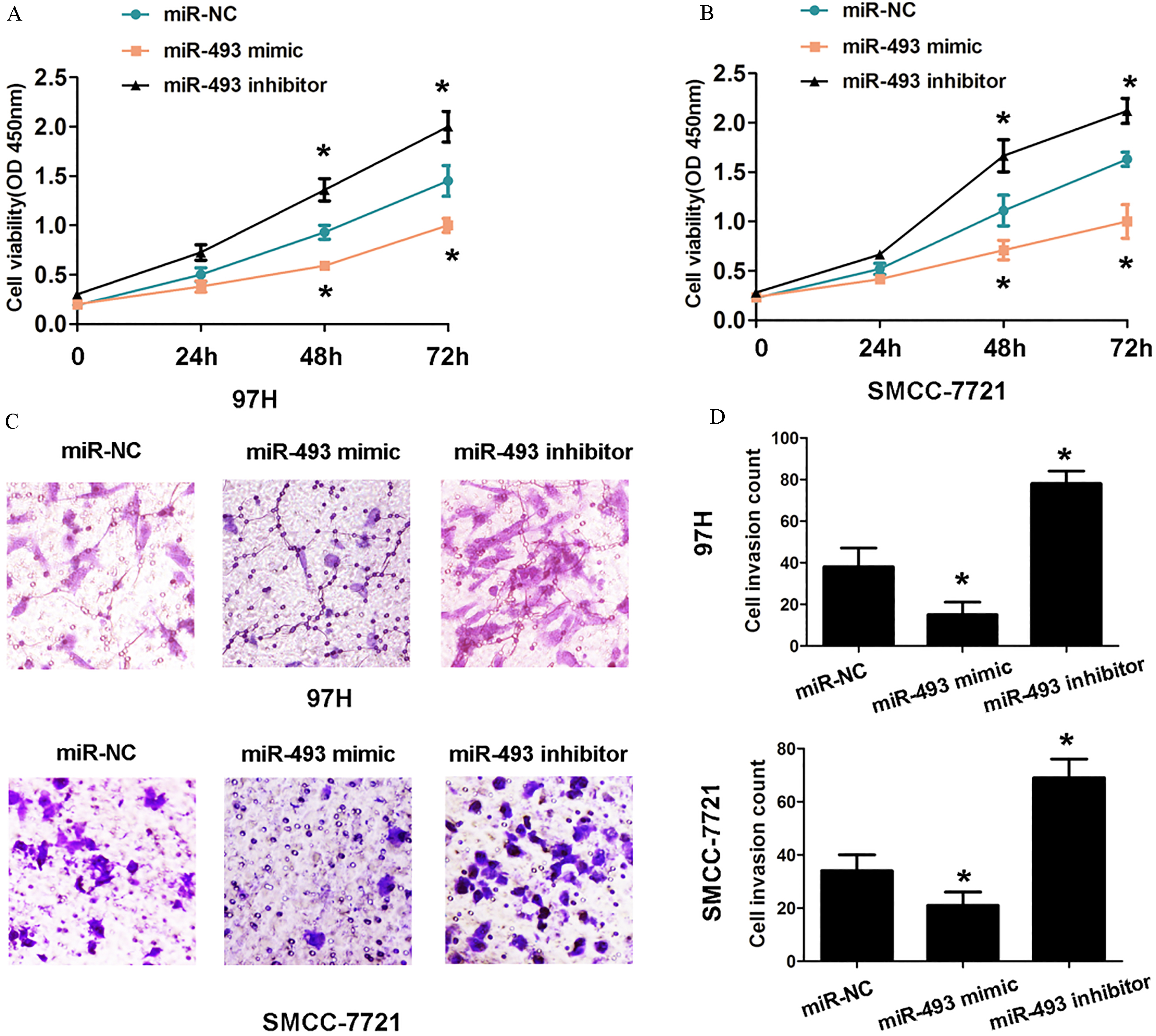
ZFX was a target of miR-493. (A) ZFX was shown was a potential target of miR-493 by searching online software miRanda. The wild type (WT) ZFX 3’ untranslated region (UTR) or mutant type (MUT) was cloned into pMIR-Report vectors. (B) HEK 293T cells were cotransfected with 100 ng WT or MUT pMIR-Report vector and miR-493 mimic or miR-NC. At 48 h post-transfection, luciferase activity was detected by a Dual-Luciferase Reporter Assay system. (C) and (D) The relative expression of ZFX mRNA and protein expression was detected using qRT-PCR and western blot analysis in 97H cells. (E) and (F)The relative expression of ZFX mRNA and protein expression was detected using qRT-PCR and western blot analysis in SMCC-7721 cells. 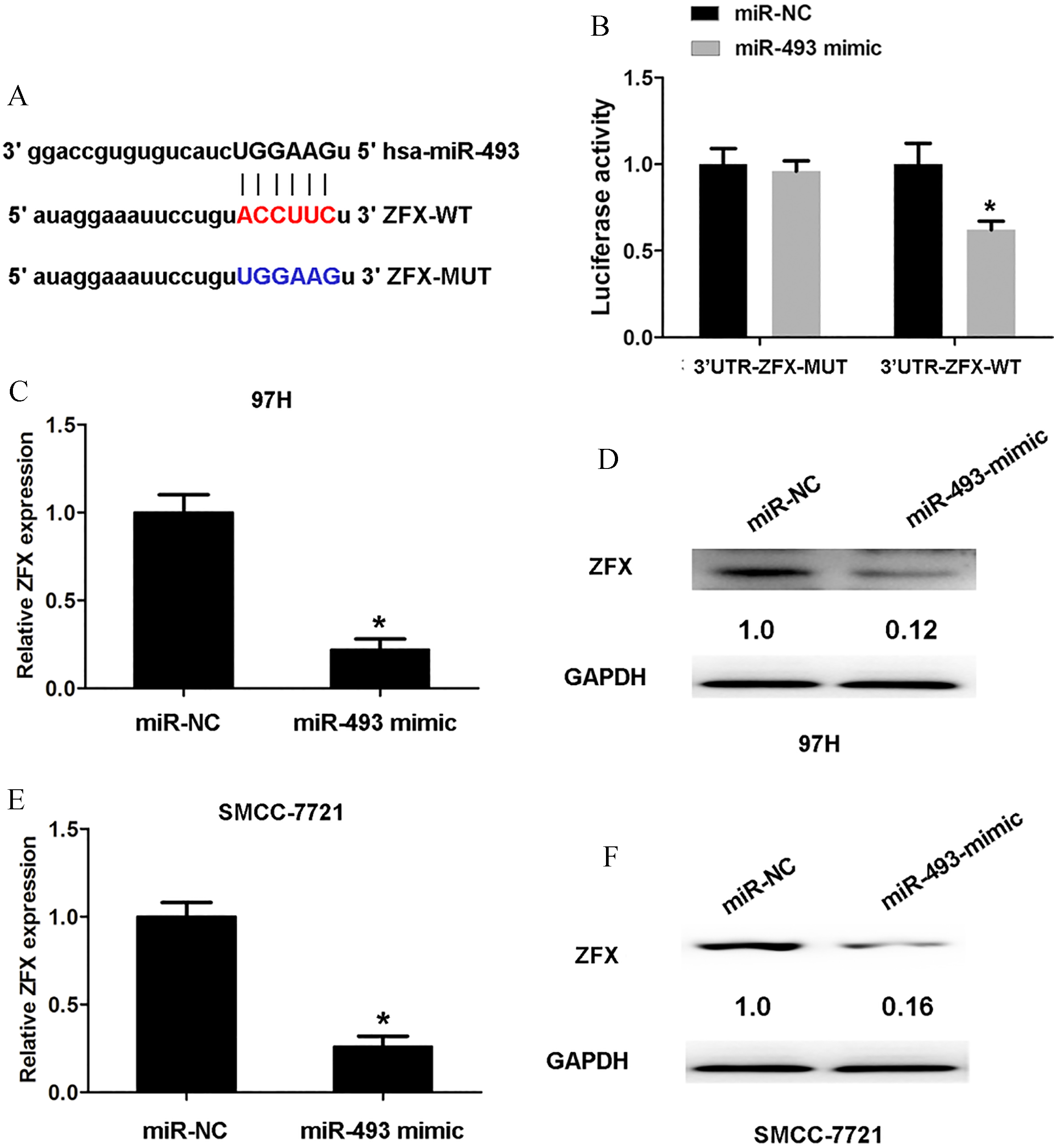
We performed gain-and-loss function assays to detect the effects of miR-493 expression on cell proliferation and invasion in 97H and SMCC-7721 cells (the lowest and highest expression of miR-493 in two cells). Results showed that transfection of miR-493 mimic in 97H and SMCC-7721 cells suppressed cell proliferation, however, transfection of miR-493 inhibitor promoted cell proliferation ability, compared to control groups by CCK8 assay analysis at 48 and 72 h (
MiR-493 negatively regulated ZFX expression in HCC cells. (A) The expression of ZFX was examined in 58 cases of HCC tissues and adjacent normal tissues using qRT-PCR analyses. (B) The expression of ZFX was examined in human HCC (LM3, MHCC97, Huh7 and SMMC-7721 cells) and one normal hepatic epithelial cell line (LO2, control) using qRT-PCR analyses. (C) miR-493 expression was negatively associated with ZFX expression in HCC tissues using Pearson correlation analysis (
An online prediction software Miranda (www. microrna.org) was applied to search for potential target genes of miR-493. We found that ZFX is a potential target of miR-493 (Fig. 3A). The 3’-UTR (wild-type) of ZFX and 3’-UTR mutated-type of ZFX were subcloned into pMIR-reporter luciferase vectors. Results from a luciferase assay demonstrated that the luciferase activity of ZFX 3’-UTR-WT luciferase vector in HEK 293T cells was suppressed by transfection of miR-493 mimic, but was not changed for the mutated ZFX 3’-UTR luciferase vector, compared to miR-NC group (Fig. 3B). The mRNA and protein expression was detected when 97H or SMCC-7721 cells were transfected with miR-493 mimic or miR-NC using qRT-PCR and western blot analysis. Results showed that miR-493 mimic inhibited ZFX expression in 97H and SMCC-7721 cells compared to miR-NC groups, respectively (Fig. 3C and E). Thus, these results indicated that ZFX was a direct downstream target of miR-493.
MiR-493 inhibits cell proliferation and invasion by negatively regulating ZFX
We examined the mRNA expression of ZFX in HCC tissues and adjacent normal tissues using qRT-PCR analyses. Results showed that ZFX expression was significantly higher in HCC tissues than adjacent normal tissues (Fig. 4A). Consistently, ZFX was higher expression in HCC cell lines than in LO2 cells (Fig. 4B). Lower miR-493 expression showed a negative association with higher ZFX expression using Pearson correlation analysis (
Discussion
Recent studies have verified the crucial regulating roles of miRNAs in HCC cell proliferation, invasion and metastasis [13]. MiR-493, a tumor suppressor, has been identifies in some tumors. MiR-493 suppresses the proliferation and invasion of gastric cancer cells by targeting RhoC [14]. MicroRNA-493 suppresses tumor growth, invasion and metastasis of lung cancer by regulating E2F1 [15]. Tumor suppressor microRNA-493 decreases cell motility and migration ability in human bladder cancer cells by downregulating RhoC and FZD4 [16]. In HCC, MicroRNA-493 suppresses hepatocellular carcinoma tumorigenesis through down-regulation of anthrax toxin receptor 1 (ANTXR1) and R-Spondin 2 (RSPO2) [11]. In the study, we identified that miR-493 was downregulated in HCC tissues and cells. Furthermore, we found that miR-493 expression associated with tumor size and vascular invasion in HCC patients. Lower miR-493 expression also showed a poor disease free survival and overall survival time. Thus, our results implied that miR-493 expression was lower in HCC and related to HCC prognosis. Furthermore, we demonstrated that miR-493 functioned as tumor suppressor to reduce cell proliferation and invasion.
Zinc finger X-chromosomal protein (ZFX) functioned as an oncogene to be involved in tumor progression. Such as, Zinc finger X-chromosomal protein predicts a poor prognosis and promotes cellular malignant potential in gallbladder cancer [17]. High expression of Zinc-finger protein X-linked promotes tumor growth and predicts a poor outcome for stage II/III colorectal cancer patients [18]. Overexpression of ZFX confers self-renewal and chemoresistance properties and promotes cell proliferation in hepatocellular carcinoma [19]. In the study, luciferase reporter activity assay demonstrated that ZFX was a target of miR-493. Our results showed that miR-493 expression was negatively related to ZFX expression in HCC tissues and overexpression of miR-493 inhibited ZFX expression. Moreover, we demonstrated that overexpression of ZFX promoted cell proliferation and invasion compared with control groups. However, cotransfection of pcDNA3.1-ZFX and miR-493 mimic rescued the effects. These results indicated that miR-493 inhibited cell proliferation and invasion by regulated ZFX expression. ZFX acts as a transcriptional regulator in multiple types of human tumors according to previous study. The mechanism by which ZFX influences transcriptional regulation has not been determined. In this future, we hope to explore the downstream targets of miR-493/ZFX in HCC.
In conclusion, we demonstrated that miR-493 expression was lower in HCC and lower miR-493 expression predicted poor prognosis of HCC. In vitro, we demonstrated that miR-493 inhibited cell proliferation and invasion by targeting ZFX. Thus, our results implied that miR-493 may be a potentially therapeutic target for HCC.
Footnotes
Acknowledgments
The study was supported by National Science Foundation (Grant No: 81360274) and National 973 Project (Grant No: 2015CB755402).