
Abstract
BACKGROUND:
MicroRNAs (miRNAs) have been demonstrated to play an important role in the development and progression of various types of cancer including glioblastoma (GBM).
OBJECTIVE:
The aim of this study was to investigate the expression pattern and prognostic significance of serum miR-203 in patients with GBM.
METHODS:
miR-203 extracted from cell culture medium and serum samples was detected by real-time PCR. The correlation between serum miR-203 expression as well as clinicopathological characteristics and patient survival was determined.
RESULTS:
The expression level of miR-203 was remarkably reduced in the GBM cells and their culture medium. Serum miR-203 expression was significantly decreased in GBM patients compared with low grade glioma (LGG) patients and healthy controls. In addition, serum miR-203 discriminated GBM patients from LGG patients and healthy subjects. Chi-squared analysis showed that a significant correlation was found between low serum miR-203 expression and larger tumor size as well as lower Karnofsky Performance Scale scores. Patients with lower serum miR-203 suffered poorer overall survival (OS) and progression free survival (PFS). Multivariate analysis indicated that low miR-203 expression is an independent prognostic factor for poor OS in GBM patients.
CONCLUSIONS:
These data demonstrate that serum miR-203 expression might serve as a potential prognostic indicator of GBM.
Introduction
Glioblastoma multiforme (GBM), originating from glial cells, is the most common histological subtype and lethal form of primary brain tumor [1]. Despite great advances in standard therapy (surgery, radiotherapy, and chemotherapy) over the past 30 decades, the median survival time of GBM patients remains poor, ranging from 12–14 months [2]. Further studies are necessary in order to identify novel and clinical useful biomarkers for early detection and prognostic prediction of this malignancy.
MicroRNAs (miRNAs) are endogenous small non-coding RNA molecules of 19–22 nucleotides in length [3]. They regulate gene expression at the post-transcriptional level by binding to the 3’untranslated region (3’UTR) of target mRNAs, resulting in translational inhibition or mRNA degradation. Over 50% of annotated human miRNAs are located at fragile sites which are associated with cancer initiation and development [4]. Numerous studies have shown that these small RNA molecules function as oncogenes or tumor suppressors in cancer. Therefore, examining miRNAs levels in the biological clinical samples might help explore novel molecular biomarkers that improve the therapeutic efficacy of GBM. A five-miRNA signature including miR-222, miR-132, miR-129, miR-145 and miR-20a was associated with clinical outcome and chemoresistance in GBM patients with MGMT promoter methylation [5]. Upregulation of miR-663 suppressed the proliferation and invasion of GBM cells both in vitro and in vivo, and PIK3CD was identified as a downstream target. In addition, GBM patients with lower miR-663 levels suffered an unfavorable clinical outcome than those higher miR-663 expression, indicating miR-663 acted as a tumor suppressor in GBM [6].
Previous studies have shown that miR-203 is important for cell proliferation, differentiation, apoptosis, neuropathic pain, development [7, 8, 9]. More importantly, miR-203 is aberrantly expressed in many types of cancer and it seems to play an important in the carcinogenesis process [10, 11, 12]. miR-203 has been reported to function as a tumor suppressor in GBM [13]. However, whether miR-203 levels were deregulated in the serum samples from GBM patients and its potential clinical significance remained unknown. The aim of current study was to investigate the prognostic value of serum miR-203 in GBM.
Materials and methods
Cell culture
The GBM-derived cell lines T98G, A172 and U251, LGG cell lines CHG5 (derived from oligodendrogliom), SHG44 (derived from astrocytoma) and the normal brain cell line HBMEC were maintained in DMEM with 10% FBS, 100 U/mL penicillin, and 100
Patients
We included 70 patients with GBM, 30 patients with LGG (14 patients with astrocytoma, 10 patients with oligodendroglioma and 6 patients with mixed glioma) and 30 healthy controls in this study. All 10 patients with oligodendroglioma have 1p/19q co-deletion. None of the patients had received chemotherapy, radiotherapy or surgery before blood samples collection. Complete clinical information such as age, gender, Karnofsky Performance Scale score, tumor size, extension of resection, family history of cancer, IDH1 mutation status and follow-up time was available for all the patients. Serum samples from thirty healthy volunteers without any known comorbidities were used as normal controls. All patients and healthy donors signed an informed consent form for participation in the study and for the use of their biological samples. Fast venous blood samples of all patients were collected before receiving any kind of therapy. Samples were immediately separated by centrifugation and then stored at
Total RNA isolation and reverse transcription- quantitative polymerase chain reaction (RT-qPCR)
Total RNAs were extracted from the cells, culture medium and serum samples using mirVana miRNA isolation kit (Life Technologies, Carlsbad, CA, USA) according to the manufacturer’s protocols. RNA concentration and purity were measured using a NanoDrop 2000 spectrophotometer (Thermo Fisher Scientific, Waltham, MA, USA). The integrity of total RNA was checked by running the samples on a denaturing 15% polyacrylamide gel. RNA was reversed transcribed into complementary DNA using the PrimeScript RT Master Mix (Takara Biotechnology Co., Ltd., Dalian, China). Amplification of cDNA was used by SYBR Premix DimerEraser (Takara Biotechnology Co., Ltd.) and qRT-PCR was run on the ABI 7500 Fast Real-Time PCR system (Applied Biosystems, Foster City, CA, USA). The cycling conditions were as follows: 95
The expression level of miR-203 in glioma cells and their culture medium.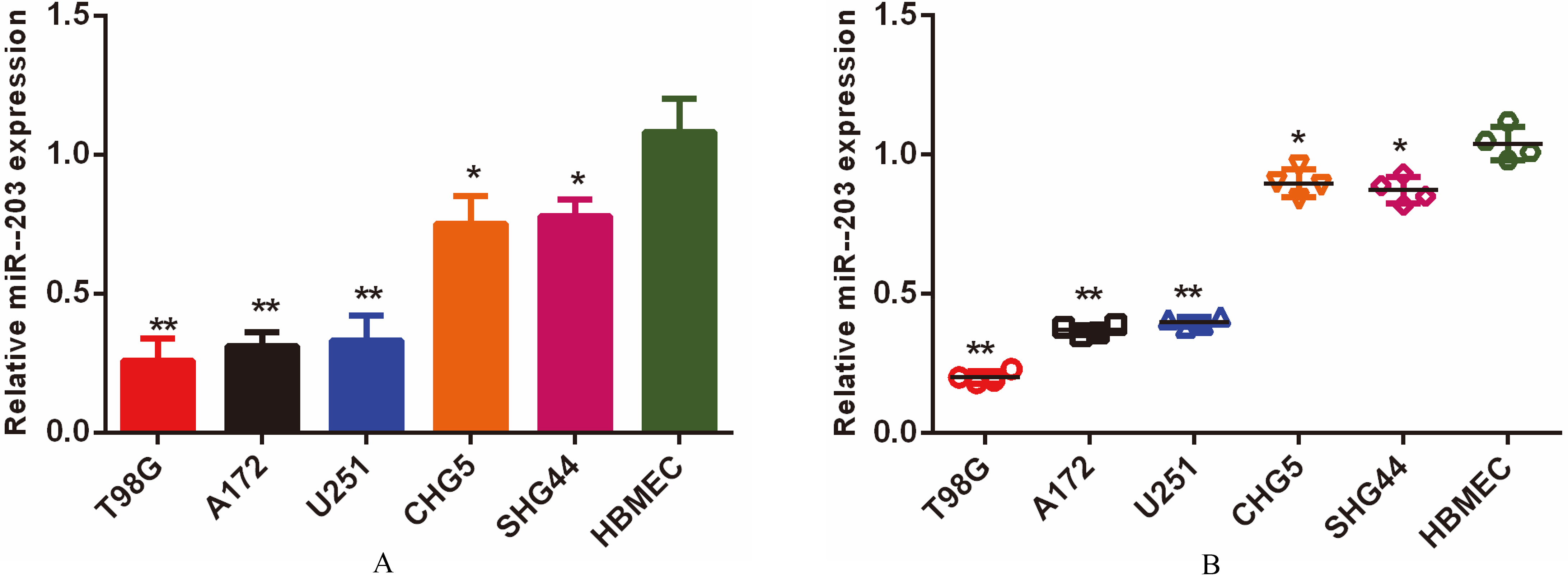
Statistical analyses were performed by MedCalc 5.0 (MedCalc Software, Mariakerke, Belgium) and GraphPad Prism 6.0 (GraphPad Software Inc., La Jolla, CA, USA) Kruska-Wallis test was performed to compare the expression of miR-203 levels in the the cell lines, serum samples and culture medium. The association between clinicopathological parameters and serum miR-203 levels was assessed using the Chi-square test. The diagnostic performance of serum miR-203 for GBM was evaluated by the receiver operating characteristic (ROC) curve analysis. Accuracy is measured by the area under the curve (AUC). Kaplan-Meier method and log-rank test were used to evaluate the correlation of overall/progression free survival and the expression of serum miR-203. A Cox regression model was used for multivariate analysis of prognostic variables. A
Results
miR-203 was downregulated in the culture medium of GBM cells
We compared the expression levels of miR-203 among glioblastoma cell lines, LGG cell lines and normal brain cell line as well as the culture medium from these cell lines. Our real-time PCR analysis showed that miR-203 levels were most downregulated in glioblastoma cell lines. Its level was progressively increased from LGG cell lines to the normal brain cell line (
Serum miR-203 differentiated GBM patients from LGG patients and healthy control subjects
The expression levels of serum miR-203 were compared among GBM patients, LGG patients and healthy control subjects. Serum miR-203 levels were most underexpressed in patients with GBM (
Serum miR-203 levels were reduced in the patients with GBM.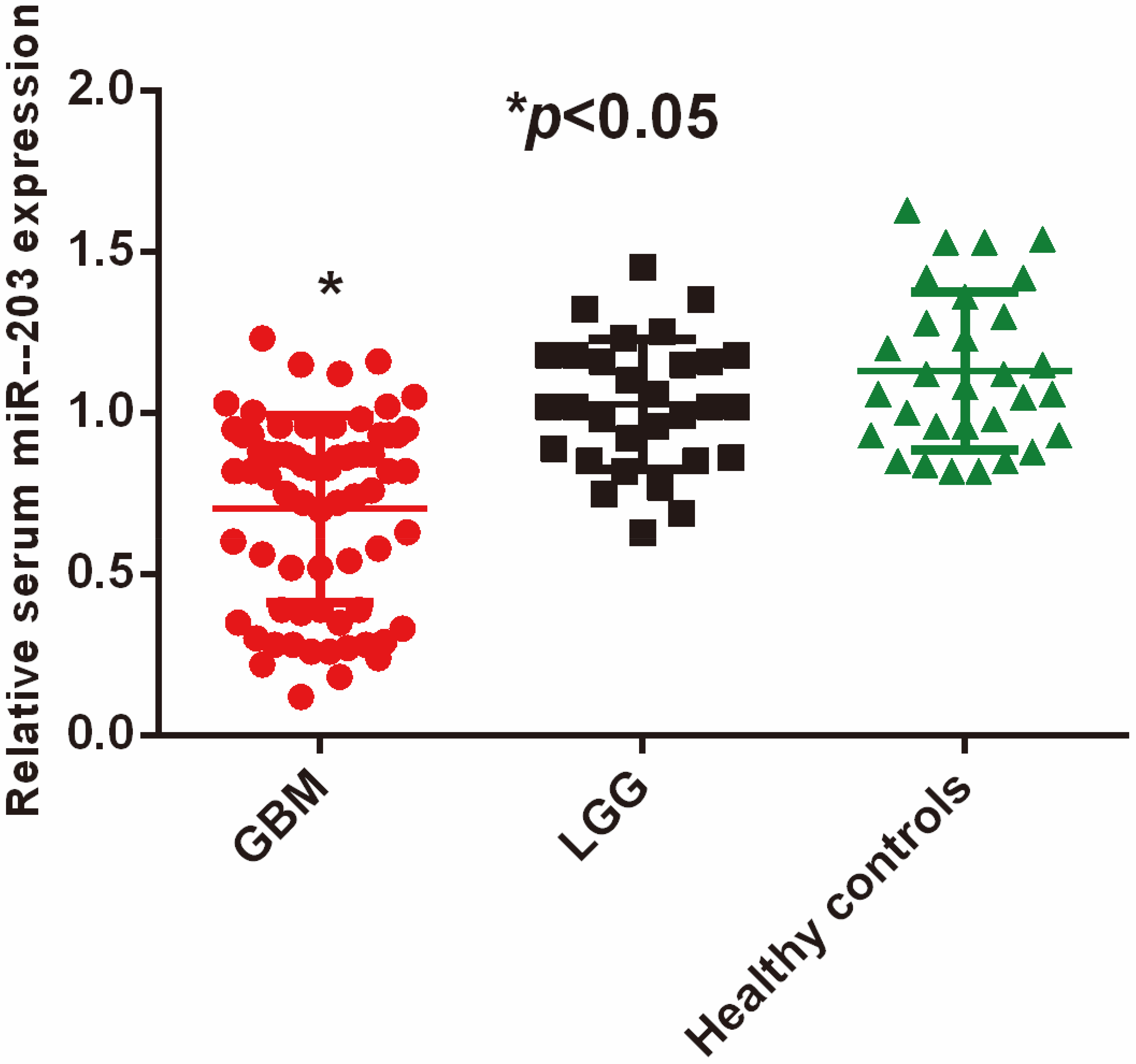
The diagnostic performance of serum miR-203 for GBM.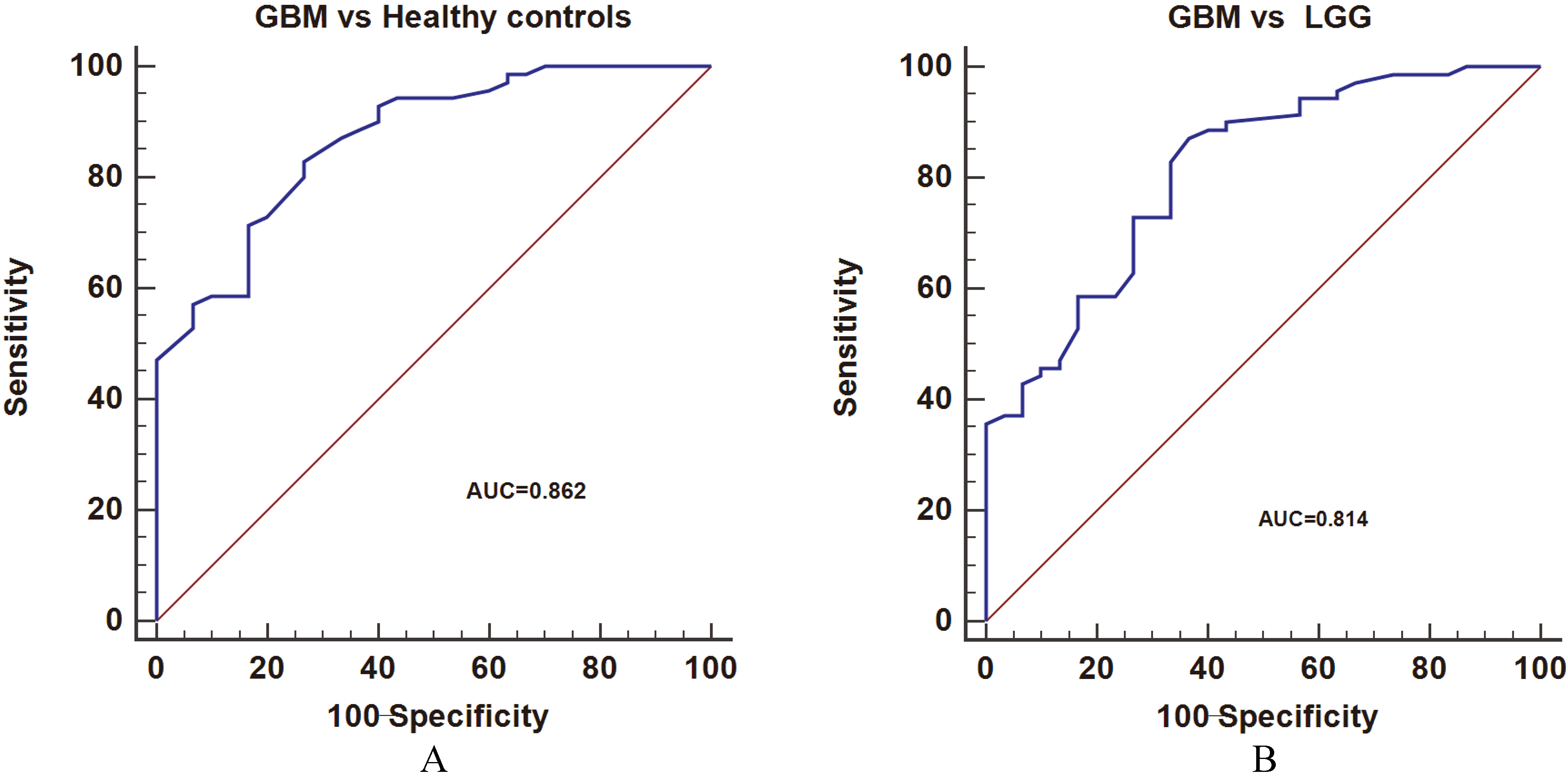
ROC analysis was performed to determine the diagnostic value of serum miR-203 for GBM. The results showed that serum miR-203 discriminated GBM patients from healthy controls (AUC
The ROC curve was used to find out the optimal cut off value to divide the GBM patients into two groups. Twenty-six GBM patients were in the low serum miR-203 group while 44 in the high serum miR-203 group. A positive correlation was found between low serum miR-203 levels and larger tumor diameter (
Association between serum miR-203 expression and clinicopathological variables of patients with glioblastoma
Association between serum miR-203 expression and clinicopathological variables of patients with glioblastoma
Kaplan-Meier curves and log rank tests were used to evaluate the prognostic significance of serum miR-203 in GBM. Survival analysis demonstrated that GBM patients in the low serum miR-203 group had poor OS rates compared to patients in the high serum miR-203 group (
The association between serum miR-203 levels and the survival of patients with GBM.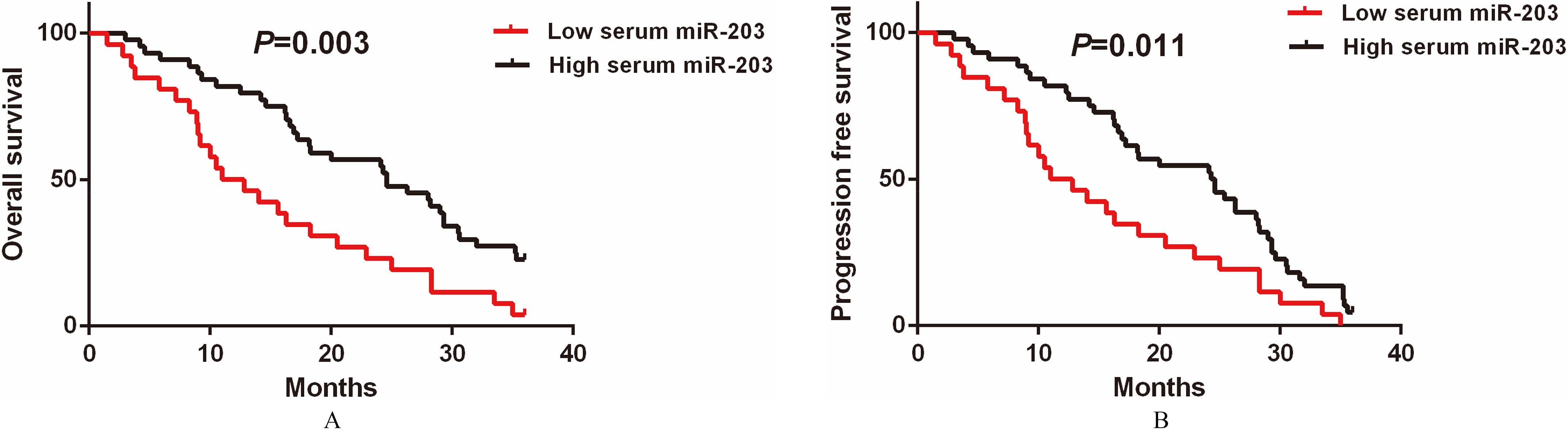
Multivariate analysis revealed that low preoperative KPS scores (HR
Multivariate Cox regression analyses of OS in patients with GBM
Our study showed that decreased miR-203 expression levels were not only found in glioblastoma cells and their culture medium, but also in the serum samples from the patients with GBM. One possible reason was that miR-203 function as a tumor suppressor and it might be downregulated in the glioblastoma cells. Thus glioblastoma cells were unable to generate enough amount of miR-203 into extracellular environment, leading to its reduction in the culture medium and serum samples. Then the ROC analysis revealed that serum miR-203 was able to differentiate GBM patients from LGG patients as well as healthy controls, indicating serum miR-203 has potential diagnostic value. Liquid biopsy has been becoming an attractive approach which provides important clinical information for diagnosis, prognosis, and prediction of response or resistance [14]. The levels of miRNAs in serum are stable, reproducible, and consistent, thus these properties make them become excellent biomarkers for many human diseases [15, 16]. As serum miR-203 levels might be also deregulated in other types of cancer or other diseases, we have to combine other clinical indicators for the diagnosis of GBM. One potential pitfall of current study is that the therapy is an important factor that have effects on the prognosis of GBM. The GBM patients in this study received similar therapeutic approach. However, it is very difficult to ensure that all the patients received the same therapeutic approach due to the complex of clinical situation and individual differences. In the future, we should try to follow up those patients who refused to receive treatment or those receive the same treatment to minimize the effects of therapy on the clinical outcome of GBM.
Serum miR-203 levels were correlated with unfavorable clinical parameters and survival rates. Also, deceased serum miR-203 was demonstrated to be an independent risk factor for GBM. These data suggest that reduced serum miR-203 is strongly associated with poorer prognosis of GBM. To the best of our knowledge, this was the first study to evaluate the potential clinical significance of serum miR-203 in GBM. Our results further corroborated that miR-203 played a tumor suppressive role in GBM, and its downregulation promote the tumorigenesis. Similarly, ectopic expression of miR-203 inhibited the epithelial-mesenchymal transition (EMT) process of glioblastoma cells and sensitized them to chemotherapy. EMT and chemoresistance were promoted when miR-203 was suppressed [17], suggesting that miR-203 is closely associated with the progression and therapeutic responses of GBM. Upregulation of miR-203 could significantly increase the radiation sensitivity of GBM cell lines by regulating various signaling pathways associated with DNA damage repair, prosurvival signaling, and EMT process [17]. A recent study found that overexpression of miR-203 not only could effectively suppress the features of stemness in glioblastoma stem cells, but also inhibited their proliferation and increased apoptosis [18]. It is therefore miR-203 might be a promising therapeutic target for the treatment of GBM. MGMT promoter methylation status is an important prognostic factor for GBM. One possible limitation is that we do not examine the MGMT promoter methylation status of the enrolled GBM patients. We will explore the association between MGMT promoter methylation status and serum miR-203 expression levels in the future study.
miR-203 was also deregulated in other types of cancers. For instance, miR-203 levels were found to be underexpressed in polyps and colorectal cancer (CRC). miR-203 might regulate the carcinogenesis process of CRC by modulating DNMT3b-ABCG2 pathway [19]. The expression of miR-203 was significantly downregulated in melanoma tissues compared to the controls. In addition, reduced miR-203 was associated with worse clinicopathological variables and shorter overall survival time. Furthermore, miR-203 was an independent prognostic factor for melanoma [20]. Similarly, reduced miR-203 was also correlated with worse clinical outcome in patients with cholangiocarcinoma [21]
Our data demonstrate that serum miR-203 levels are significantly downregulated in GBM patients and reduced serum miR-203 is associated with poor prognosis of GBM. Taken together, these findings indicate that serum miR-203 might serve as a promising diagnostic and prognostic biomarker for GBM.
Conflict of interest
We deny any conflict of interest.
Footnotes
Acknowledgments
This study was supported by the Medical and Health projects of Yichang City (No: A17-301-15).