
Abstract
BACKGROUND:
AGR2 expression is associated with luminal breast cancer. Overexpression of AGR2 is a predictor of poor prognosis. Several studies have found correlations between AGR2 in disseminated tumor cells (DTCs) in breast cancer patients.
OBJECTIVE:
This study aims to determine the correlation between anterior Gradient2 (AGR2) expression with the incidence of distant metastases in luminal breast cancer.
METHODS:
This study was an observational study using a cross-sectional method and was conducted at Wahidin Sudirohusodo Hospital and the network. ELISA methods examine AGR2 expression from blood serum of breast cancer patients. To compare the AGR2 expression in metastatic patients and the non-metastatic patient was tested with Mann Whitney test. The correlation of AGR2 expression and metastasis was tested with the Rank Spearman test.
RESULTS:
The mean value of AGR2 antibody expression on ELISA in this study was 2.90 ± 1.82 ng/dl, and its cut-off point was 2.1 ng/dl. Based on this cut-off point value, 14 subjects (66.7%) had overexpression of AGR2 serum ELISA, and 7 subjects (33.3%) had not. The mean value AGR2 was significantly higher in metastatic than not metastatic, 3.77 versus 1.76 (p < 0.01). The Spearman rank test obtained a p-value for the 2 tail test of 0.003 (p < 0.05), which showed a significant correlation of both, while the correlation coefficient of 0.612 showed a strong positive correlation of AGR2 overexpression and metastasis.
CONCLUSIONS:
AGR2 expression is correlated with metastasis in Luminal breast cancer.
Introduction
Breast cancer is the most common malignancy and a leading cause of cancer death in women worldwide, including Indonesia [1]. Approximately 70% of breast cancer are luminal breast cancer. Luminal is the main indicator of potential response to endocrine therapy [2].
Transcriptomic screens in breast cancer have identified a protein named anterior gradient-2 (AGR) as a potential novel oncogene overexpressed in Luminal Breast Cancer [3]. AGR2 is a protein in mammals belonging to the family of Protein Disulfide Isomerase (PDI). AGR2 can be found in the Endoplasmic Reticulum (RE) and the extracellular membrane [4]. In the RE, AGR2 functions to reduce, oxidize and isomerize disulfide bonds of proteins [5]. AGR2 plays a role in reducing RE stress, promoting cancer cell metastases, and stimulating cell survival and proliferation [4]. AGR2 gene is coexpressed with ER and is a direct target of ER [5]. AGR2 mediates the pro-survival pathway of the Luminal Breast Cancer and is involved in the Estrogen Receptor pro-oncogenic signaling pathway [5].
Increased primary tumor AGR2 expression was associated with decreased breast cancer survival [6]. Therefore, the presence of AGR2 in the primary tumor is a possible prognostic indicator of poor patient outcomes in breast cancer. Barraclough DL, et al. found an association of AGR2 expression with poor survival in patients with stage I and II breast cancer [7]. AGR2 is not only found in primary tumors, AGR2 as a secreted protein is also found in the blood, so it is useful for detecting circulating tumor cells in a patient’s peripheral blood [8].
Expression of Cathepsin B and D [9], MMP3, and MMP9 are induced by AGR2 [10]. Cathepsin B/D, MMP3, and MMP9 are proteases that degraded the extracellular matrix. AGR2 disrupts apicobasal polarity by relocating E-cadherin,beta-catenin, and laminin v from the basal part into the cell cytoplasm. AGR2 controls Epithelial-Mesenchymal-Transition (EMT) by inducing expression of Zeb1, Vimentin, and E-cadherin [10].
The objective of this study was to determine whether there was a correlation between the expression of AGR2 and the incidence of distant metastases in luminal breast cancer.
Methods
Setting and subjects
It was an observational cross-sectional study. The samples were obtained from Wahidin Sudirohusodo Hospital Makassar, a top referral hospital in the east of Indonesia, from October 2019 to January 2020. Female breast cancer patients with invasive carcinoma mammae, luminal subtype were included in the study. The AGR2 expression was detected using ELISA from blood serum. Data were collected, managed, analyzed, and presented in table and narration form, and then we compared them with the result of other studies.
ELISA
Store samples serum to be assayed within 24 hours at 2–8 °C. Samples should be collected into a serum separator tube. Coagulate the serum by leaving the tube undisturbed in a vertical position overnight at 4 °C or room temperature for up to 60 minutes—centrifuge at approximately 1000 × g for 15 min. Analyze the serum immediately.
Serum AGR2 concentrations were measured using the AGR2 ELISA kit (Abexxa Ltd, Cambridge, UK), according to the manufacturer’s instructions. This kit is based on sandwich enzyme-linked immunosorbent assay technology. An antibody specific to AGR2 is pre-coated onto a 96-well plate. The standards and samples are added to the wells, incubated, and washed with wash buffer. A biotin-conjugated antibody specific to AGR2 is used for detection. TMB substrate is used to visualize HRP activity. HRP catalyzes TMB to produce a blue color product that changes into yellow after adding the stop solution. The intensity of the color yellow is proportional to the AGR2 amount bound on the plate. The O.D. absorbance is measured spectrophotometrically at 450 nm in a microplate reader, and then the concentration of AGR2 can be calculated.
Ethical clearance
The Ethical Commission has approved this study of Health Study, Medical Faculty, Hasanuddin University, with the registry number 1008/UN4.6.5.31/PP36/2019 (Register: UH19090697).
Result
Characteristics
The twenty-one enrolled female patients with luminal breast cancer diagnosed and treated at Wahidin Sudirohusodo General Hospital met the study’s inclusion criteria. Their ages ranged from 39 to 69 years, with a mean age of 51.1 ± 8.7 years. All twenty-one cases were invasive breast carcinoma. The obtained histopathologic grading was a low grade in 3 cases (14.3%), moderate grade in 13 cases (61.9%), and high grade in 5 cases (23.8%).
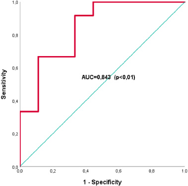
The results of the AGR2 examination in this study showed values between 0.10–7.02 ng/dl with a mean of 2.90 ± 1.82. To get the AGR2 cut-off value in this study, we performed a ROC analysis for AGR2 expression. After a ROC analysis of the AGR2 expression, we found that the ROC curve had a significant Area Under Curve (AUC) value of 0.843. This shows that the AGR2 cut-off value can be used to predict the presence of distant metastases. The ROC curve coordinate table is used to determine the AGR2 cut-off. From the ROC curve coordinate table, the AGR2 value of 2.1 ng/dl has a high sensitivity value of 91.7% and sufficient specificity, amounting to 66.7%. Based on these results, the cut-off value = 2.10 was used in this study, where the AGR2 value ≥2.10 ng/dl was considered overexpression, while the AGR2 value <2.10 ng/dl was considered not overexpression. This study found 14 subjects (66.7%) had AGR2 overexpression, and 7 subjects did not overexpression (33.3%). In this study, there were 12 subjects (57.1%) with metastases disease. The characteristics of the samples are presented in Table 1, and the ROC analysis and ROC coordinate curve are shown in Fig. 1 and Table 2.
Characteristics
Characteristics
Source: Primary data.
Comparison of the AGR2 expression in metastatic patients and non-metastatic patients are shown in Table 3. The mean value of AGR2 expression in metastatic breast cancer patients was 3.77 ± 1.72, whereas that of non-metastatic patients was 1.76 ± 1.28. The mean value of AGR2 was significantly higher in metastatic breast cancer patients than those without metastatic (p-value = 0.009; p < 0.05).
AGR2 ROC analysis*.
ROC coordinate curve**
Source: Primary data, *ROC coordinate curve, **ROC analysis.
Comparison of the AGR2 expression in metastatic patients and non-metastatic patient
Source: Primary data, *Mann–Whitney U test.
The correlation between AGR2 expression and distant metastases of breast cancer is demonstrated in Table 4. There was a significant correlation between AGR2 expression with distant metastases of breast cancer (p = 0.994; p > 0.05). The correlation coefficient of 0.612 shows a strong correlation between AGR2 expression and distant metastasis. It shows that the higher the value of AGR2 expression, the more likely to occur metastatic.
Correlation between AGR2 expression and distant metastases of breast cancer
**Correlation is significant at the 0.01 level (2-tailed). Source: Primary data, *Spearman rho.
In this study, ages varying between 39–69 years, with a mean of 51,1 ± 8,7 years. The highest age distribution is in the age group <50 years, this is in line with this study, based on the American Cancer Society in Breast Cancer Fact and Figures [11]. The western lifestyle shift in our society is one reason for the increase in breast cancer population at a younger age.
We found that the incidence of breast cancer with metastasis was higher than that of early-stage breast cancer (57.1% compared to 42.9%). In line with this study, based on data from the Ministry of the Health Republic of Indonesia, 60–70% of breast cancer patients present in advanced stages at the time of diagnosis [12]. It is because much alternative medicine in our society is misleading many of our cancer patients.
The AGR2 examination of 21 luminal breast cancer patients in this study showed the AGR2 value was between 0.10–7.02 ng/dl, with a mean of 2.90 ± 1.82. In this study, the Mann–Whitney test was performed and found the mean of AGR2 expression was significantly higher in metastatic breast cancer patients than those without metastatic, which was 3.77 compared to 1.76 (p-value = 0.009 (p < 0.01)). In this study, the cut-off point AGR2 serum ELISA was 2.10 ng/dl. According to this cut-off point, the AGR2 value of ≥2.10 ng/dl was considered overexpression, while the AGR2 value of <2.10 ng/dl was considered not overexpression.
A correlation test with the Spearman rank was obtained to find out the correlation of AGR2 expression with an incidence of distant metastasis in luminal breast cancer. We found a significant correlation between AGR2 expression and distant metastases (p = 0.003; p < 0.05). This test shows a strong positive correlation between AGR2 expression and metastasis (correlation coefficient + 0.612). A significantly strong positive correlation between AGR2 expression and distant metastases in this study is in line with the results obtained from previous studies. This is in line with the role of AGR2, both intracellular, which prices proteases such as Cathepsin B/D, and extracellular AGR2, which induces EMT and degradation of extracellular matrix through its induction of MMP3, MMP9, N-cadherin, and Vimentin, and inactivation of E-cadherin.
A study conducted by Hrstka R et al. found that overexpression of AGR2 has a significant relationship with recurrence and metastasis in breast cancer [3]. Likewise, studies conducted by Baraclough DL et al.; identified the correlation of AGR2 with poor survival in patients with early-stage breast cancer [7]. In the same study, Baraclough DL et al. obtained AGR2 as a significant prognostic independent indicator [7]. In another study, HE Innes et al. reported the significance of the AGR2 protein in causing metastases in breast cancer patients treated with hormonal therapy [13]. Lacambra et al., in their study, is examining AGR2 expression in KGB metastases. Another study of Lacambra found higher AGR2 expression in metastatic breast cancer than in primary tumors [14].
Hrstka R et al. found the role of AGR2 in regulating cellular interactions at the location of metastases and promoting the colonization of cancer cells at distant sites [3]. These studies confirm the role of AGR2 as a gene that plays an important role in the occurrence of metastasis. However, this is different from the study conducted by Fritzsche et al. Fritzsche et al. found an association between overexpression of AGR2 and a better prognosis in stage I breast cancer [15]. Several other studies regarding the prognostic value of AGR2 expression in other cancer cases, such as studies conducted by Fritzsche et al. regarding AGR2 expression in non-small cell lung cancer [16] and a study conducted by Reiner et al., regarding the significance value of AGR2 in ductal pancreatic adenocarcinoma [17], also failed to get a significant relationship between AGR2 expression and prognosis.
Although AGR2 is a normal resident of the endoplasmic reticulum, the AGR2 protein can be found in the extracellular space (eAGR2). AGR2 in the extracellular space is due to AGR2 being a form of secreted protein (not an accumulation due to cell lysis). Clarke et al., in their study of getting AGR2 as a secreted protein, both in human and rat mammary epithelial cells [18]. Chen et al., in their study, reported the existence of eAGR2 as a secreted protein [19].
eAGR2 is a microenvironmental regulator. eAGR2, as a soluble protein, is secreted into the interstitial space and interacts with the extracellular matrix. As an active functional protein, eAGR2 can make tumor cells invasive and metastatic [10]. eAGR2 will interact synergistically by inducing and strengthening the IGF-1 signaling pathway (which is also secreted in the interstitial space) for cell proliferation, migration, and EMT by the role of ERα in the cell membrane. In this microenvironment, eAGR2 will also interact with MUC-1, acting as a receptor of eAGR2 [20]. Fessart, et al. in their study, found that eAGR2 damages the adhesion of epithelial cells and controls the Epithelial-Mesenchymal Transition (EMT), promotes invasion, and will decrease its expression if intracellular AGR2 is inhibited [10]. Dumartin L et al., in their study, found that AGR2 expression induced an increase in the levels of Cathepsin B (CTSB) and Cathepsin D (CTSD), known to play a role in the dissemination of cancer cells [9]. In research conducted by Fletcher GC et al., the interaction between AGR2, C4.4a, Dystroglican 1 (DAG1), and laminin extracellular provides an opportunity for cancer cells to migrate and metastasize [21].
Conclusion
We found a significant correlation between AGR2 expression and distant metastases. A strong positive correlation between AGR2 expression and metastasis shows that the higher the value of AGR2 expression, the more likely it is metastatic. These results suggest that AGR2 expression plays a role in metastatic. AGR2 expression has a substantial correlation with metastatic. This is the first study about AGR2 on breast cancer that examined AGR2 expression cut off on blood serum with ELISA Assay. Our study may suggest our cut-off point 2.10 ng/dl of AGR2 serum ELISA as a cut-off point to determine the expression of AGR2 on blood serum.
Footnotes
Conflict of interest
The authors declare that they have no conflict of interest.