
Abstract
The presence of parkinsonism features in primary progressive aphasia (PPA) is a subject of ongoing research. These features are usually more pronounced in the advanced stages of the disease, particularly in the non-fluent/agrammatic subtype, and are exceptionally rare in the logopenic variant (lvPPA). Here we report a case of a 63-year-old man presenting as language impairment, predominantly naming and word-finding difficulties, emerged alongside a left-sided internal tremor. Neurological examination revealed bilateral, left-side predominant rigidity, bradykinesia, and resting tremor. Notably, anosmia and constipation were present. Language assessments showed preserved single-word comprehension, object knowledge, and a minimal apraxia of speech, as well as sentence repetition issues. Neuroimaging and biomarker analysis supported a diagnosis of primary progressive logopenic aphasia with amyloid pathology co-existing with prominent and early parkinsonism. This case underlines the intricate relationship between language disorders, parkinsonism, and amyloid pathology in lvPPA.
Keywords
INTRODUCTION
Primary progressive aphasias (PPA) are neurodegenerative diseases typically characterized by an early and selective language impairment. According to the diagnostic criteria by Gorno-Tempini in 2011, three main variants have been described, based on clinical features, along with neuroimaging, neuropathological, and genetic data: non-fluent/agrammatic aphasia (nfvPPA), semantic aphasia (svPPA), and logopenic aphasia (lvPPA) [1]. lvPPA typically presents with word finding difficulty, phonemic paraphasia, sentence repetition deficits and, as the disease progresses, impaired sentence comprehension, in addition to lack of frank agrammatism and motor speech. lvPPA is considered one of the potential focal and early onset presentation of Alzheimer’s disease (AD), although other pathological profiles have also been reported, including Lewy body dementia, TDP-43, and tau [1–3]. A meta-analysis suggests that the presence of amyloid-β (Aβ) is more common in individuals diagnosed with lvPPA among PPA variants [4]. Furthermore, postmortem lvPPA cases reveal AD pathology as the predominant finding [4]. However, there is limited data regarding the association between lvPPA and parkinsonism. A recent case report describing a patient with lvPPA and coexisting anosmia and mild parkinsonism suggested possible connections between these symptoms [5]. Another report documented an individual initially diagnosed with idiopathic Parkinson’s disease (PD) who later developed symptoms of logopenic progressive aphasia that eventually progressed to generalized dementia, with pathological biomarkers indicating AD [6]. Growing evidence suggests that α-synuclein plays a significant role in the pathophysiology of AD. Elevated levels of α-synuclein have been found in AD, potentially contributing to various pathological processes, including the facilitation of Aβ oligomerization, tau phosphorylation, activation of kinases, disruption of tau-tubulin interactions, and the formation of tau aggregates [7]. In this manuscript, we report a potential relationship between lvPPA, parkinsonism, and amyloid pathology, highlighting the complexity of neurodegenerative disorders and the importance of exploring various pathological profiles in the context of lvPPA.
CASE PRESENTATION
A 63-year-old right-handed lawyer with a medical history of arterial hypertension, post-surgical hypothyroidism, and mild bilateral sensorineural hearing loss presented at our hospital with a two-years history of slowly progressive difficulties in word-finding occurring during legal disputes, accompanied by a sense of “mental fog”. His wife, as his assistant, noted similar issues emerging while transcribing legal documents dictated by him. These language-related challenges had a slight impact on occupational activities, consistent with a global Clinical Dementia Rating (CDR) score of 0.5. The patient reported a three-year history of anosmia and constipation. In the last two months, he also complained of a left-sided internal tremor. No other clinical features, such as REM sleep behavior disorder, orthostatic hypotension, bladder dysfunction, or hallucinations, were reported. There was no family history of neurodegenerative disorders. The neurological examination revealed a fixed gaze, reduced blinking, bilateral left-sided predominant rigidity, bradykinesia, and resting tremor (Movement Disorders Society-Unified Parkinson Disease Rating Scale (MDS-UPDRS-III: 14). The patient scored 29 out of 30 on Mini–Mental State Examination (MMSE). Clinical data of the patient are detailed in Table 1. An extensive neuropsychological evaluation did not show deficits in memory, executive functions, attention, visuospatial abilities, and social cognition, as presented in Table 2. Mild difficulties were observed during the immediate recall, which were most likely due to anxiety (as shown in the subtest of the Hospital Anxiety and Depression Scale in Table 2); however, it did not affect the overall performance in the related domain. The language examination was conducted collaboratively by a speech-language pathologist and a neurologist during personal narrative and medical interviews initially, revealing normal prosody and speech rate, along with pauses for retrieving words and tip-of-the-tongue phenomena. Screening for Aphasia in NeuroDegeneration (SAND) [8] revealed impaired sentence repetition primarily characterized by word omission or substitution, especially the final words in longer sentences, yet providing semantically appropriate renditions of repeated sentences. Additionally, phonological errors were noted. Single-word comprehension and object knowledge were preserved and there were no signs of dysarthria. During picture description, the patient exhibited occasional pauses, some lasting more than 6 seconds, due to difficulties in word retrieval, circumlocutions, and repaired sequences, while maintaining a normal number of correct information units, cohesion and coherence. Furthermore, there was a limited use of subordinate clauses with a normal verb inflection and verb argument structure. A month after the first examination, to assess agrammatism, a battery for the analysis of language deficits in aphasia [9] was conducted, including auditory and visual grammaticality judgments, and controlled written picture descriptions. Results showed well-formed complex structures sentences in absence of morphological, syntactic, and grammatical errors. Apraxia of Speech Rating Scale revealed slight apraxia of speech, primarily characterized by speech initiation difficulty and false starts/restarts, with lesser prominence of phonetic impairment characterized by distorted sound substitutions and additions, particularly with increased utterance length and speech rate [10, 11]. The calculation test from the aphasia assessment developed by Capasso et al. [12] revealed errors in complex calculations, specifically those involving multiple digits. Language assessment is summarized in Table 3. MRI showed small white matter T2/FLAIR hyperintense lesions due to chronic small vessel disease (Fazekas Scale 1) (Fig. 1a,b). 123I-FP-CIT dopamine transporter SPECT revealed reduced presynaptic dopamine transporter levels in the putamen and caudate, with left-side predominance and right-side predominance, respectively, as reported in Fig. 1c. Cardiac I-123 MIBG scintigraphy indicated an early and late H/M ratio of 1.8, compatible with absence of sympathetic denervation. Flutemetamol Amyloid PET showed predominant Aβ amyloid accumulation in the left temporal region (Fig. 1f). 18F-FDG PET imaging demonstrated hypometabolism in both parietal lobes, most prominently on the left, and in the left lateral temporal area (Fig. 1d, e). Comprehensive laboratory tests and cerebrospinal fluid (CSF) analysis were unremarkable, except for CSF Aβ42 levels of 235 pg/ml (normal value > 500 pg/ml) and an Aβ42/Aβ40 ratio of 0.06 (cut-off>0.5), with normal Aβ40, tau, phospho-tau181 39.90 pg/ml (normal value < 56.50 pg/ml), total-tau/Aβ42, and phosphor-tau181/Aβ42 ratios. Genetic testing for frontotemporal dementia (FTD) was negative for C9orf72 repeat expansions, GRN, and MAPT mutations. An oral administration of levodopa (100 mg three times a day), significantly improved parkinsonism signs (MDS-UPDRS-III: 9), with no clear impact on speech-related difficulties.
Clinical data of the patient
MMSE, Mini–Mental State Examination; CDR, Clinical Dementia Rating; MDS-UPDRS, MDS Unified Parkinson Disease Rating Scale; H& Y, Hoehn & Yahr.
Neuropsychological evaluation (language assessment detailed separately)
/, not applicable.
Language assessment and key observations
SAND, Screening for aphasia in NeuroDegeneration; B.A.D.A., Batteria per l’analisi dei deficit afasici.
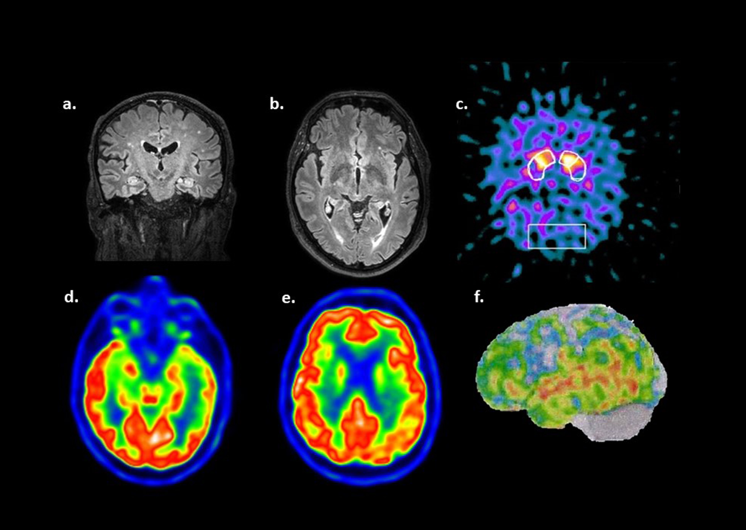
Cerebral MRI images (a, b). Fluid-attenuated inversion recovery (FLAIR) shows hyperintense lesions due to chronic small vessel disease, Fazekas Scale 1. Hippocampus and basal ganglia do not show abnormalities. 123I-FP-CIT dopamine transporter SPECT (c) revealed an inhomogeneous presynaptic dopamine transporter reduction in the putamen and caudates, with left-side and right-side predominance respectively. 18F-FDG PET imaging (d, e) demonstrated hypometabolism in both parietal lobes, most prominently on the left, and in the left lateral temporal area. [18F] Flutemetamol Amyloid PET showed predominant Aβ amyloid accumulation in the left temporal region (f).
DISCUSSION
This case illustrates an uncommon but valuable interaction between different clinical manifestations, requiring a comprehensive evaluation to uncover the underlying pathology, which can provide useful prognostic information and therapeutic implications. In our case, the most prominent and progressively worsening deficit observed two years after the disease onset primarily consisted in language difficulties. These language impairments moderately impacted the patient’s daily life functioning, especially during work-related activities, and were not accompanied by other cognitive deficits, such as issues with episodic memory, visual memory, visuoperceptual abilities, or behavioral disturbances. Thorough investigations ruled out other potential causes of aphasia, including non-degenerative neurological conditions or medical disorders, aligning with the clinical presentation of PPA [1].
In detail, the patient exhibited difficulty retrieving single words during spontaneous speech and naming, along with challenges in repeating sentences and phrases due to phonemic errors, word omission or substitution, particularly at the end of longer sentences. Phonological errors and slight apraxia of speech (AOS) were also noticeable. Importantly, single-word comprehension and object knowledge remained intact, in the absence of frank agrammatism or dysarthria. These language features potentially meeting criteria by Gorno-Tempini for both nfPPA and lvPPA, reflecting a mixed clinical presentation of PPA [1]. Although we diagnose patients as discrete syndromes based on the original consensus criteria, these syndromes present several overlaps that complicate their clinical differentiation. For example, while intact motor speech is typically considered non-core criteria for lvPPA, the presence of a motor speech disorder does not necessarily exclude an lvPPA diagnosis [1]. A study by Duncan et al. assessing AOS found that approximately two-thirds of fifteen lvPPA patients displayed some degree of AOS [13]. The extensive assessment of quantitative and qualitative errors obtained through the aforementioned tests, as well as in connected speech during picture description, personal narrative, and medical interviews by both the speech-language pathologist and neurologist revealed a prominent logopenic component. This was further supported by imaging findings indicating left lateral and parietal temporal involvement, leading to a diagnosis of lvPPA according to Gorno-Tempini criteria [1, 14].
AD pathology has been observed to be the most prevalent underlying pathology in the context of the lvPPA, with 86% of lvPPA cases showing Aβ positivity, according to a meta-analysis. Moreover, post-mortem examinations of lvPPA cases consistently reveal AD pathology as the predominant finding. Notably, the prevalence of Aβ positivity in non-logopenic variants showed an upward trend with advancing age [4]. Therefore, at the age of 63, as seen in the current case, the prevalence is relatively low. In addition, in the present case, 18F-FDG PET imaging revealed hypometabolism, primarily in the left lateral temporal area and both parietal lobes, with the left side being the most significantly affected. This pattern of hypometabolism was consistent with the typical anatomical damage observed in lvPPA subjects with AD pathology. This FDG-PET phenotype is often associated with AD alone, referred to as the so-called endophenotype [15, 16]. According to the ATN system, the presence of CSF and PET amyloid positivity (A+) in this case categorizes the patient within the AD continuum. CSF p-tau levels nearing the upper limit of the normal range (T-), alongside with the FDG-PET endophenotype (N+) and the typical AD phenotype in this lvPPA case, suggest a probable primary diagnosis of AD and concomitant suspected non-Alzheimer’s pathological changes. At the same time, the possibility that the specific cut-off values utilized might not have accurately identified the CSF p-tau181 patient as pathological has to be taken into account, emphasizing the importance to consider a continuum trajectory of biomarkers [16].
The prevalence of parkinsonism as part of the PPA spectrum remains a subject of ongoing research. In the natural course of PPA, individuals with the nfvPPA frequently exhibit parkinsonism, which become more prominent over time, often developing three years or more into the disease course [17]. In contrast, the development of parkinsonism is less common in the context of lvPPA and is exceptionally rare in the svPPA [17]. In a recent case report, lvPPA was observed in a 69-year-old patient presenting mild memory complaints, concurrent mild parkinsonism (MDS-UPDRS-III: 4), and anosmia, suggesting potential connections between these manifestations [5]. In our case, these symptoms manifested at a younger age, except for memory deficits, and motor symptoms were more severe, requiring treatment with levodopa. Interestingly, parkinsonism was present in approximately 25% of cases of lvPPA with mutations in the GRN gene, especially in instances of non-amyloid status [3]. In our case, genetic testing was negative for this mutation, as implied by amyloid positivity in CSF and PET. Some authors have observed that in AD, significant losses of DAT sites occur primarily in the nucleus accumbens and are not observed to the same extent in the caudate or putamen, differently from PD [18]. AD with parkinsonism exhibits a different pattern of DAT site loss, more prominent in the rostral striatum compared to AD or PD groups [18]. Furthermore, Ditter et al. demonstrated that among 20 neuropathologically confirmed AD brains, eleven cases (55%) displayed PD-related changes, including Lewy body formation, neuronal loss, and gliosis of pigmented nuclei [19]. A case report presenting with PPA along with parkinsonism showed both AD and prominent Lewy body pathology at autopsy, in line with Lewy body pathology co-occurred with AD, also known as the Lewy body variant of AD [20, 21]. In this case, neither fluctuation, cognitive impairment, visual hallucinations, nor REM-behavioral disorder were present. Preservation of cingulate metabolism compared to parieto-occipital regions is observed in DLB, although this feature is considered non-specific for distinguish patients with young-onset, non-amnestic dementia [22]. In our case, considering the age, clinical features, and the predominant involvement of the left lateral temporal area in proportion to the parietal lobes and the other regions, diagnosis of DLB appears less likely. The neurologic examination of this patient showed the presence of asymmetrical parkinsonism, consisting in left-sided bradykinesia, rigidity, and resting tremor. These findings, along with anosmia, are commonly seen in synucleinopathies. However, there is growing evidence suggesting that α-synuclein plays a significant role in the pathophysiology of AD. In fact, elevated levels of α-synuclein in AD are believed to potentially contribute to various pathological processes, including the facilitation of Aβ oligomerization, tau phosphorylation, activation of kinases, disruption of tau-tubulin interactions, and the formation of tau aggregates [7]. The presence of inhomogeneous reduced presynaptic dopamine transporter levels in the putamen and caudate, as well as significant motor improvement levodopa treatment, was suggestive of PD in the current case. However, the diagnosis of PPA ruled out the Movement Disorder Society Clinical Diagnostic Criteria for PD, also reinforced by the absence of sympathetic denervation [23].
Given the predominant left-side rigidity, bradykinesia, resting tremor, and slight apraxia of speech, corticobasal syndrome deserves consideration. However, in this case, there was no limb dystonia or myoclonus, nor orobuccal or limb apraxia present. The sustained response to levodopa and the absence of other higher cortical features do not align with the typical characteristics of corticobasal syndrome, for either a probable or possible diagnosis according to Armstrong criteria [24]. Additionally, the lack of consistent lateralization of degeneration in cortical and subcortical structures, as evidenced by imaging modalities such as cerebral MRI, 18F-FDG PET, and 123I-FP-CIT dopamine transporter SPECT, makes the diagnosis less likely. Moreover, the absence of abnormal ocular movements, postural instability, falls, gait disturbances, and axial rigidity further diminishes the likelihood of progressive supranuclear palsy according to current diagnostic criteria [25]. This is also supported by the fact that typical but non-sensitive radiological markers were not identified.
In summary, this case highlights the complex interplay between language impairments in the context of the logopenic variant of PPA, amyloid pathology, and parkinsonism. While lvPPA appears to be the most likely diagnosis, the co-occurrence of parkinsonism with clinical features suggestive for synucleinophaty could suggest a co-pathological process with amyloid pathology. The rarity of lvPPA with early parkinsonism emphasizes the importance of thorough multidisciplinary evaluation and the need for advanced diagnostic tools to elucidate underlying pathologies accurately. Nevertheless, lvPPA can exhibit asymmetrical early parkinsonism, bearing similarities to the clinical features seen in PD and atypical parkinsonism, offering insight into the spectrum of clinical phenotypes.
AUTHOR CONTRIBUTIONS
Martina Caccamo (Data curation; Investigation; Methodology; Writing – original draft; Writing – review & editing); Daniele Urso (Conceptualization; Data curation; Investigation; Methodology; Supervision; Writing – original draft; Writing – review & editing);
Alfredo Gabriele Nanni (Writing – review & editing)’ Valentina Gnoni (Writing – review & editing); Alessia Giugno (Writing – review & editing); Alessandra Vitulli (Data curation; Investigation; Writing – original draft; Writing – review & editing); Davide Vilella (Investigation; Writing – review & editing); Chiara Zecca (Writing – review & editing); Maria Teresa Dell’Abate (Writing – review & editing); Antonio Anastasia (Writing – review & editing); Roberto De Blasi (Writing – review & editing); Alessandro Introna (Writing – review & editing); Giancarlo Logroscino (Conceptualization; Data curation; Methodology; Supervision; Writing – review & editing).
Footnotes
ACKNOWLEDGMENTS
We would like to thank the patient and his family for allowing us to use their medical information for scientific publication.
FUNDING
This work has been supported with the founding of Regione Puglia and CNR for Tecnopolo per la Medicina di Precisione. D.G.R. n. 2117 of 21.11.2018 (CUPB84I18000540002) - C.I.R.E.M.I.C. (Research Center of Excellence for Neurodegenerative Diseases and Brain Aging) - University of Bari “Aldo Moro”.
CONFLICT OF INTEREST
The authors have no conflict of interest to report.