
Abstract
In an attempt to determine whether or not the virus of equine encephalomyelitis reported by Meyer, Haring and Howitt 1 spreads by way of an initial blood stream invasion and secondary penetration of the meningo-choroid plexus or by axonal propagation as in poliomyelitis, 2 - 5 the distribution of the infective agent in the tissues of guinea pigs following the intranasal instillation of the virus was studied.
Several series of small guinea pigs were each given 2 cc. of a 20% saline suspension of California virus dropped into the nares. Three animals were killed by bleeding from the heart at each of the different periods of time as shown in Table I. No blood was removed at the twelfth hour, however. The tissues were removed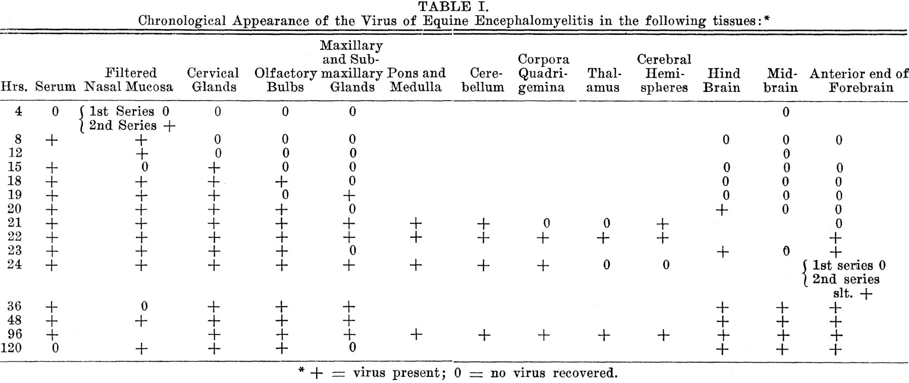 aseptically, pooled from the 3 animals, ground with saline and injected intracranially into guinea pigs. The nasal mucosa was extracted in saline and passed through a Seitz filter before inoculation. The serum was given both into the brain and subcutaneously. Several guinea pigs were allowed to live until prostration as controls on the viability of the virus. They all succumbed to the disease.
The results as given in Table I show an immediate and earlier invasion of the blood stream for this experiment than that previously reported 6 for guinea pigs. The virus was constantly present in the serum after the eighth hour and was almost always recovered from the nasal mucosa. Its constant presence in this tissue, however, may have been due to the amount of blood necessarily obtained from this region. It was then recovered from the cervical and salivary glands, the olfactory bulbs and subsequently from the cerebral nerve tissues beginning with the hind brain. After the 36th hour, coincident with the rise in temperature, the virus was constantly present in all of the tissues examined, except for the disappearance from the blood after defervescence and the subsequent prostration of the animal.
Get full access to this article
View all access options for this article.