
Abstract
In several hundred autopsies of guinea pigs with generalized tuberculosis, produced chiefly by subcutaneous, intraocular, intratesticular and intraspinal inoculation of tubercle bacilli, we have failed to discover any true tubercle formation in the intestines, although hypertrophy of the lymph nodes, particularly those in the cecum, do occur. Similarly, feeding guinea pigs with suspensions of virulent tubercle bacilli for a long time, or with food contaminated with fly specks containing the organisms, was unsuccessful in producing intestinal lesions.
On the other hand, inoculation of a virulent human strain (H 37) into the left ventricle of 23 normal pigs, either in single doses of 34,000 and upwards, or in three repeated doses of 30,000 to 5,000,000 organisms, resulted in 17 animals developing definite tubercles in the lymph follicles of the gut. The cecum is most frequently involved, and next in order the ascending colon and ileum. In a few cases, feeding contaminated sputum did not alter the typical lesion. Grossly, the lymphoid follicles are enlarged, and 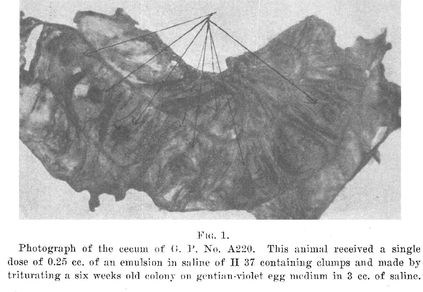
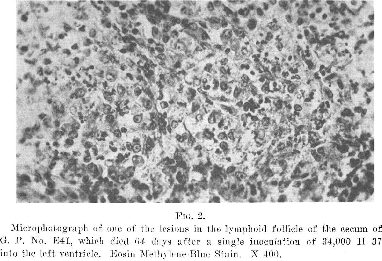
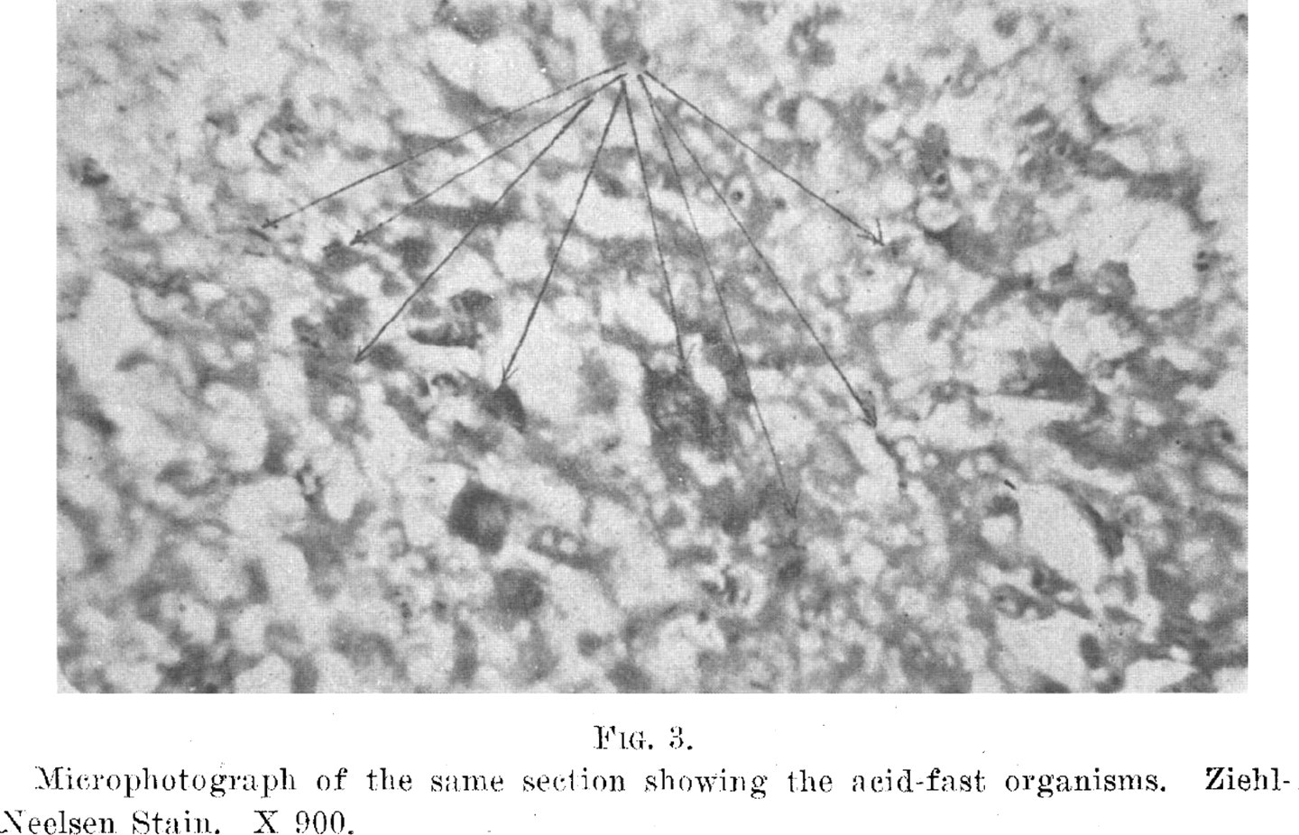
Microscopically, the lesions occur in the submucosa, especially in the lymphoid tissue of the cecum. The process is essentially a mononuclear infiltration, and cells with 2 and 3 nuclei are found. Polymorphonuclears, lymphocytes and nuclear dehris make up the other constituents, while acid-fast organisnis are to hle found in the lesions, often in large nunililers, either extra-cellular or phagocytosetl. This lesion in every respect resemldes the tubercles formed in normal animals in contradistinction to reinfected animals.
Get full access to this article
View all access options for this article.