
Abstract
Japanese cedar pollinosis is the predominant seasonal allergic rhinitis in Japan, and it has increased in prevalence during the past 10 years. Sublingual immunotherapy (SLIT) is considered a safe and effective treatment for pollinosis. Micro-RNAs (miRNAs) are a class of short single-stranded RNA molecules that posttranscriptionally silence gene expression and may mediate allergic immune responses. The aim of this study was to investigate the miRNA alteration in asymptomatic subjects sensitized to Japanese cedar pollen under prophylactic SLIT under part of a randomized, double-blind, placebo-controlled, multiple-center trial. Analysis was undertaken in 15 asymptomatic subjects sensitized to Japanese cedar pollen–specific IgE (ImmunoCAP class ≥2) who participated in 2013. The SLIT group (n = 6) received standardized Japanese cedar pollen extract and the placebo group (n = 9) received an inactive placebo for 5 months covering the cedar pollen season. Changes in serum miRNAs were measured by real-time quantitative polymerase chain reaction to determine whether SLIT had effects on profiles of circulating miRNA. Seven subjects in the placebo group developed pollinosis symptoms, whereas no subjects in the SLIT group did (p = 0.007). Serum hsa-miR-223 was significantly up-regulated in postseason compared with preseason samples. The hsa-let-7b was significantly more down-regulated in postseason than in preseason samples from the placebo group; however, no significant differences were observed in those from the SLIT group. A significant decrease in circulating let-7b was also observed in the subjects who developed symptoms. Prophylactic SLIT was effective in preventing the development of pollinosis. Alterations in miRNA expression occurred in asymptomatic, sensitized subjects during cedar pollen season.
Pollinosis is thought to be an adaptive immune response that manifests as a type 1 allergic reaction. 4 In response to antigen entry into the mucous membrane, IgE antibodies are produced in the nasal mucosa and regional lymphatic tissues. A strong rationale for improving the efficacy of allergen-specific immunotherapy by reducing the incidence and severity of adverse reactions mediated by IgE has been reported. 5 Allergen-specific sublingual immunotherapy (SLIT) is clearly efficacious and is associated with a favorable safety profile. The safety and efficacy of SLIT in treating allergic rhinitis associated with major aeroallergens are well documented in multiple largescale placebo-controlled clinical studies. 6 Several studies showed that the ratio of serum-specific IgE to total IgE (sIgE/tIgE) could predict the clinical response to SLIT.7,8 However, the certain specific molecules associated with allergic pathogenesis under SLIT remain unknown.
The current literature offers intriguing data to support a role for epigenetics in the development and persistence of allergic diseases such as asthma and allergic rhinitis.9–11 Epigenetic regulation, including micro-RNA (miRNA)–mediated regulation, may in part mediate the complex gene-by-environment interactions that can lead to allergic diseases. The miRNAs may be extremely important to pathogenesis of many allergic diseases in humans, and they also may be tractable targets for novel anti-inflammatory therapies. 12 Here, we analyze data and samples from a randomized, double-blind, placebo-controlled, trial study of SLIT to examine miRNA expression in asymptomatic subjects sensitized to Japanese cedar pollen. The key role of miRNAs in regulating homeostatic immune architecture and acquired immunity has been nicely reviewed by Lu and Rothenberg. 13 Therefore, we chose 14 miRNAs that were described in their review and used real-time quantitative polymerase chain reaction (RT-qPCR) to measure serum levels of each miRNA both before and after the Japanese cedar pollen season.
Methods
Study Participants
A total of 17 participants were recruited for part of a randomized, double-blind, placebo-controlled, multiple-center trial study of asymptomatic subjects sensitized to Japanese cedar pollen during the 2013 pollen season. Although each participant had IgE specific to Japanese cedar pollen of at least class 2, none of the subjects had ever developed pollinosis symptoms during a pollen season. Diagnosis of Japanese cedar pollinosis is based on the criteria described in the article by Uekusa et al. 14 Fluorescent enzyme immunoassays (CAP-FEIAs; Phadia, Tokyo, Japan) were used to measure Japanese cedar—sIgE titers and tIgE in serum both before and after the study. Participants who were pregnant or breast-feeding, as well as patients who suffered from chronic rhinosinusitis, were excluded. All of the subjects we recruited did not change their residence for several years. For 2 of the 17 subjects who participated in the study, blood samples were unable to be obtained after the treatment. Thus, the analysis was conducted with data from a total of 15 subjects. This study was approved by Mie University School of Medicine Ethical Committee (no. 2283). Written informed consent was obtained from each subject before the study began.
Clinical Protocols
The SLIT group (n = 6) received standardized Japanese cedar pollen extract (Torii Pharmaceutical Co. Ltd., Tokyo, Japan), 15 and the placebo group (n = 9) received an inactive placebo. The protocol consisted of treatment with graded courses of the extract in 50% glycerol, followed by maintenance therapy. 16 Briefly, the extract was graded into two strengths: 200 or 2000 Japanese allergy units (JAU)/mL. Starting in early December 2012, the subjects received increased doses with each vial every day, beginning with 0.2 mL of the 200-JAU/mL vial, and increasing until reaching the maintenance dose of 1.0 mL of the 2000-JAU/mL vial after 2 weeks. From the 3rd week, each of the six participants received the maintenance dose of 1.0 mL of the 2000-JAU/mL vial daily until the end of April 2013. The vaccine was taken sublingually, kept in place for 2 minutes without a retention reagent, and then swallowed. Each of the nine subjects in the placebo group received inactive 50% glycerol in physiological saline on the same schedule.
Clinical Symptoms
Each subject completed a pollinosis diary to record nasal and eye symptoms and the use of symptomreducing drugs. Subjects were judged to have developed symptoms based on both their pollinosis diary and a nasal provocation test performed at the end of April. The amount of pollen scattered from Japanese cedar (Cryptomeria japonica) in Tsu, Mie Prefecture was 11098 grains/cm2 during the 2013 pollen season.
Blood Sampling and Measurement of Total and Antigen-Specific Immunoglobulin Titer
Peripheral blood was obtained from each subject both before and after the treatment period from December 2012 to April 2013. Serum was isolated from 10 mL of venous blood by centrifugation at 600 × g for 10 minutes at room temperature. CAP-FEIA was used to measure serum levels of C. japonica 1 (Cry j 1)-sIgE titers. The tIgE in serum was also measured by CAP-FEIA.
RNA Extraction from Serum
Separated serum samples were centrifuged at 16,000 × g for 10 minutes at 4°C to remove any residual cellular nucleic acids attached to cell debris. Serum samples were kept at ~80°C until small RNA extraction. The miRNeasy Serum/Plasma Kit (Qiagen, Hilden, Germany) was used to isolate small RNA from 200 μL of serum according to the manufacturer's instructions. A synthetic Caenorhabditis elegans miR-39 miRNA mimic (Qiagen) and carrier RNA (0.94 μg; MS2 bacteriophage total RNA; Roche Applied Sciences, Indianapolis, IN) were added to each serum sample before RNA extraction. Isolated RNA was eluted with 15 μL RNase-free water.
Measurement of Serum miRNA Levels
SYBR Green RT-qPCR assays were used to assess miRNA expression in duplicate with each serum sample. The miScript Reverse Transcription Kit (Qiagen) was used according to the manufacturer's instructions to synthesize cDNA from 5 μL of serum RNA containing miRNA. The miScript SYBR Green PCR kit (Qiagen) along with the miScript Universal Primer and the miRNA-specific forward primers were performed in the ABI Step One Plus RT-PCR System (Applied Biosystems, Foster City, CA). SDS software 2.2.2 (Applied Biosystems) was used to determine the threshold cycle (Ct) value. We examined miRNA expression levels among 15 paired serum samples (preseason and postseason) as follows: 14 target human miRNAs (let-7a, let-7b, let-7c, let-7d, miR-21, miR-146a, miR-146b, miR-155, miR-365, miR-375, miR-223, miR-142-5p, miR-142–3p, and miR-193b), five candidate reference miRNAs (miR-93, miR-103a, miR-191, miR-423, and miR-425), the exogenously added cel-miR-39, and snRNA RNU6B. For 10 of the 30 samples, miR-365 was under the limit of detection.
Statistical Analysis
Before statistical tests, the normality of the data was examined. Parametric and nonparametric data between the two groups were analyzed for differences by Student's t-test and Mann–Whitney's U test, respectively. Differences in proportions between two or more groups were analyzed by χ 2 -test or Fisher's exact test. Two-group comparisons of ΔCt value between preand postseason were performed using Student's paired t-test. A value of p = 0.05 was considered statistically significant. SPSS Version 19 (SPSS, Inc., Chicago, IL) for Windows was used to perform all statistical analyses.
Results
Clinical Effects
The demographic characteristics of the 15 subjects before treatment are shown in Table 1. The participants suffered from tremendous cedar pollens in 2013, especially in March (10,833 grains/cm2, 97.6% of the total cedar pollen counts). Seven of the nine subjects in the placebo group and no subjects in the SLIT developed symptoms during pollen season. The proportion of subjects who developed pollinosis was statistically lower for the SLIT group than for the placebo group (p = 0.007; Fisher's exact test). To assess the factors that might influence this clinical effect, we compared age, sex ratio, and sIgE/tIgE ratio between the SLIT group and the placebo group; however, no significant differences were observed in the preseason (Table 1).
Clinical data of 15 participants

p = 0.01.
Fisher's exact test.
Student's t-test.
Mann–Whitney U test.
Specific IgE to Japanese cedar pollen; ImmunoCAP raw value (UA/mL), mean.
NS = not significant; sIgE = specific IgE; SLIT = sublingual immunotherapy; tIgE = total IgE.
Stability of Endogenous miRNAs in Human Serum
Consensus was not reached concerning which miRNA should be used as an internal control for qPCR analysis of serum samples. Therefore, we checked the endogenous control of serum miRNA according to RefFinder online tool of the EST database. 17 Specifically, we tested eight different candidate miRNA standards—exogenously added cel-miR-39, snRNA RNU6B, the individual values of five reference miRNAs (miR-93, miR-103a, miR-191, miR-423-5p, and miR-425), and the combined average of these five miRNAs—to identify a standard that could be used to normalize expression values of target miRNAs. Table 2 shows the ranking of the eight tested candidate miRNA standards. The recommended comprehensive ranking order was as follows: (1) the average of five reference miRNAs, (2) miR-425, (3) miR-423-5p, (4) miR-93, (5) miR-103a, (6) miR-191, (7) cel-miR-39, and (8) RNU6B. Therefore, we used the average of the five reference miRNAs as an endogenous reference measure to normalize miRNA levels.
Stability in expression of potential reference miRNAs
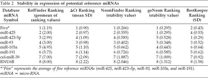
“Five” represents the average of five reference miRNAs (miR-425, miR-423–5p, miR-93, miR-103a, and miR-191). miRNA = micro-RNA.
Changes of Serum miRNA Levels between before and after Pollen Season
The miR-223 levels were significantly (p = 0.017) higher in postseason than in preseason samples (Fig. 1). However, no significant difference was observed for any other miRNA between preseason and postseason samples (data not shown).

Change in serum miR-223 expression for each participant. The value of micro-RNA (miRNA) in preseason samples was designated as 1 for each subject, and the fold changes in miRNA value in postseason samples were then determined. Statistical significance was determined with a Student's paired t-test using ΔCt values adjusted with the reference value. Placebo group, closed circles and solid lines; sublingual immunotherapy (SLIT) group, open circles and dotted lines.
Serum miRNA Changes by SLIT and Development of Pollinosis Symptoms
Neither SLIT nor development of pollinosis was significantly associated with a change in any miRNA level other than that of let-7b (data not shown). Interestingly, let-7b levels decreased significantly in the placebo group (Fig. 2 A; p = 0.004) but not in the SLIT group (Fig. 2 B; p = 0.811). Serum let-7b levels of subjects who had developed pollinosis were significantly lower in the postseason than in the preseason (Fig. 3 A; p = 0.007), whereas no significant differences were evident between preseason and postseason in subjects with no symptoms (Fig. 3 B; p = 0.935).

Comparison of expression levels of let-7b between preseason and postseason among the (A) placebo and (B) sublingual immunotherapy (SLIT) groups. The value of micro-RNA (miRNA) in preseason samples was designated as 1 for each subject, and the fold changes in miRNA values in postseason samples were then determined. Statistical significance was determined with a Student's paired t-test using ΔCt value adjusted with the reference value. Placebo group, closed circles and solid lines; SLIT group, open circles and dotted lines.

Comparison of expression levels of let-7b in subjects (A) with symptoms and in those (B) without symptoms. Statistical significance was determined with a Student's paired t-test using ΔCt value adjusted with the reference value. Placebo group, closed circles and solid lines; sublingual immunotherapy (SLIT) group, open circles and dotted lines.
Discussion
In this study, no subjects in the SLIT group developed symptoms, whereas seven of the nine subjects in the placebo group exhibited symptoms during pollen season. This immunotherapy may function via IgG4, which is an IgG isotype; might act as a “blocking antibody” by competing for the same epitopes as sIgE5,18; and has an anti-inflammatory role in immunologic responses. 19 Wachholz and colleagues 20 showed that allergen-specific IgG antibodies induced by immunotherapy can inhibit the formation of allergen-IgE complexes. In addition, IgG4 was observed to be increased after SLIT treatment among allergic rhinitis patients. 21 We speculate that SLIT-induced production of allergen-specific IgG4 may lead to the prevention of systemic increases of IgE and IgE-dependent allergic responses.
To the best of our knowledge, this is the first report of miRNA alterations in asymptomatic subjects sensitized to Japanese cedar pollen via prophylactic SLIT. The miRNAs are potentially critically important in the pathogenesis of allergic inflammation. The role of miRNAs in relation to the immune system has been extensively studied since the discovery of miRNAs in mammalian cells ~10 years ago.22 In our study, we showed that serum miR-223 was significantly higher in postseason than in preseason samples. Herberth et al.23 suggested that high miR-223 expression is associated with lower regulatory T-cell numbers. IL-10–producing regulatory T cells have been implicated in preventing Th2 responses to allergens.24 Yamanaka et al.25 showed that the percentages of IL-10–producing regulatory T cells are lower in patients with untreated cedar pollinosis than in healthy control subjects. Additionally, miR-223 is strongly correlated to the eosinophil level in the esophagus. 26 Activated eosinophils can express FcεRI, which may participate in the production of IgE and the amplification of IgE responses. These findings indicated that miR-223 may play a role in the regulation of allergic inflammation responses.
According to Chavali et al., 27 most miRNAs act complementarily to regulate multiple mRNAs. They analyzed miRNA and mRNA expression data from CD4+ T cells from patients with seasonal allergic rhinitis and found that a combination of two complementary miRNAs, miR-223 and miR-139–3p, could be targeted to alter the release of type 2 helper T-cell (Th2) cytokines. In this study we found that serum miR-223 levels were affected by the exposure to allergen, suggesting the relation to Th2-type inflammation.
Significant down-regulation of let-7b was observed in the placebo but not in the SLIT group after pollen season. Furthermore, a significant decrease of let-7b was observed in the study participants who developed symptoms. We anticipated that the decrease in serum let-7b would be associated with development of pollinosis and that SLIT might partially prevent allergic responses via let-7b action. Kumar et al. 28 showed that levels of let-7 family members, including let-7b, were reduced in lungs of mice with asthma after ovalbumin challenges and down-regulation of let-7 family members enhanced Th2 responses. Our result was consistent with the data of Kumar et al., although the specimens and allergens were different. On the other hand, Collison et al. 12 found that let-7b was up-regulated in airway smooth muscle of mice challenged with housedust mites. Given the discrepancies reported in the literature, future studies are needed to better define the role of let-7b in allergic inflammation.
Our preliminary study showed that there were no significant differences in levels of serum miR-223 and let-7b between the healthy volunteers and sensitized asymptomatic subjects out of pollen season (data not shown). In this study, we found that miR-223 was significantly up-regulated in pollen season and let-7b was down-regulated in subjects developing pollinosis. Therefore, we speculate that the changes of miR-223 would reflect the exposure to antigen in the pollen season. Changes of let-7b during cedar pollen season in asymptomatic, sensitized subjects may play a role in the pathogenesis of allergic rhinitis. Although there were some limitations in our study, including a small sample size and a lack of pathway studies, our results may shed a light on the role of miRNAs in pollinosis development. Future studies are needed involving larger numbers of participants following extension of SLIT protocol (dose and duration).
Footnotes
Acknowledgments
The authors thank Dr. Yoshitaka Okamoto from Chiba University for assisting with the research. They also thank the participants who provided blood samples for the study.