
Abstract
The presence of frontal cells poses unique challenges when using endoscopic approaches. This study describes the use of a balloon dilation system as an aid for functional endoscopic sinus surgery (FESS) to access the frontal sinus in cases that would traditionally require open approaches. We present a case series of four patients with chronic rhinosinusitis refractive to medical management who underwent FESS with the aid of a balloon dilation system at a tertiary referral center. All patients had variant forms of frontal sinus anatomy. Surgical techniques will be described and use of the balloon system will be reviewed. All patients (aged 13–68 years) successfully underwent fontal sinusotomies with the assistance of a balloon dilation system, which was used in a variety of ways: to dilate the narrow infundibulum of a high intersinus septal cell, to remove an anteriorly located type III frontal sinus cell, to expand the natural frontal ostium in the presence of excessive agger nasi pneumatization, and to remove a type IV frontal sinus cell. All patients were spared an osteoplastic flap or trephination, and there were no intraoperative complications. No postoperative bleeding, infection, or cerebral spinal fluid leaks were reported. Balloon dilation in combination with standard frontal sinus dissection techniques may be beneficial for a select group of patients with complex frontal anatomy. In this series of patients, the balloon dilation system was used as a tool during FESS and eliminated the need for open approaches.
The presence of frontoethmoid cells in 20% of patients additionally contributes to the pathogenesis of frontal CRS by further narrowing the FSOT. 6 Historically, reestablishment of frontal sinus drainage in these situations has been done one of several ways: through an intranasal approach, an external approach, or by completely obliterating the sinus. 2 With the presence of some frontoethmoid cells, external approaches such as trephination and osteoplastic flaps have been used to reestablish drainage of the frontal sinus. 3
Recently, the balloon dilation system was introduced as a tool to enlarge the sinus ostia in select patients with CRS. The balloon system has been recognized for its minimally invasive nature as well as its ability to preserve mucosa while dilating the maxillary sinus ostium. 7 Used intraoperatively, either by itself or in combination with endoscopic sinus surgery, the balloon's use has resulted in reports of both reduced bleeding and operative time. 8
We propose that, when combined with standard functional endoscopic sinus surgery (FESS) in appropriate situations, the balloon dilation system may have usefulness in accessing a tightly confined frontal recess, particularly in the setting of difficult bony anatomy. In the present study, we describe several methods in using the balloon catheter as an aid for FESS to treat complex frontal sinus disease.
Methods
After approval by the University of North Carolina Institutional Review Board, we reviewed four patients with chronic frontal rhinosinusitis refractive to maximal therapy who underwent FESS with the balloon dilating catheter system (Acclarent, Inc., Menlo Park, CA) from 2010 to 2013. All patients had radiographic evidence of FSOT obstruction in the presence of complex frontal sinus cell anatomy. Balloon dilating catheter systems were used in combination with the endoscopic approach to the frontal sinus (Table 1).
Demographics, radiographic findings, and indications for balloon sinuplasty

CT = computed tomography; FSOT = frontal sinus outflow tract.
The ability to access the frontal sinus using the balloon dilating catheter system during FESS was assessed. During each reported case, a sinus guide catheter was inserted under direct visualization with a rigid angled endoscope. A lighted flexible guide wire was then passed through the catheter and threaded into the narrowed region of concern. The position of the wire was confirmed by positive transillumination of the frontal sinus as well as direct endoscopic examination. A sinus balloon catheter was then passed over the guide wire into the frontal recess and advanced to the desired location. The balloon was then inflated up to 10 atmospheres of pressure in the targeted space. The balloon was deflated and removed, and endoscopic inspection was used to assess for successful dilation. If necessary, balloon inflation was repeated until the appropriate removal of the frontal cell or other obstruction. The crushed bony ledges of the cells were then removed using standard endoscopic techniques. Loose mucosa was removed with a microdebrider.
Results
All patients (aged 13–68 years) underwent frontal sinusotomies with the assistance of the balloon dilation system with postoperative symptomatic improvement sustained throughout all postoperative visits and without the need for repeat operation. The mean length of follow-up was 13 months, with one patient lost to follow-up 2 weeks postoperatively. The balloon was used for a variety of purposes: to dilate the narrow infundibulum of a high intersinus septal cell, to crush and remove type III and type IV frontal cells to dilate FSOTs, and to find the natural frontal ostium in the presence of challenging anatomy. All patients were spared from undergoing an osteoplastic flap and there were no intraoperative complications. Patients were discharged on the same day without postoperative bleeding, infection, or cerebral spinal fluid leak.
Case I: High Intersinus Septal Cell
A 31 year-old male patient with a history of asthma, seasonal allergies, and frequent sinus infections presented with a 3-month history of right frontal headaches, which were associated with nasal obstruction and purulent rhinorrhea. Computed tomography revealed a very large hourglass-shaped intersinus septal cell measuring 15 mm in the anterior-posterior dimension and 44 mm in the superior-inferior dimension, with a narrow 2-mm hourglass infundibulum (Fig. 1). The patient was taken to the operating room for a Draf IIB frontal sinusotomy, which included removal of the ipsilateral frontal sinus floor from the lamina papyracea to the nasal septum. The high intersinus septal cell was identified by direct endoscopic visualization and was found to be difficult to reach with standard instrumentation. The opening of the intersinus septal cell was then appreciated, and a lighted guide wire was inserted into the cell. A 6-mm-diameter, 24-mm-long balloon catheter was then inserted over the guide wire into the ostium and inflated several times to 10 atmospheres of pressure. A ball probe was then used to further fracture the frontal sinus cell, and the balloon was again inserted lateral to the infundibulum and used to fracture the anterior wall of the septal cell. Endoscopic visualization was subsequently used to confirm that the frontal sinus was maximally opened.

Intersinus septal cell—sagittal and coronal views.

Anteriorly displaced type III cell—sagittal and coronal views.
Case II: Anteriorly Located Type III Frontal Sinus Cell
A 13-year-old male patient with asthma and anterior drainage had radiographic findings of a large, partially opacified type III sinus cell extending through the ostium into the true right frontal sinus and causing obstruction of the frontal recess (Fig. 2). The patient was taken to the operating room for bilateral FESS. On the right, the anterior ethmoid cells were identified endoscopically and removed with a curette. A lighted sinus guide wire was placed into the frontal recess and advanced into the frontal sinus under direct visualization. The 6-mm-wide, 24-mm-long balloon catheter was advanced over the wire and positioned adjacent and posterior to the frontal cell, and then inflated to 10 atmospheres of pressure until the cell was crushed anteriorly. Further removal of bone and mucosa was performed. The frontal sinus was then noted to be maximally opened and the fontal sinus cell was removed. The patient was symptomatically improved at 2 weeks postoperatively; however, he was subsequently lost to follow-up.
Case III: Bilateral Extensively Pneumatized Agger Nasi Cells
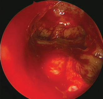
A 68-year-old male patient with allergic disease and nasal polyps presented for operative evaluation. His computed tomography scans revealed extensive pneumatization of agger nasi cells bilaterally, causing symptomatic obstruction of the FSOT (Fig. 3). The patient underwent bilateral total ethmoidectomy, but the frontal anatomy still proved to be quite difficult to maneuver, and the frontal recess could not be visualized past the large agger nasi cell. The lighted guide wire was brought into the operating field to help identify the natural outflow of the frontal sinus. A 6-mm-wide, 24-mm-long balloon was then threaded over the guide wire into the frontal recess and advanced into the ostium of the sinus until it was felt to juxtapose the agger nasi cell. The balloon was inflated twice to 10 atmospheres of pressure to fracture the posterosuperior wall of the agger nasi cell and dilate the frontal ostium. This allowed for passage of endoscopic instruments into the frontal sinus, and the ostium was further enlarged using the standard instrumentation (Fig. 4).

Excessively pneumatized agger nasi cell—sagittal and coronal views.

Endoscopic view of maximally opened frontal sinus after removal of excessively pneumatized agger nasi cell.
Case IV: Type IV Frontal Sinus Cell and Intersinus Septal Cell
A 60-year old male patient with a 1-year history of posterior drainage was found to have radiographic findings of a type IV frontal sinus cell on the right (Fig. 5). Additionally, an excessively pneumatized intersinus septal cell was appreciated and found to be in communication with the frontal sinus. The patient was taken to the operating room for bilateral FESS. On the right, the type IV frontal cell was identified and a lighted guide wire was passed into the frontal sinus and advanced between the medial edge of the frontal cell and the frontal sinus. The guide wire's placement was confirmed with transillumination and a balloon dilating catheter was inserted over the guide wire and positioned at the medial edge of the frontal cell. The balloon was subsequently inflated to 10 atmospheres, released, brought inferiorly, and inflated to 10 atmospheres of pressure again, crushing the frontal cell wall laterally. The balloon was deflated and removed, remaining bony fragments were removed, and further was dissection performed. The ostium of the intersinus septal cell was subsequently visualized in the right frontal sinus and was enlarged with standard instruments. The frontal sinus was then noted to be maximally opened and the frontal sinus cell was removed (Fig. 6).

Type IV frontal sinus cell with intersinus septal cell—sagittal and coronal views.
Endoscopic view of maximally opened frontal sinus after removal of type IV frontal sinus cell.
Discussion
The balloon dilation system was approved by the U.S. Food and Drug Administration in 2005 and since then, >50,000 sinus procedures have been performed using the device. 9 In the frontal sinus, the advantages of using the balloon device include a potential for a less invasive approach to the frontal sinus, shorter operative time, and less morbidity than more aggressive open approaches. 10 The frontal recess, especially, can be extremely challenging, and some authors have even suggested that the area demands correct surgical technique more so than any other area in the paranasal sinuses. 2 Additionally, disease most often recurs in the frontal recess after FESS, because endoscopic bone removal in the frontal sinus results in tissue trauma and inflammation, and disruption of mucosa increases the risk of stenosis. 3
The balloon dilation system may aide in accessing the frontal sinus by dilating and compressing underlying bone and mucosa, helping to limit the risk of iatrogenic damage. 8 Multicenter studies have shown successful intraoperative dilation in 96.9% of sinus ostia widened by balloon manipulation with an 82% 6-month patency rate for the frontal sinuses. 10 Conversely, opponents of the balloon system state that the technique leaves behind infected, diseased bone, and that the crushing of this osteitic tissue increases the risk of recurrent inflammation and delayed mucocele formation. 11
However, balloon dilation, in combination with standard frontal sinus dissection techniques, may alleviate some of the concerns of opponents by removing crushed bone and redundant mucosa. Furthermore, the balloon dilating catheter may be beneficial for select groups of patients with complex disease and anatomy by enlarging narrow, difficult-to-access regions and to allow for access with endoscopic instruments. A recent prospective study of 195 sinus ostia dilated with balloons showed that patients who underwent balloon dilation during endoscopic sinus surgery showed significant symptom improvement as well as radiographic confirmation of disease resolution 2-years postoperatively, showing the durability of the hybrid procedure. 12
In our series of patients, the balloon system was used successfully, in conjunction with FESS, to relieve FSOT obstruction that was a direct result of frontoethmoid air cells and allowed for the definitive removal of diseased, osteitic bone. Our study shows that a hybrid FESS balloon dilation procedure is effective in both adult and pediatric patients with frontal CRS who have anatomic FSOT obstruction. Furthermore, it provides the surgeon a less invasive access into the frontal sinus than trephination or the use of an osteoplastic flap. Our study is limited by its observational nature, small sample size, and short follow-up period, but it shows that the balloon offers great promise as an adjunct to FESS. Prospective studies with larger cohorts and longer follow-up periods are warranted to compare surgical outcomes in groups with and without balloon dilation to establish the dilating system's full scope as an instrument for frontal sinus surgery.
Conclusion
Our observational study implies that balloon dilation, in combination with standard frontal sinus dissection techniques, may be beneficial for select groups of patients with complex disease and anatomy. The balloon system has the advantage of enlarging narrow cavities that would otherwise be extremely difficult or inaccessible with standard frontal instruments. When paired with FESS, the hybrid procedure allows for both FSOT dilation and definitive removal of diseased tissue, while sparing patients from the more invasive options of trephination or osteoplastic flap.
Footnotes
The authors have no conflicts of interest to declare pertaining to this article