
Abstract
Most autoimmune diseases predominantly affect females. Many of these diseases occur in women who have the potential to become pregnant or wish to plan a pregnancy. The potential for fetotoxic effects of immunosuppressive medications that are commonly used to treat systemic autoimmune diseases must be weighed against the need for control of disease activity during pregnancy and the postpartum period, since active disease can be an independent risk factor for adverse pregnancy outcomes. Although far from conclusive, most data concerning the safety of medications for use during pregnancy come from case series and observational studies. It is often necessary to continue treating patients throughout pregnancy and lactation in order to control the activity of the underlying disease. The aim of this paper is to review the evidence regarding the safety of the most commonly used medications in rheumatic disease during pregnancy or lactation and to enable practitioners and patients to make informed decisions regarding treatment during this period in a woman's life.
 Medscape: Continuing Medical Education Online
This activity has been planned and implemented in accordance with the Essential Areas and policies of the Accreditation Council for Continuing Medical Education through the joint sponsorship of Medscape, LLC and Future Medicine Ltd. Medscape, LLC is accredited by the ACCME to provide continuing medical education for physicians.
Medscape, LLC designates this educational activity for a maximum of
Learning objectives
Upon completion of this activity, participants should be able to:
Describe the potential adverse effects of nonsteroidal anti-inflammatory drugs (NSAIDs) for the pregnant woman and her fetus
Identify the optimal corticosteroid to use during pregnancy
Describe the immunosuppressive therapies available for women with autoimmune lupus disease
Identify the appropriate immunosuppressive medications for pregnant women with inflammatory bowel disease
Describe appropriate treatment for newly diagnosed breast cancer during pregnancy
Systemic autoimmune diseases commonly affect women during childbearing years. With advances in therapies for these diseases, many patients have sustained periods of disease quiescence, less progressive disability and may be more likely to consider childbearing and child rearing. When treating women of childbearing age with chronic medications, it is imperative that the potential effects of medication use on the developing fetus are considered, whether a future pregnancy is planned or an unanticipated pregnancy is discovered. The goals of treatment are to control maternal disease activity while simultaneously maximizing the potential for a safe and successful outcome for the fetus. Since randomized controlled trials of medications for pregnant or lactating women are not routinely performed, most of the data available are based on animal data, individual case reports and observational cohorts. With extremely few exceptions, no medications are deemed to be without any risks to the expectant mother or developing fetus. However, some medications may elicit fewer safety concerns than others and may be considered safer alternatives for the successful control of maternal disease. Greater knowledge of the risks and benefits of commonly used immunosuppressive drugs during pregnancy and lactation will enable improved preconceptional counseling for women who desire a future pregnancy and improved counseling and management of patients who inadvertently become pregnant while undergoing disease-modifying therapy and wish to continue the pregnancy, as well as improving the management of patients with increased disease activity during an established pregnancy.
The US FDA has a classification system for pregnancy risk that ranges from A (studies demonstrating safety in animals and humans) to X (absolutely contraindicated in pregnancy) [101]. Drugs are given a classification at the time of approval, often based primarily on animal studies, and are generally not updated as new evidence becomes available. Therefore, the current classification system may not be the most accurate resource for managing medication exposures during pregnancy. A narrative summary of potential risks and benefits of the commonly used medications for the treatment of autoimmune diseases is presented below.
NSAIDs
It is unclear if NSAIDs have an effect on fertility. Both cyclooxygenase (COX)-1 and COX-2 have been demonstrated to be involved in ovulation and implantation in animal studies [1]. In humans, there have only been small case report series describing infertility caused by NSAID use owing to inhibition or delay in the luteinized follicle [2,3]. Other reports have suggested that NSAID use may increase the risk of miscarriage. In a prospective cohort study by Li et al. with 1055 pregnant women in the Kaiser Permanente population, use of NSAIDs or aspirin around the time of conception and/or during pregnancy increased the risk of miscarriage [4]. The hazard ratio for NSAID use versus no NSAID use in this study was 1.8 (95% CI: 1.0–3.2) but increased to 5.6 (95% CI: 2.3–13.7) if taken during the week of conception and increased to 8.1 (95% CI: 2.8–23.4) if taken for more than 1 week. Paracetamol use did not affect risk. By contrast, another study examined pregnancy outcomes among aspirin users compared with nonusers and did not find a difference in miscarriage between the two groups (risk ratio: 0.92; 95% CI: 0.71–119). Based on these data, it is suggested that patients should discontinue the use of NSAIDs or use them sparingly while trying to conceive, especially if there is difficulty with conceiving.
The effect of NSAID use on the fetus is dependent upon the trimester. A number of cohort studies looking at the teratogenicity risk of NSAID use during the first trimester have not found an increased risk of congenital malformations [5]. It should be noted that selective COX-2 inhibitors were not studied and pregnancy outcomes have not been well observed. However, there is concern for adverse fetal outcomes with late-term NSAID use, namely constriction and/or premature closure of the ductus arteriosus. Both COX-1 and COX-2 are expressed in the smooth muscle and endothelial cells and when inhibited, constriction occurs. The majority of studies reviewing this have noted it as a late-term manifestation occurring primarily after the 27th week. Therefore, NSAIDs should be used sparingly during the first and second trimesters and stopped completely during the third trimester of pregnancy.
The NSAIDs that the American Academy of Pediatrics (A AP) consider to be compatible with breastfeeding include ibuprofen, indomethacin, diclofenac, naproxen, piroxicam, ketorolac and tolmetin [6]. These recommendations are based on observational studies looking at adverse effects on the infant and/or based on expert opinion. Owing to the fact that NSAIDs are weak acids, usually only a small amount will enter into breast milk, and this is often less than 5% of a therapeutic infant dose [7]. Ibuprofen has an especially low level in breast milk and a short half-life, and is therefore considered to be a reasonable choice as an analgesic for the lactating mother.
Corticosteroids
Corticosteroids vary structurally and will affect the mother and fetus differently. β-methasone and dexamethasone have fluorine at the 9a position and are not well metabolized by the placenta. They will cross the placenta and have direct effects on the fetus. However, most other corticosteroids are metabolized in the placenta by 11-β-hydroxysteroid dehydrogenase to inactivated forms, leaving less than 10% of the active drug to reach the fetus [8]. If the goal is to treat the mother, prednisone is the most ideal glucocorticoid since a very small amount of active drug will enter the fetal circulation. If the goal is to treat the fetus, β-methasone and dexamethasone are better options and are commonly used in women who are at risk for preterm delivery in order to reduce the risk for respiratory distress and cerebral hemorrhage in the preterm infant. Some recent reports suggest that β-methasone may be preferred to dexamethasone since it may offer better long-term neurodevelopmental outcomes for the fetus. [9,10].
Corticosteroids carry a risk of elevated blood pressure, steroid-induced hyperglycemia and osteopenia. These risks are pertinent in a pregnant woman since pregnancy requires tight blood pressure control and may induce insulinresistance (and often frank gestational diabetes), as well as osteopenia or osteoporosis. These issues can be exacerbated by steroids; therefore, proper monitoring and calcium and vitamin D supplementation should be employed.
Nonfluorinated corticosteroids do not appear to put the fetus at risk for congenital abnormalities; however, there have been a few reports of an increased risk of cleft lip and/or palate, which are first trimester risks. A meta-analysis investigating birth defects after maternal exposure to corticosteroids reported a 3.3-fold increase in the odds ratio of cleft lip and/or palate after first trimester exposure [11]. Subsequent studies have reported mixed results – some showing similar results and some showing no difference. However, if there is an increase risk, it should be noted that cleft palates occur at a rate of approximately one in 1000 individuals in the general population and, thus, the risk with steroid use would increase this to three in 1000. This small increase in risk should be weighed against the need to treat active disease in the mother. There have also been rare isolated reports of neonatal cataracts and adrenal suppression in infants after corticosteroid use, which may be dose-dependent [5].
The pharmacokinetics of prednisolone, the active form of prednisone, have been studied in breast milk [11,12]. Levels measured in the milk are typically less than 0.1% of the total prednisolone dose ingested by the mother, which is less than 10% of the infant's endogenous cortisol production (when small-to-moderate doses are being used). Ost et al. evaluated a patient taking up to 80 mg of daily prednisolone and calculated that the prednisolone ingested from the milk would add, at most, 10% to the endogenous corticosteroid production of the infant, which may have little clinical significance [12]. Peak steroid levels in breast milk occur approximately 2 h after a dose is taken and decline rapidly [11]. These small-to-moderate amounts of corticosteroids do not appear to have adverse effects on the developing infant; however, exposure may be minimized if nursing is performed 3–4 h after the dose is taken.
Hydroxychloroquine
Despite initial animal studies demonstrating toxicity to retinal and auditory structures in animal teratogenicity studies of high doses of chloroquine [13], data have accumulated in support of the safe use of hydroxychloroquine (HCQ; up to 400 mg daily) during human pregnancy [5]. There have been a number of observational studies and a recent meta-analysis demonstrating no increased risk of congenital abnormalities or increased risk of miscarriage using the typically prescribed dose of 200–400 mg HCQ daily [14–16]. Clowse et al. not only reported the safety of HCQ, but also found that HCQ use was associated with decreased disease activity among lupus patients [17]. A single placebo-controlled, randomized, double-blinded study has been performed to assess the role of HCQ during pregnancy in lupus patients [18]. Again, the data demonstrated no congenital malformations associated with HCQ. All cases of disease flare-up during pregnancy and preeclampsia were in the placebo group. HCQ crossed the placenta and the concentration in cord blood was approximately equal to maternal concentrations, but excretion into breast milk was very low (<0.2 mg/kg/day) and this level is not thought to be toxic [19]. If women remain concerned about antenatal exposure to HCQ and wish to eliminate it from the maternal system prior to conception, it must be remembered that complete elimination may take 6 months. HCQ appears to be a safe option for conception, pregnancy and lactation.
Methotrexate
Methotrexate is an FDA category X medication that is contraindicated during pregnancy and is not considered safe in lactation. There are known risks of fetal abnormalities in humans associated with methotrexate use. These risks have been observed in women being treated for cancer or when methotrexate was unsuccessfully used to terminate a pregnancy. However, there are also reports of fetal abnormalities when methotrexate has been used at doses prescribed in the rheumatology setting [20,21]. In a recent systematic review, methotrexate-exposed pregnancies using doses of 5–25 mg/week in the first trimester were analyzed. The number of methotrexate-exposed pregnancies was 101. There were 19 miscarriages (23% of pregnancies), 55 live births (66% of pregnancies) and five of the babies had neonatal malformations (5% of pregnancies) [22]. Methotrexate is the first-line DMARD and is the drug of choice, especially as initial therapy, for the treatment of rheumatoid arthritis (RA). Therefore, all women of childbearing potential should be strongly counseled and advised to use reliable forms of contraception while taking methotrexate. Furthermore, the patient and practitioner should have a discussion regarding the use of alternative agents when initiating treatment, especially if pregnancy is desired in the near future or the practitioner feels that the patient's methods of contraception are unreliable. The same restrictions are also true for males. If a woman inadvertently becomes pregnant, she should discontinue the medication immediately, be counseled concerning the risks of congenital abnormalities and be referred immediately to a genetic counselor in order to discuss her options.
If a woman who is already being treated with methotrexate desires pregnancy, several issues need to be addressed during preconception counseling. One question that arises is for how long the drug should be discontinued before it is safe to conceive. Unfortunately, there is not a set guideline for this and, thus, practice is largely based on the known pharmacokinetics of the drug. Dalrymple et al. recently studied the pharmacokinetics of oral methotrexate in patients with RA [23]. The median times for methotrexate polyglutamate concentrations to become undetectable in red blood cells ranged from 4 to 10 weeks (also showing wide interpatient variability of elimination in adults with RA). Methotrexate is also widely distributed in tissues and can persist in the liver for up to 4 months after exposure [24]. Therefore, it is our practice to recommend discontinuing methotrexate at least 3 months prior to conception in order to allow adequate time for elimination of the drug. Methotrexate is also a derivative of aminopterin, which is a folate antagonist. Rheumatologists commonly prescribe folic acid supplementation during methotrexate therapy; however, folic acid supplementation should be continued during the preconception period, even after methotrexate is stopped, and should be continued throughout pregnancy. It is recommended that all pregnant women take at least 800 μg of folic acid daily in order to reduce the risk of neural tube defects. Closure of the neural tube occurs during the 5th week of pregnancy (3 weeks after conception), so early supplementation is essential.
Although the amount of methotrexate excreted in breast milk is very low, it is unknown how this may affect a young infant and, therefore, methotrexate is considered to be unsafe until more data become available.
Leflunomide
It is known that leflunomide causes fetal anomalies in animals, primarily CNS and skeletal anomalies. When leflunomide was given to rats at only 1% of the human dose, it resulted in fetal anomalies, decreased birth weight and increased mortality in the offspring after birth [25]. Based on available animal data, the FDA has categorized leflunomide as category X; this indicates that clear associations between the drug and fetal abnormalities have been identified and that the risks of use in pregnant humans outweigh any potential benefits. Managing elimination of leflunomide in a woman who has become pregnant while taking leflunomide or desires pregnancy is neatly outlined by Brent in his article reviewing the reproductive risks of leflunomide, largely based on manufacturer and FDA recommendations [26]. The level that is believed to be safe in humans is 0.03 mg/l (i.e., ~10 half-lives). If a woman treated with leflunomide wants to become pregnant, the medication should be stopped and cholestyramine should be administered in order to rapidly decrease the blood levels to this level. Cholestyramine of 8 g taken three-times daily reduces the half-life of the active metabolite from up to 96 days to approximately 1 day. Therefore, a 10–11-day regimen of 8 g cholestyramine three-times daily is recommended. This approach is certainly conservative but was suggested by the FDA and is the manufacturer's recommendation. Plasma levels of the drug should be verified to be less than 0.03 mg/l by two separate tests performed 2 weeks apart. If levels still remain above 0.03 mg/l, the regimen should continue to be administered and women should use effective contraception until levels are below 0.03 mg/l. Based on the natural half-life of the drug (up to 96 days, as mentioned previously) it may take up to 2 years to reach plasma active metabolite levels of 0.03 mg/ml owing to individual variations; therefore, levels should be checked (and, if necessary, cholestyramine administered) in any patient who has been treated with leflunomide in the previous 2 years. This practice is also supported by a panel of 29 experts who participated in a consensus workshop on antirheumatic drugs during pregnancy and lactation [5]. Data from the Organization of Teratology Information Services (OTIS) Collaborative Research Group prospectively evaluated 64 women with RA who were treated with leflunomide (95% of whom were treated with cholestyramine early in their pregnancy), 108 women with RA who were not treated with leflunomide and 78 healthy pregnant women. The rate of major structural defects in the exposed group was 5.4%, which was insignificant when compared with both comparison groups (4.2 and 4.2%, respectively; p = 0.13). Although the study group was small, these results are encouraging and suggest that these defects may be preventable with appropriate and early intervention [27].
It is unknown how leflunomide exposure may affect a young infant and, therefore, lactation is considered to be unsafe in women treated with leflunomide; this is likely to remail the case until more data become available.
Sulfasalazine
Fertility in women does not appear to be affected by sulfasalazine use; however, it can lead to reduced fertility in men owing to oligospermia and reduced sperm motility. Fortunately, this is not an irreversible event; spermatogenesis should return to normal approximately 2 months after cessation of the drug. Men wishing to conceive a child with a partner should be counseled to discontinue the medication 3 months prior to conception [28]. It is also important to counsel men to explain that, although fertility may be reduced, the use of sulfasalazine is not considered to be an effective form of contraception. Abnormal or reduced spermatogenesis is due to the sulfasalazine metabolite, sulphapyridine.
In contrast to its effects on men, sulfasalazine is among the preferred DMARDs for pregnant women who require ongoing disease-modifying therapy. Much of the data regarding pregnancy outcomes with sulfasalazine exposure are derived from the inflammatory bowel disease (IBD) literature. A recent meta-analysis of 2200 pregnant women with IBD (642 women received sulfasalazine or related agents) found no statistically significant differences for congenital anomalies (odds ratio: 1.16; 95% CI: 0.76–1.77) or other adverse pregnancy outcomes [29]. These data confirm several other reports of population-based studies published over the past several decades [30,31]. Congenital abnormalities discussed in isolated case reports have not been observed in systematic population-based studies.
Two additional considerations are worth noting. The first is the theoretical risk that sulfasalazine may cause folate deficiency since it inhibits dihydrofolate reductase and the cellular uptake of folate [32]. Pregnant women require at least 800 μg of folic acid replacement, which should be continued, and perhaps increased, during sulfasalazine use. Folic acid deficiency has been linked to neural tube defects, which can occur 5 weeks into pregnancy (or as early as 3 weeks after conception), so this should be part of the preconception planning discussions. The second consideration is with regard to late-term use. Sulfapyridine, a metabolite of sulfasalazine, can cross the placenta, displace bilirubin from albumin and possibly lead to neonatal jaundice [33]. Although there are no reports of this occurring after in utero exposure to sulfasalazine, one may consider discontinuing sulfasalazine use during lactation in a preterm or jaundiced baby for 1–2 months.
Except in the setting of prematurity, hyperbilirubinemia or other acute stresses, sulfasalazine is considered safe during lactation. Sulphapyridine levels in breast milk were found to be approximately 30–60% of those in the mother's serum, but should not pose a risk to a healthy full-term infant [34].
Anti-TNF agents
There are few data regarding the safety of TNF inhibitors in pregnancy. There have been a few studies looking at infliximab, primarily in the gastroenterology literature. In one report evaluating 58 pregnancies with first trimester exposure, five live-born infants in the series had complications, two were structurally normal but had complicated neonatal courses and three had structural or developmental complications (one had developmental delay, one had tetralogy of Fallot and one had intestinal malrotation) [35]. Another retrospective evaluation of ten pregnant women with infliximab exposure demonstrated no structural abnormalities; however, three children were born prematurely [36]. Soluble TNF receptors (e.g., etanercept) do cross the placenta in mice but do not appear to impair fetal development [37]. There are a few reports of human data demonstrating embryotoxicity, teratogenicity or pregnancy loss. In 14 women who were prescribed etanercept during pregnancy, no malformations were reported [21]. A report from the British Society of Rheumatology Biologics Register (BSRBR) found no structural or developmental abnormalities in 17 pregnancies and the rate of miscarriage was comparable to that of the general population [38]. The same registry reported no adverse outcomes in three patients who were treated with adalimumab. Chambers et al. reported on 32 pregnancies from the OTIS registry with TNF exposure, demonstrating no increase in fetal malformations, miscarriage or preterm birth [39]. Orozco et al. analyzed 84 pregnancies with TNF exposure (27% continued through pregnancy). A total of 93% of those studied had live births and most were full-term. There were two malformations, which is not an increased rate compared with the general population [40]. Carter et al. reported very different findings. The report outlined 41 children with 61 congenital anomalies after TNF inhibitor exposure based on data collected from FDA reports. A total of 56% of the anomalies fell into the vertebral, anal atresia, cardiac, trachea, esophageal, renal and limb defects (VACTERL) category; the most common anomalies were cardiac and urinary and there were two cases of Down syndrome [41]. Unfortunately, there is no information regarding the total number of patients exposed to TNF antagonists and, thus, the rate of malformations. In other words, we cannot say with certainty if these are related to the drug or are in line with the rate observed in the general population. Furthermore, many individuals in the field of rheumatology have criticized the definition of VACTERL used in the Carter paper. Cumulatively, the data appear to indicate that exposure to anti-TNF agents in pregnancy does not seem to increase the risks of miscarriage, preterm delivery or congenital malformations. However, most available data reflect preconceptional and early first trimester use in pregnant women with RA or Crohn's disease. Current recommendations for TNF use during pregnancy suggest that conception and early pregnancy are not adversely affected by TNF exposure, and that the potential benefits of continued TNF inhibition during the later stages of pregnancy may outweigh the harm in women with persistent high disease activity [42].
There are no published data regarding lactation and TNF use. There are a few case reports looking at levels of both etanercept and infliximab in the milk of lactating women, which appear to be very low [43,44]. However, the effects of these drugs on a young infant are unknown. Theoretically, because these drugs are proteins, they are probably digested in the GI tract and not absorbed in whole systemically. Yet again, physicians should use caution and should counsel women on this lack of information regarding appropriate use.
Azathioprine
Azathioprine has been used during pregnancy for many years in the management of solid organ transplantation, IBD, malignancy and rheumatic diseases. Azathioprine undergoes hepatic metabolism to 6-mercaptopurine (6-MP) and further metabolism to 6-thiouric acid (an inactive metabolite). Azathioprine and its metabolites (6-MP and 6-thiouric acid) are known to cross the placenta. In one study, radioactively labeled azathioprine (35S-azathioprine) was administered to humans and the ratio of radioactivity in fetal blood compared with maternal blood was measured. In maternal blood, 27–41% of the administered radioactivity was present as 6-thiouric acid, 14–27% as 6-MP and 21–28% as azathioprine. In fetal blood, the majority of radioactivity present (48–63%) was due to thiouric acid (the inactive metabolite). The majority of radioactivity in amniotic fluid and in the placenta was also due to thiouric acid [45]. Numerous studies of fetal outcomes in pregnant patients treated primarily for organ transplantation or IBD have consistently demonstrated a lack of a specific pattern of congenital malformations, although sporadic malformations have been reported [5]. However, most studies confirm an association between azathioprine use and intrauterine growth restriction and preterm delivery [46]. A recent study of 476 women taking azathioprine during early pregnancy for a variety of indications further supports the increased risk of preterm and low birth weight infants without a statistically significant increase in rates or patterns of congenital malformations [47]. It remains unclear whether preterm delivery and lower birth weight are manifestations of the severity of the underlying disease that mandates azathioprine use whether they or are a direct effect of the medication itself. In women with active autoimmune diseases requiring at least moderate immunosuppression, azathioprine remains a good choice and is certainly felt to have fewer adverse affects on a pregnancy than methotrexate, mycophenolate mofetil (MMF) or cyclophosphamide.
Use of azathioprine during lactation has been examined in several very small studies [46]. Levels of mercaptopurine in breast milk have been either undetectable or very low in most instances. One study of mercatopurine excreted in the breast milk of eight lactating women on maintenance azathioprine found that the majority of excretion occurs within 4 h of ingestion and that less than 10% of peak maternal milk values are observed by 6 h after ingestion. The authors estimate that exposed infants may ingest mercaptopurine at very low levels (<0.008 mg/kg/day) [48]. No adverse events have been reported in breast-fed infants who are exposed to maternal azathioprine [46]. Based on these data, it is felt that azathioprine can be taken during lactation without a high risk to the infant.
Mycophenolate mofetil
Mycophenolate mofetil, which is a reversible inhibitor of inosine monophosphate, was FDA approved in 1995 for use in solid organ transplantation. It is being used with increasing frequency for the treatment of autoimmune conditions including lupus nephritis. MMF has been demonstrated to be teratogenic in experimental animals, and increasing data from case reports and registry studies support its teratogenicity in humans [49–52]. MMF readily crosses the placenta. Exposure to MMF during embryogenesis possibly leads to an increased rate of spontaneous abortions [44] and an estimated 22–26% rate of congenital malformations [49,50]. A distinctive MMF embryopathy has been identified as the “EMFO tetrad: Ear (microtia and auditory canal atresia); Mouth (cleft lip and palate); Fingers (brachydactyly fifth fingers and hypoplastic toenails; and Organs (cardiac, renal, CNS, diaphragmatic and ocular)” [49]. Based on these data, the FDA has recently included a Black Box Warning on the package insert discussing teratogenicity as a concern with the use of MMF in women of childbearing potential [102]. Use of reliable contraception is mandatory for women of childbearing potential. Women taking MMF who wish to become pregnant should discontinue the drug at a minimum of 6 weeks prior to conception. In cases where ongoing immunosuppression is required in order to maintain the mother's health, azathioprine is often considered to be a safer alternative. There are no data regarding the excretion of MMF into breast milk or its effect if it is ingested by infants. Therefore, lactation is also contraindicated while using MMF.
Cyclophosphamide
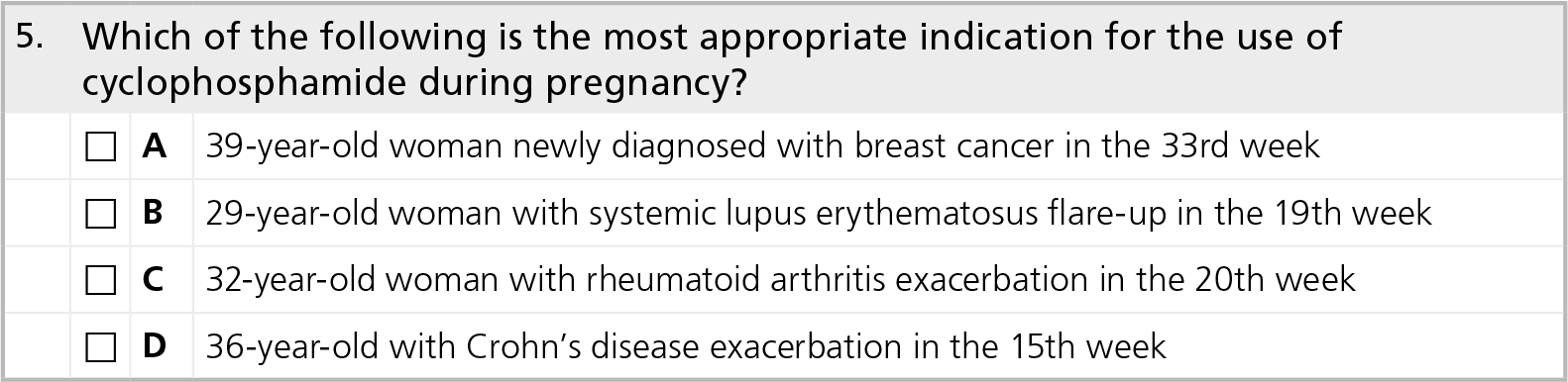
Cyclophosphamide is an alkylating agent that is commonly used for the treatment of malignancies, as well as the treatment of lupus nephritis and various forms of vasculitis. Antenatal exposure to cyclophosphamide early in pregnancy in several animal species and humans is associated with a severe embryopathy that includes craniosynostosis, facial anomalies, distal limb defects and developmental delay [53]. While absolutely contraindicated in the first trimester of pregnancy, cyclophosphamide can be used in the latter half of pregnancy in situations where the life and health of the expectant mother is at risk, most commonly in cases where breast cancer is diagnosed during pregnancy. In one study, three pregnant women diagnosed with breast cancer between weeks 14 and 20 of gestation were reported on; all three women received a combination of cyclophosphamide and doxorubicin shortly after diagnosis. All three women delivered healthy infants without fetal abnormalities [54]. By contrast, Clowse et al. reported on four lupus patients who were treated with cyclophosphamide during pregnancy. Two patients who were inadvertently exposed to cyclophosphamide at conception for the treatment of lupus nephritis experienced spontaneous abortions at weeks 9 and 13 of gestation. Two additional patients were treated with cyclophosphamide at gestational weeks 20 and 22 for severe multiorgan lupus flare-ups, and both suffered fetal demise within 7 days of introduction of cyclophosphamide, despite normal ultrasounds prior to treatment [55]. The severity of underlying maternal disease is a likely confounder for these fetal deaths.
Based on these data, cyclophosphamide is contraindicated in early pregnancy and should be used with caution in severely ill women during the second half of pregnancy, although outcomes may still be poor for both the mother and fetus. Similarly, lactation is contraindicated during cyclophosphamide use.
Future perspective
It is hard to imagine that randomized controlled trials of disease-modifying or immunosuppressive medication use during pregnancy will ever be commonplace. However, as more women achieve adequate control of underlying disease and improved daily function, there are likely to be increasing numbers of women who desire pregnancy and look forward to raising children. The rise of pregnancy-related registries that incorporate complete data regarding medication exposure, coupled with data regarding the severity and activity of underlying disease, will enable a more comprehensive understanding of the risk and benefit analysis of medication use during pregnancy.
Executive summary
When counseling women with autoimmune diseases regarding medication use during pregnancy, it is important to balance the potential adverse effects of medication use with the potential adverse effects of untreated, active disease during pregnancy.
Most data regarding antenatal exposure to disease-modifying or immunosuppressive therapies are based largely on observational data and may be confounded by the severity of the underlying autoimmune disease.
Hydroxychloroquine can be used safely during pregnancy, and studies have demonstrated an increase in adverse pregnancy outcomes associated with flare-ups of underlying systemic lupus erythematosus in pregnant women who discontinue therapy.
Methotrexate, leflunomide and mycophenolate mofetil should not be used during pregnancy owing to unacceptable risks of teratogenicity.
Hydroxychloroquine, corticosteroids, sulfasalazine and azathioprine may be safer alternative agents to control underlying disease during pregnancy.
Although there does not appear to be a safety signal regarding the use of TNF inhibitors in early pregnancy, few data are available regarding the safety of continued use of TNF inhibitors during the later stages of pregnancy. However, the benefits of better disease control may outweigh the potential risks of ongoing therapy in some cases.
 Immunosuppressive medications during pregnancy & lactation
To obtain credit, you should first read the journal article. After reading the article, you should be able to answer the following, related, multiple-choice questions. To complete the questions and earn continuing medical education (CME) credit, please go to www.medscapecme.com/journal/wh. Credit cannot be obtained for tests completed on paper, although you may use the worksheet below to keep a record of your answers. You must be a registered user on Medscape.com. If you are not registered on Medscape.com, please click on the New Users: Free Registration link on the left hand side of the website to register. Only one answer is correct for each question. Once you successfully answer all post-test questions you will be able to view and/or print your certificate. For questions regarding the content of this activity, contact the accredited provider,


Footnotes
The authors have no relevant affiliations or financial involvement with any organization or entity with a financial interest in or financial conflict with the subject matter or materials discussed in the manuscript. This includes employment, consultancies, honoraria, stock ownership or options, expert testimony, grants or patents received or pending, or royalties.
No writing assistance was utilized in the production of this manuscript.
CME Author
Désirée Lie, MD, MSEd
Clinical Professor, Family Medicine, University of California, Irvine, Orange, CA, USA; Director of Research and Patient Development, Family Medicine, University of California, Irvine, Medical Center, Rossmoor, CA, USA Disclosure: Désirée Lie has disclosed the following relevant financial relationship:
Served as a nonproduct speaker for: “Topics in Health ” for Merck Speaker Services.
Authors and Disclosures
Amy B Elliott, MD
Division of Immunology and Rheumatology, Stanford University School of Medicine, Palo Alto, CA, USA Disclosure: Amy B Elliott has disclosed no relevant financial relationships.
Eliz a F Chak ravarty, MD, MS
Division of Immunology and Rheumatology, Stanford University School of Medicine, Palo Alto, CA, USA Disclosure: Eliza F Chakravarty has disclosed no relevant financial relationships.
Editor
Elisa Manzotti, Editorial Director, Future Science Group, London, UK.
Disclosure: Elisa Manzotti has disclosed no relevant financial relationships.