
Abstract

Scheibe RJ, Gros G, Parkkila S, et al. Expression of membrane-bound carbonic anhydrases IV, IX, and XIV in the mouse heart. J Histochem Cytochem. 2006;54(12):1379–91. doi:10.1369/jhc.6A7003.2006
In this article, two errors have come to the authors’ attention. These errors pertain to Figure 5, image E, and Figure 7, image A. The corrected composite Figures 5 and 7 are presented here.
In Figure 5, a new image unit (E-G) representing the green and red channels, as well as their respective overlay, has been included. In Figure 7, image A has been replaced with the appropriate image of preimmune staining.
The authors emphasize that these corrections do not affect the overall message of these figures or the article.
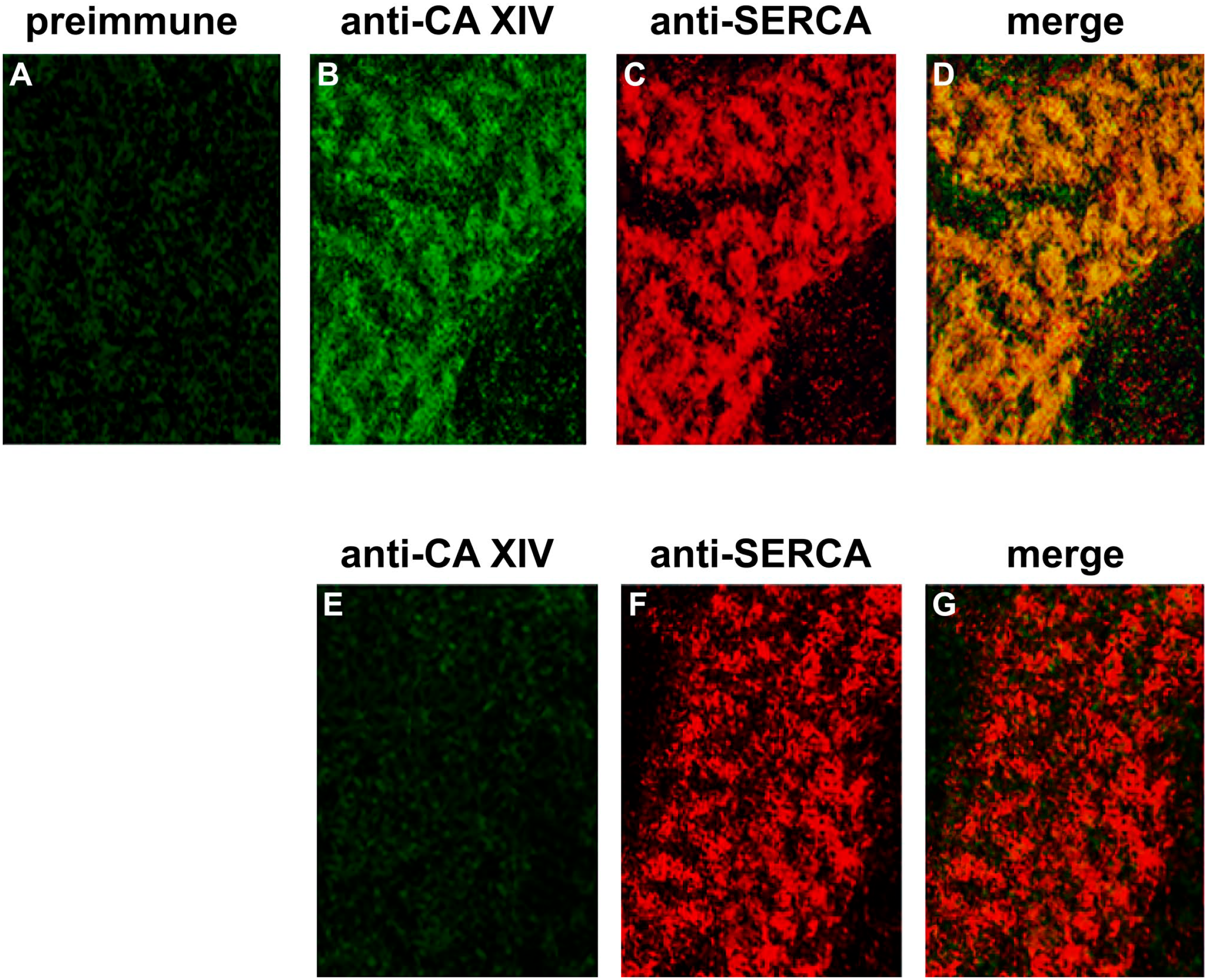
Double-immunofluorescence staining of CA XIV and anti-SERCA2 in adult cardiomyocytes of mouse. (A–D) Cardiomyocytes of wildtype mouse. (A) Incubation with preimmune serum and anti-rabbit IgG/FITC. (B) Anti-mouse CA XIV/FITC. (C) Anti-mouse SERCA2/TRITC. (D) Merge of B and C. (E–G) Cardiomyocytes of CA XIV KO mouse. (E) Anti-mouse CA XIV/FITC. (F) Anti-mouse SERCA2/TRITC. (G) Merge of E and F. Confocal laser scanning microscopy.
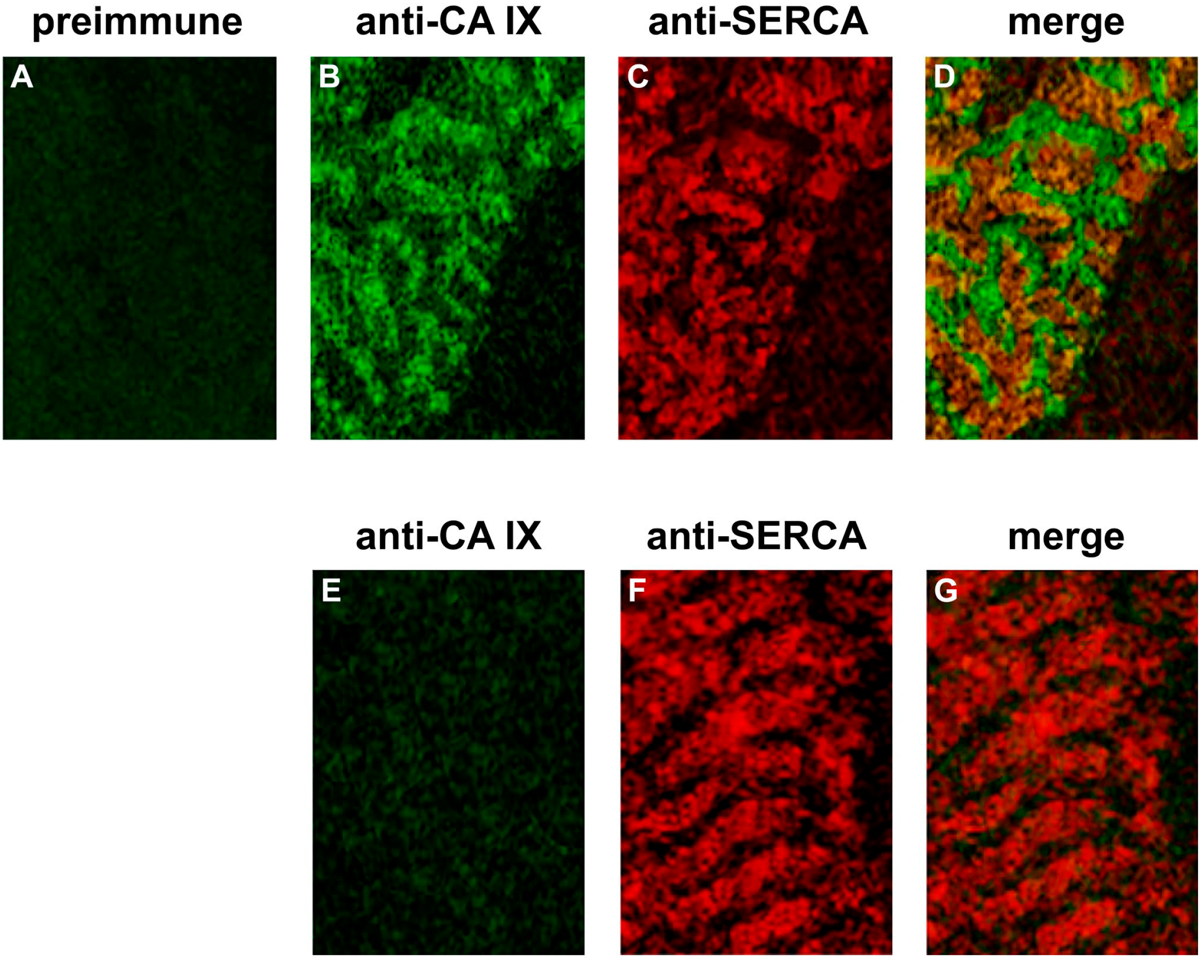
Double-immunofluorescence staining of CA IX and SERCA2 in adult cardiomyocytes of mouse. (A–D) Cardiomyocytes of wild-type mouse. (A) Incubation with preimmune serum and anti-rabbit IgG/FITC. (B) Anti-mouse CA IX/FITC. (C) Anti-mouse SERCA2/TRITC. (D) Merge of B and C. (E–G) Cardiomyocytes of CA IX KO mouse. (E) Anti-mouse CA IX/FITC. (F) Anti-mouse SERCA2/TRITC. (G) Merge of E and F. Confocal laser scanning microscopy.