
Abstract
Clinical and pathologic findings for the spontaneous poisoning by Sida carpinifolia in cattle are described in this study. A survey on field cases of S. carpinifolia in cattle was carried out on farms of Alto Vale do Itajai, State of Santa Catarina, southern Brazil. Sixteen affected animals were clinically evaluated and 9 were subjected to postmortem examination. The main clinical signs consisted of marching gait, alert gaze, head tremors, and poor growth. Histologic and ultrastructural lesions consisted of vacuolization and distension of neuronal perikarya, mainly from Purkinje cells, and of the cytoplasm of acinar pancreatic and thyroid follicular cells. Clinical signs and lesions varied from mild to severe. Improvement of the clinical signs was observed in cattle after a period of up to 90 days without consuming the plant; however, residual lesions, mainly characterized by axonal spheroids and absence of Purkinje neurons in some areas of the cerebellum, were observed in these cases. It is concluded that the natural chronic consumption of S. carpinifolia was the etiologic cause of storage disease in cattle in this study.
Introduction
Tremorgenic diseases in cattle are common in southern Brazil and usually are caused by phytotoxins. 36 Plants that affect the central nervous system (CNS) are a meaningful group in Brazil. Within this group are plants that cause, among other clinical signs, head and neck tremors and thus are called tremorgenic plants. This group is represented by Phalaris angusta; 17, 34 Solanum fastigiatum var. fastigiatum; 31 Ipomea spp. I. sericophylla, 4 I. riedelli, 4 and I. carnea; 2 Turbina cordata; 10 Sida carpinifolia; 7, 13, 16, 19, 25, 33 and the fungi Claviceps paspali 32 and Aspergilus clavatus. 24
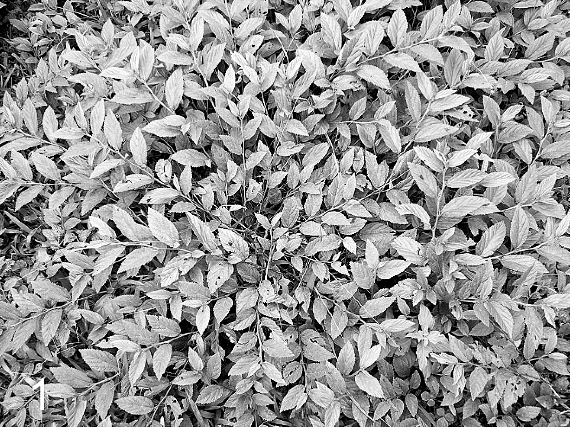
S. carpinifolia, of the Malvaceae family, is a weed plant common in humid and shaded places, flourishing especially in pastures and wastelands. 23 This plant is responsible for producing lysosomal storage disease in goats, 7, 13, 19 ponies, 25 sheep, 33 and cattle. 16 The main clinical signs in goats, sheep, and cattle are incoordination with dysmetria and head tremors associated with frequent falls. Necropsy does not reveal significant gross lesions. Microscopically, the main lesions are limited to the CNS and are characterized by neuronal cytoplasmic vacuolization, mainly in Purkinje and glial cells. 7, 13, 16, 19, 33 Interestingly, a chronic disease characterized by head and neck tremors, alert gaze, uncoordinated gait, and underdevelopment has been observed in ranches located at the region of Alto Vale do Itajaí, State of Santa Catarina, southern Brazil. Signs of consumption of large amounts of S. carpinifolia by cattle were observed on all ranches for which the disease was diagnosed. This study aimed to characterize the epidemiology; clinical signs; and gross, microscopic, and ultrastructural lesions of S. carpinifolia poisoning in cattle in southern Brazil.
Materials and Methods
Outbreaks occurred on 6 farms (Nos. 1–6) in 4 districts located in Alto Vale do Itajaí region, State of Santa Catarina, southern Brazil. Sixteen bovine specimens spontaneously poisoned were clinically evaluated, from which 9 were chosen for postmortem examination (Nos. 1–9). The clinical examination consisted of evaluation of posture, behavior, spontaneous and induced movement, and the head raising test 30 (HR test). The HR test consists of raising the head of the animal for 60 seconds and then abruptly releasing it.
Bovine Nos. 1, 5, 6, 7, and 8 were removed from the pasture infested by S. carpinifolia and euthanatized, whereas bovine Nos. 2, 3, 4, and 9 were transferred to a pasture without the presence of the plant and euthanatized after a period of 90, 90, 120, and 150 days, respectively, without consuming S. carpinifolia.
Tissue samples, including the CNS, of animal Nos. 1–9 were fixed in neutral formalin, embedded in paraffin, and stained with hematoxylin and eosin (HE). Samples of cerebellum, pancreas, and thyroid of animal No. 6 were collected for electron microscopy in glutaraldehyde at 2%, buffered in sodium cacodylate acidulate, postfixed in osmium tetroxide at 1%, and dehydrated in crescent solutions of ethanol to be embedded in Epon. Thin fragments were stained with uranyl acetate and lead citrate.
Results
The disease occurred on small ranches where S. carpinifolia (Fig. 1) infested the paddocks; in some cases, that species was the predominant vegetation. The owners of the spontaneously poisoned animals reported that, after a period of S. carpinifolia consumption, animals developed a preference for grazing the plant, even when other pasture was available. The morbidity rate varied from 7% in outbreak No. 5 to 90% in outbreak No. 3. The mortality rate was low and usually happened electively by euthanasia. Animal No. 6 was euthanatized after falling into a ditch, not being able to rise again, whereas animal No. 8 was euthanatized after staying recumbent for 2 days. The main neurologic signs observed in the spontaneous poisoning cases caused by S. carpinifolia consisted of alert gaze with frequent ear movements, continuous head–neck shaking, incoordination with reeling gait, hypermetria, frequent falls, and wide stance. In the most severe cases, animals lay down frequently and had difficulties rising. At HR test, animals presented exacerbation of the clinical signs, and the most severely affected fell to the ground. Animal Nos. 2, 3, 4, and 9 showed slight, moderate, moderate, and severe neurologic signs, respectively; however, after withdrawal of S. carpinifolia for a period of 90, 90, 120, and 150 days, most clinical signs faded away; nevertheless, animals remained small in stature, even after weight gain. A single animal (No. 8) out of the 16 clinically evaluated showed normal bone development. All other ailing animals were thin and showed underdevelopment characterized by small body size and a comparatively oversized head. Animal owners reported that intermittent diarrhea was frequently observed. At postmortem examination, no important lesions were found in any organ or tissue.
Sida carpinifolia
The main microscopic lesion observed consisted of a variable degree of fine granular vacuolization in the perikarya of neurons in the central and peripheral nervous systems, thyroid follicular cells (Fig. 2), and acinar pancreatic cells. Because of this vacuolization, cells had a foamy and swollen appearance. The most affected neuronal cells were the Purkinje cells in the cerebellum (Fig. 3), with less intensity observed in neurons from other regions of the CNS, including caudate nucleus, basal nuclei, cerebellar cortex, hippocampus, thalamus, hypothalamus, midbrain, bulb, and spinal cord. In the peripheral nervous system, vacuolization was observed in trigeminal ganglion neurons and in the myenteric plexus.
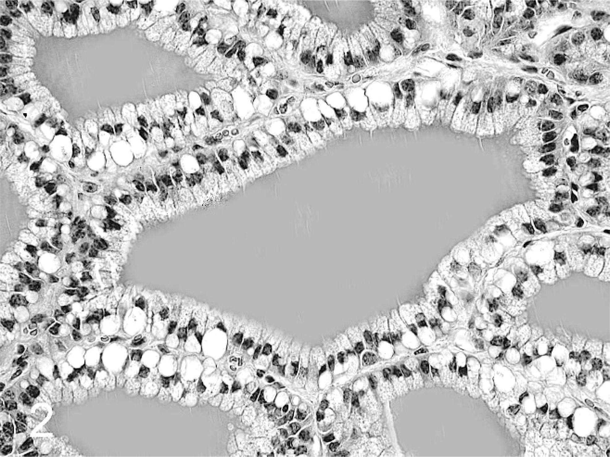
Thyroid; animal No. 8. Fine granular vacuolization in the cytoplasm of follicular cells. HE.
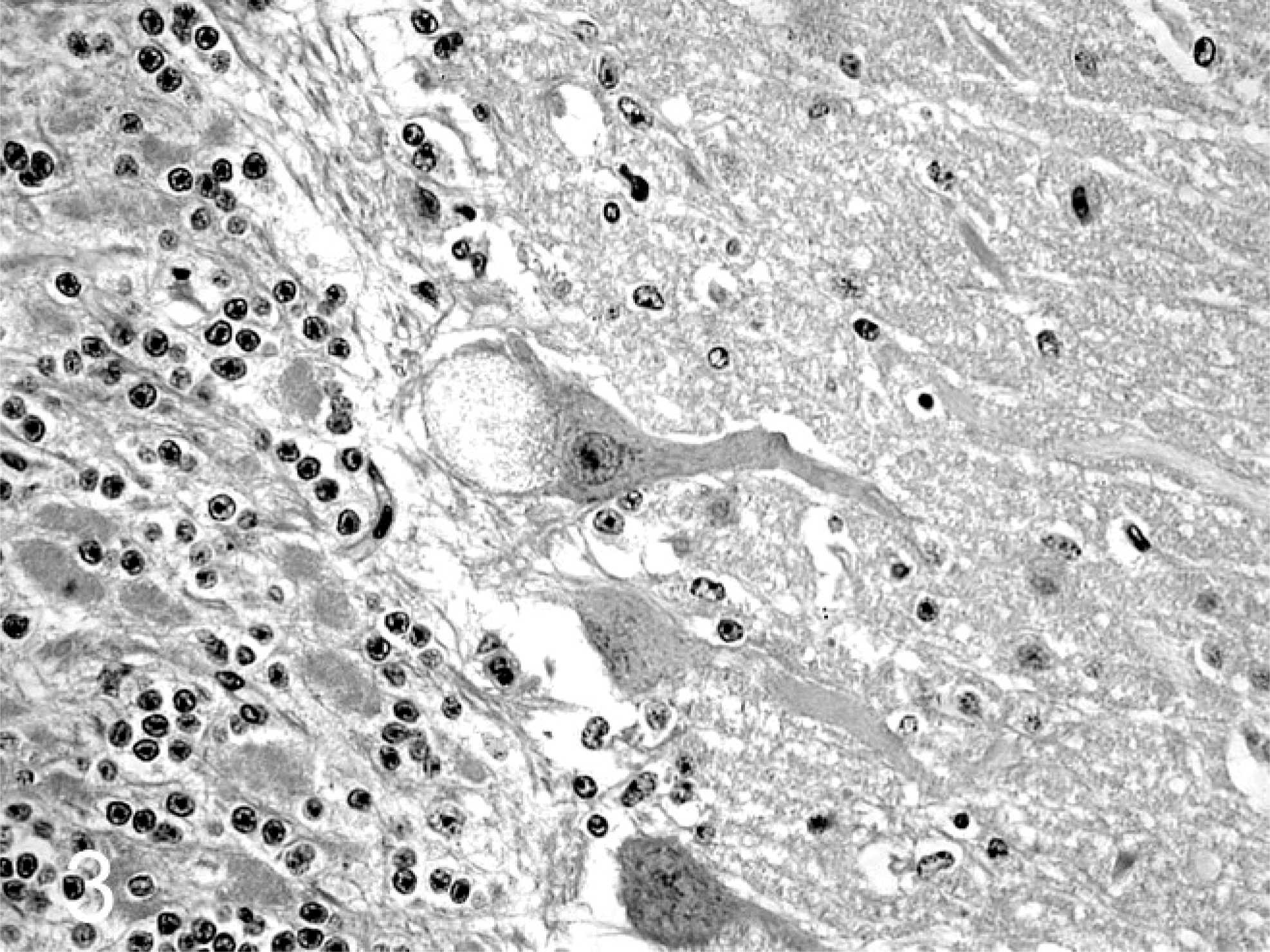
Cerebellum; animal No. 6. Fine granular vacuolization in Purkinje perikarya. HE.
Animal Nos. 1, 5, 6, 7, and 8 showed moderate to severe clinical signs and were euthanatized. Animal Nos. 1 and 5 showed moderate to severe vacuolization in some Purkinje cells and slight vacuolization in the neurons of the granular layer of the cerebellum and ventral horn of the spinal cord. Accentuated vacuolization was found in acinar pancreas cells and macrophages of lymphatic tissues. Axonal spheroids were frequently found, primarily in the granular layer of the cerebellum and in the bulb. In the cerebellum, formation of vacuoles at the molecular level was observed, occasionally with fine granular material. The histologic lesions observed in animal Nos. 6, 7, and 8 were similar to those found in animal Nos. 1 and 5, albeit with more accentuated intensities. Animal Nos. 6, 7, and 8 also showed vacuolization in the collector ductal epithelium of the salivary glands, slight vacuolization in the myenteric plexus and trigeminal ganglion neurons, and moderate to accentuated vacuolization in bulb neurons and thyroid reticular cells. The ultrastructure of perikarya in Purkinje cells, acinar pancreas cytoplasm, and thyroid follicles showed membrane-bounded vacuoles, at times with fine granular material (Fig. 4).
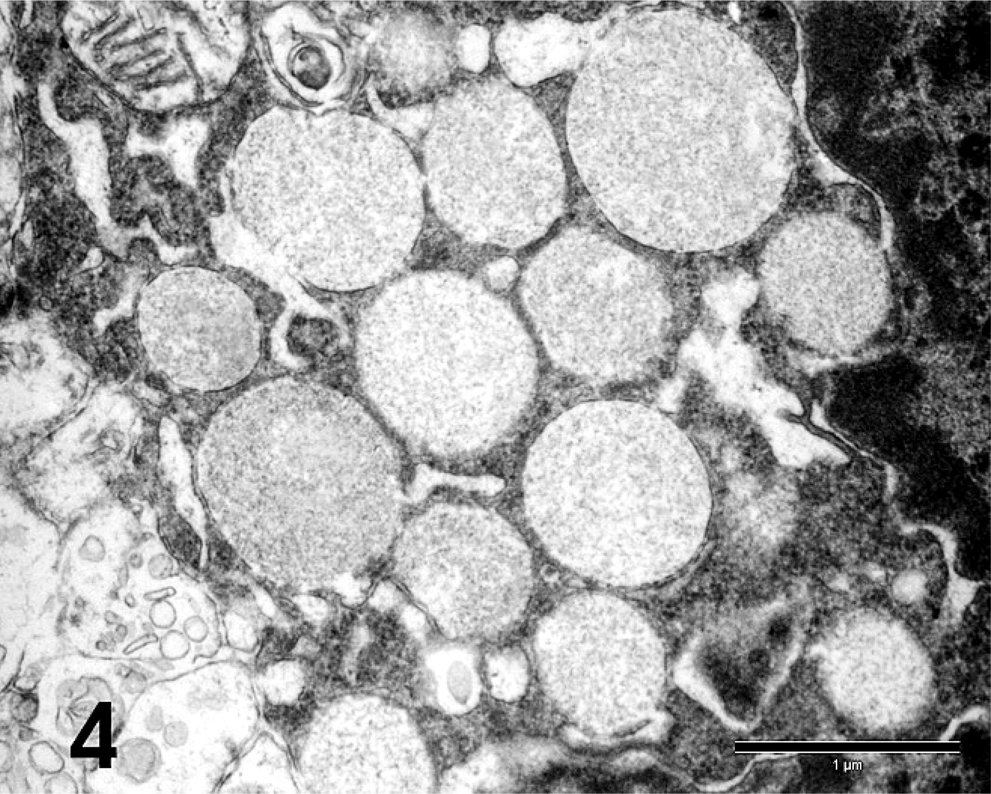
Purkinje perikarya; animal No. 6. Membrane-bounded vacuoles with fine granular material. Uranyl acetate and lead citrate. Bar = 1 µm.
In affected animal Nos. 2, 3, 4, and 9 that were denied S. carpinifolia for 90, 90, 120, and 150 days, respectively, the Purkinje cells showed no vacuolization. Some areas of cerebellum revealed lack of Purkinje cells and Bergman glial proliferation. Axonal spheroids were found at the granular layer of the cerebellum and in the bulb, being more frequent in animal Nos. 2, 3, and 4. Moreover, macrophage infiltration of low intensity was detected in the peripheral vessels in the midbrain of animal Nos. 2 and 3.
Discussion
In this study, the epidemiology, clinical signs, and microscopic and ultrastructural lesions of cattle spontaneously poisoned by S. carpinifolia were characterized. The disease was observed on small farms in which cattle ranching was not the main activity. In these ranches, S. carpinifolia was a common infestation of paddocks, being on occasion the predominant vegetation. On all ranches in which naturally occurring S. carpinifolia poisoning was seen, poor diet and hunger were the causes for intake of the plant by the animals. Another important factor was the degree of S. carpinifolia infestation in the pasture. In some ranches, the toxic plant was as dense and frequent as the cultivar of the pasture.
The neurologic signs observed in this study agree with previously reported cases of S. carpinifolia poisoning in cattle, 16 goats, 7, 13, 19 and sheep. 33 However, ponies spontaneously poisoned by S. carpinifolia mainly showed clinical signs associated with abdominal pain. 24 In this study, animal Nos. 2, 3, 4, and 9 showed moderate to accentuated clinical signs. In these animals, even after being denied the toxic plant for 90, 90, 120, and 150 days, clinical signs were only attenuated. Animals maintained small stature and underdevelopment, but weight gain resumed. Goats and sheep also showed attenuation of clinical signs after plant withdrawal. 7, 33 Despite partial or total recovery, these animals showed residual histologic lesions characterized by lack of Purkinje cells and Bergman glial proliferation in the cerebellum. Occasionally, axonal spheroids were seen at the granular layer of the cerebellum and in the bulb. Such residual lesions were also reported in goats 7 and sheep 33 that presented neurologic signs and were subject to postmortem examination after long periods without consumption of S. carpinifolia. The attenuation of neurologic signs can be explained by histologic and functional cerebellar cortex uniformity. In cases of cerebellar lesion, normal areas gradually assume the functions of areas in which cell death occurred. However, such events cannot take place if the lesion affects the cerebellar nuclei. 26 Therefore, the withdrawal of animals from paddocks infested with S. carpinifolia before they are severely affected by the disease could be an efficient prophylactic practice to avoid or minimize economic losses.
The most frequent microscopic findings were widespread, fine granular vacuolization causing a foamy cytoplasmic appearance, associated or not with swollen cells, and observed mainly in neurons, acinar pancreatic cells, and thyroid follicular cells. These cellular changes were similar to those reported previously in cattle, 16 goats, 7, 13, 19 sheep, 33 and ponies 25 poisoned by S. carpinifolia. Similar lesions were also reported in cases of poisoning by plants that contain the alkaloid swainsonine, 1, 3, 4, 9, 10, 12, 20, 22 as well as in cases of inherited alpha-mannosidosis in cattle, 21, 39 humans, 29 domestic cats, 5, 27 and the guinea pig. 28 As in the inherited disease, in the acquired form of alpha-mannosidosis, the alkaloid swainsonine inhibits lysosomal enzyme alpha-mannosidase, 6, 8, 11, 14 resulting in the storage and excretion of partially impaired oligosaccharides. In addition, swainsonine inhibits alpha-mannosidase II from the Golgi complex, modifying the synthesis and excretion of glycoproteins. 37, 38 Therefore, the enzymatic activity and excretion of oligosaccharides are different between the acquired and inherited forms of the disease. 15 Interestingly, a mononuclear perivascular infiltrate was observed in the CNS of animal Nos. 2 and 3. After examining brains from 7-year old, clinically normal cattle, Gavier-Widen et al. 18 concluded that the inflammatory infiltrate surrounding blood vessels occurred in 30% of the animals in several brain regions, which could be associated with subclinical and latent infections.
The ultrastructural findings agree with those observed in goats 13 and sheep. 33 In most cases, lysosomes were empty, sometimes with fine granular content. The lysosomal storage vacuoles are usually empty in alpha-mannosidosis because of the dissolution of the material during sample processing. 21, 35
The cattle disease characterized by head and neck tremors and body underdevelopment diagnosed in ranches in Alto Vale do Itajaí, State of Santa Catarina, southern Brazil, was related to the continuous ingestion of S. carpinifolia. This plant, when consumed by young cattle, causes body underdevelopment along with neurologic signs. The main histologic lesions caused by S. carpinifolia in bovines were mainly swollen and vacuolized Purkinje, thyroid follicular, and acinar pancreatic cells. The natural chronic consumption of S. carpinifolia was determined to be the etiologic cause of storage disease in cattle in this study.