
Abstract

Not long ago, we read an article in your journal that we found very interesting (Vet Pathol 44:842–848, 2007). The authors (Seixas et al., 2007) described a new histologic pattern in mammary tumors of female cats, the “Invasive Micropapillary Carcinoma.” Recently we diagnosed the same histological variant in a mammary tumor from a male cat.
This 14-year-old European male cat developed 1 tumor (a 1 cm × 0.5 cm × 0.5 cm nodule) in the left inguinal mammary gland. Histologically, the tumor was composed mainly of small papillary structures without stroma (Fig. 1) and small clusters of tumor cells in spaces, mimicking vascular invasion. The polarity of neoplastic cells in each nest was reversed, with the secretion border facing fibrocollagenous stroma. Neoplastic cells had moderate atypia and more than three mitotic figures per high-magnification field. Microscopic metastatic lesions in the regional lymph node had the same micropapillary arrangement as the primary tumor (Fig. 2).
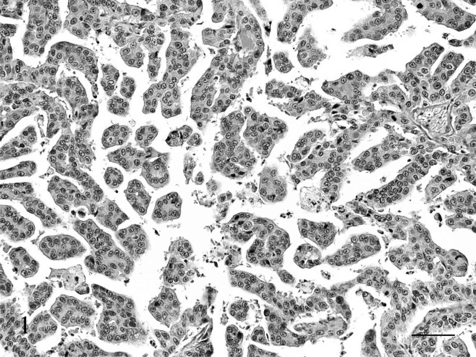
Mammary gland; male cat. Papillary structures without fibrovascular stroma. HE. Bar = 60 µm.
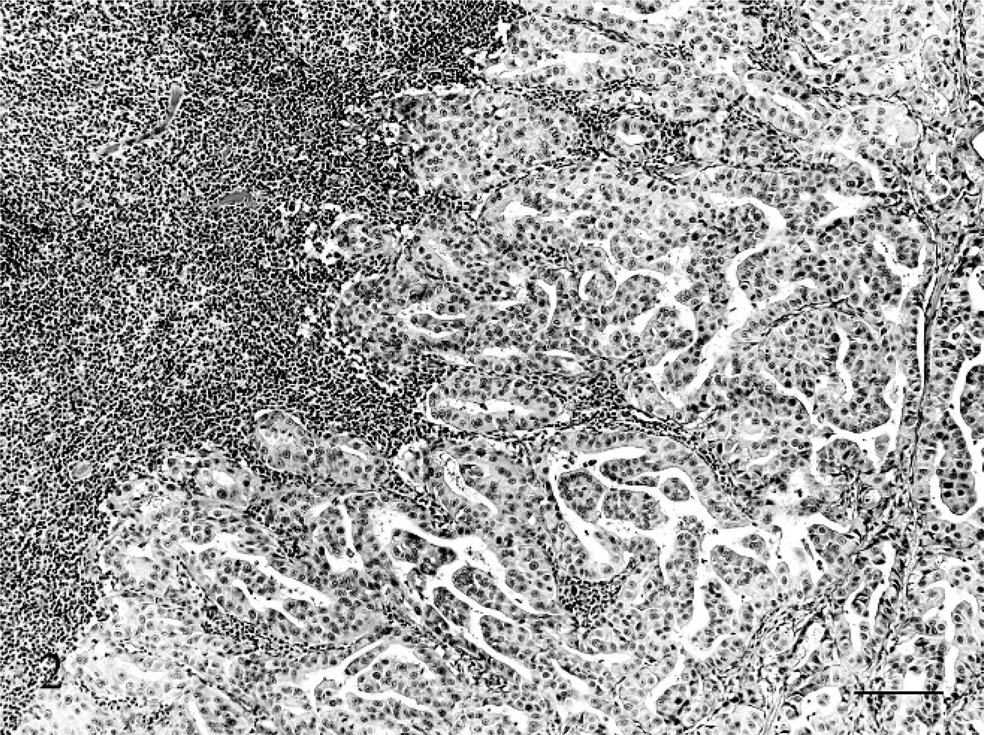
Lymph node; male cat. Lymph node metastasis with micropapillary pattern. HE. Bar = 120 µm.
Mammary invasive micropapillary carcinoma is a recognized variant of human breast cancer, described by Siriaunkgul and Tavassoli in 1993 3 and more recently described in dogs 1 and cats. 2 Feline invasive micropapillary carcinoma is a biologically aggressive tumor with decreased survival due to its tendency to invade lymphatic vessels and metastasize. 2 Mammary tumors are rare in male dogs or cats and, as far as we know, mammary invasive micropapillary carcinoma has not been reported in males of either species. In the case we report here, the histologic features and the presence of nodal metastasis indicate that micropapillary mammary carcinoma in the male cat may resemble that in female cats, with similar aggressive behavior and poor prognosis.