
Abstract

We read with interest the article recently published by Seixas et al. 6 in Veterinary Pathology on the clinicopathologic features of mammary invasive micropapillary carcinoma (IMC) in cats. In this article, the authors reported on several carcinomas of the feline mammary gland displaying histologic features that correspond to IMC of human breast.
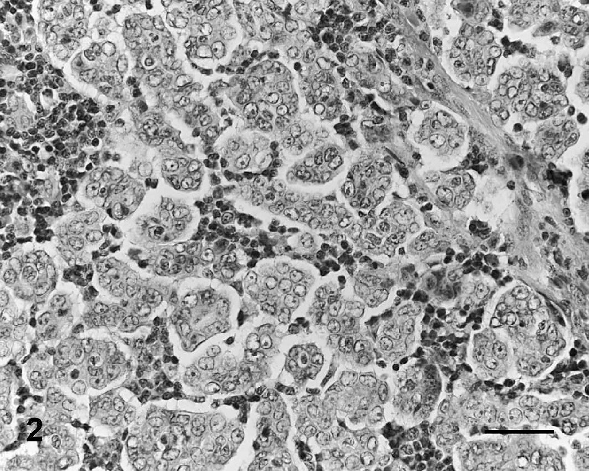
Until now, only 2 cases with this morphologic pattern were described in canine mammary gland tumors. 1 Studying a series of 296 canine mammary gland tumors, we found 9 invasive carcinomas (3%) displaying more than 50% of a micropapillary pattern, which defines an IMC. Using microscopic analysis, these tumors were shown to be characterized by papillary cell clusters surrounded by empty lacunar spaces. Papillae lacked a true fibrovascular core and were lined by polygonal cells showing intermediate- to high-grade nuclei (Fig. 1). A pure micropapillary pattern was not found, with the infiltrating micropapillary areas seen in association with tubulopapillary or solid areas. The ages of affected dogs ranged from 7 to 15 years old (mean age, 10.2 ± 2.8 years), and tumor size varied from 1 to 10 cm. Skin involvement was found in 5 cases, generally associated with extensive ulceration (n = 4). Most cases were high-grade carcinomas (3 were grade II and 6 were grade III), and lesions were always associated with stromal invasion and lymphovascular tumour emboli. The presence of regional lymph node metastases was observed in 8 out of 9 cases (Fig. 2). Follow-up was available in 5 cases: 4 bitches developed progressive disease within a 4-month period after surgery (median disease-free survival = 2 months) and died or were euthanized in this period (median overall survival = 3 months). Concerning the dogs censored for the survival analysis, one presented a history of pleural effusion associated with mammary cancer and the other presented extensive skin ulceration at the cranial mammary gland level associated with firmness, severe edema, and involvement of the extremities, without subjacent mammary nodules, which are characteristic features of canine inflammatory mammary carcinoma. 5 Both dog owners decided to euthanize the dogs immediately after cytologic/biopsy diagnosis and necropsy findings revealed distant metastasis in these cases.

Large-cell carcinoma, lung; dog. Macroscopic image of the lung, with pallor in the dorsal area of the cranial lobe and mottling of other lobes.
Large-cell carcinoma, lung; dog. Alveolar space contains neoplastic cells with ample cytoplasm and distinct borders. Note large cytoplasmatic vacuoles in some neoplastic cells (arrows). HE. Bar = 100 µm.
Invasive micropapillary carcinoma of the human breast is a subtype of invasive ductal carcinoma, was proposed as a morphologically distinct form by Siriaunkgul et al., 7 and is usually associated with lymph node metastasis and poor prognosis. 2, 3 Although the World Health Organization classification of canine mammary gland tumors is based on descriptive criteria, 4 it does not recognize the micropapillary pattern, which is included in the papillary tumor subtype. Considering that this specific invasive pattern was associated with such aggressive behavior in canine mammary tumors, similar to the observations made by Seixas et al. 6 in cats, we propose the recognition of invasive micropapillary carcinoma as a novel entity in the classification of canine mammary gland tumors.