
Abstract
Granulomatous dermatitis in horses has been linked to many etiologies, including various parasites, fungi, and bacteria. Idiopathic forms of granulomatous inflammation-producing diseases, some of which are localized to the skin, also have been reported in horses. Herein we describe a case of recurrent equine granulomatous skin disease characterized by intranuclear viral inclusions within macrophages and giant cells. The histologic changes were primarily noted in the deep dermis and included multifocal to coalescing areas of necrosis marked by histiocytic cell infiltration and presence of giant cells. Electron microscopic examination revealed intranuclear and intracytoplasmic viral particles consistent with herpesvirus. Sequence results of the polymerase chain reaction product were consistent with equine herpesvirus 2, adding another possible etiology to the list of differentials in cases of equine granulomatous skin disease.
A wide range of etiologies have been reported to cause pyogranulomatous to granulomatous inflammatory lesions localized to the deep layers of the skin in horses. Habronema and Onchocerca species of nematodes have been shown to incite this type of reaction. 6 Fungal infections, including pythiosis, phaeohyphomycosis, sporotrichosis, and histoplasmosis, can produce prolific and often deep granulomatous inflammation. 12 Similarly, botryomycosis, which has been associated mainly with Staphylococcus spp., is characterized by a chronic pyogranulomatous dermatitis and formation of microabscesses and bacterial pseudomycetoma. 10, 12 In addition, a generalized idiopathic granulomatous disease syndrome has been reported in horses. 1, 5, 14 Although generally thought of as a disseminated disease affecting many organ systems, idiopathic granulomatous disease has been described to produce only cutaneous lesions in some cases. 5 To date there have been no reports of granulomatous dermatitis associated with any herpesviruses in horses.
Herpesviruses are icosahedral, enveloped, DNA viruses with nucleocapsids approximately 100 nm in diameter that have been linked to a myriad of disease presentations in a wide range of species. 7 In horses alone, there have been multiple herpesviruses described as the etiologic agents of diseases ranging from reproductive failure and abortion in adults to respiratory disease in foals. 11 Herein we describe a case of chronic, nonresponsive granulomatous dermatitis in a horse with a suspected primary or compounding herpesviral etiology, which we believe represents a unique presentation of herpesvirus.
A formalin-fixed biopsy of skin taken from the neck of a gelding Quarter Horse estimated to be greater than 20 years of age was submitted to the University of Georgia, College of Veterinary Medicine, Veterinary Diagnostic and Investigational Laboratory (Tifton, GA) for histopathologic examination in July of 2003. The referring veterinarian reported that multiple areas of skin along the neck and on the chest were firm on palpation. These areas averaged 5–8 cm in diameter and were flush with the skin surface. Three of these areas had ulcerated, were secondarily infected, and exuding purulent material. The horse had presented similarly in June of 2001, but the problem seemingly spontaneously resolved before recurring in June of 2003. The referring veterinarian also reported some response, but incomplete recovery, when the horse was treated with Tucoprim®, an orally dosed, powdered formulation of trimethoprim and sulfadiazine (Pharmacia and Upjohn, Inc., Peapack, NJ). Routine bacterial and fungal culture of a skin swab yielded no growth. Owing to the poor response to treatment and the recurring nature of the disease, the owner elected euthanasia.
Grossly, the formalin-fixed biopsy specimen showed marked thickening, especially in the deeper dermis. The specimen was embedded in paraffin, sectioned, and stained with hematoxylin and eosin (HE), giemsa, and acid fast for examination using light microscopy. In addition, deparaffinized and rehydrated sections underwent antigen retrieval by applying immunohistochemical concentrated citrate buffer (Fisher Scientific, Middletown, VA) and heating slides in a steamer for 20 minutes. These slides were immunohistochemically stained using a Dako LSAB2 system (Dako Corporation, Carpinteria, CA) and 1 ° 25 dilution mouse monoclonal antihuman B-lymphocyte antigen (Dako Corporation, Carpinteria, CA) and 1 ° 40 dilution rabbit polyclonal antihuman CD3 (T Cell) (Zymed Laboratories Inc., South San Francisco, CA). The chromogen used was Bio-Red fast red (Biopath Labrotories, Oklahoma City, OK) and the counterstain was hematoxylin. Sections were viewed with light microscopy. For ultrastructural examination, tissue was removed from the paraffin block, deparaffinized, and rehydrated. Resulting tissue was then fixed in McDowell and Trump's modified Karnovsky's fixative, postfixed in 1% osmium tetroxide, dehydrated and embedded in epoxy resin. Sections were cut 70–90 nm thick, stained with uranyl acetate and lead citrate, and examined using a Ziess EM 900 transmission electron microscope. For molecular analysis, five 10-µm sections of the paraffin-embedded tissue were cut from the block and processed as described by VanDevanter et al. 17 for a nested polymerase chain reaction (PCR) procedure, which targeted DNA sequences from herpesviral polymerase genes. Exceptions to the protocol were that 5 µl of sample was used in the first round of PCR and all reactions were performed in a MJ Research Thermal Cycler PTC 200 with a hot bonnet (MJ Research, Inc., Incline Village, NV).
Histologically, the specimen showed massive, multifocal to coalescing areas of necrosis in the deep dermis infiltrated and surrounded by neutrophils, lymphocytes, plasma cells, and macrophages (Fig. 1). Several multinucleated giant cells with eccentrically placed nuclei often surrounded these areas. Thrombosed vessels of varying size were present in moderate frequency and some foci of necrosis were centered around these vessels. Neovascularization and fibrosis also were observed around the periphery of areas of necrosis. Large, amorphous, brightly eosinophilic intranuclear inclusion bodies, which resulted in margination of chromatin granules within these nuclei, often were found within macrophages and to a greater degree within nuclei of giant cells (Fig. 1, inset). The superficial dermis was characterized by a mild to moderate infiltration of lymphocytes, plasma cells, and eosinophils. Hair follicles and associated sebaceous and apocrine glands were unaffected, as was the epidermis. Giemsa and acid fast stains were negative for infectious agents. T and B cell immunohistochemical stains highlighted small lymphocytes and plasma cells interpreted as being within normal limits morphologically. No evidence of neoplasia was observed.
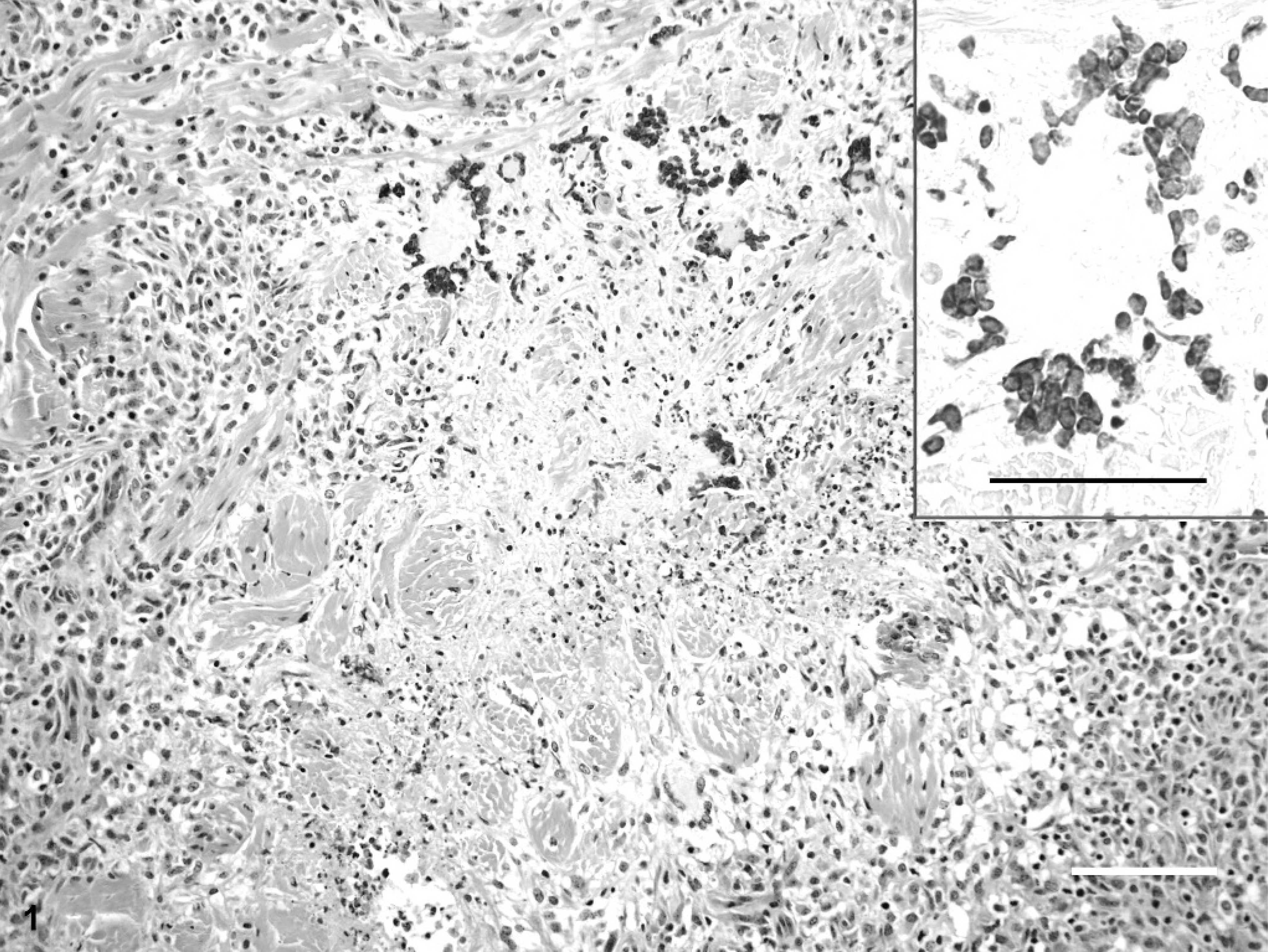
Skin; horse. Light microscopic view of the skin of a horse diagnosed with granulomatous dermatitis within the deep dermis. The inflammation was characterized by multifocal to coalescing areas of necrosis infiltrated and surrounded by neutrophils, lymphocytes, plasma cells, macrophages, and multinucleated giant cells. HE. Bar = 100 µm. Inset shows intranuclear inclusion bodies and margination of chromatin granules within the nuclei of multinucleated giant cells. HE. Bar = 50 µm.
Ultrastructurally, icosahedral virus particles with a maximum diameter of 100 nm, consistent with herpesvirus nucleocapsids, were found within the nuclei and cytoplasm of giant cells (Fig. 2). The herpesvirus nucleocapsids were indistinct, apparently owing to inadequate primary fixation or to degenerative changes prior to fixation (Fig. 2, insets). The moderately electron-dense viral capsids were consistently indistinct and separated from the moderately to intensely electron-dense nucleoid by a narrow electron-lucent space. Some virus particles lacked nucleoids.
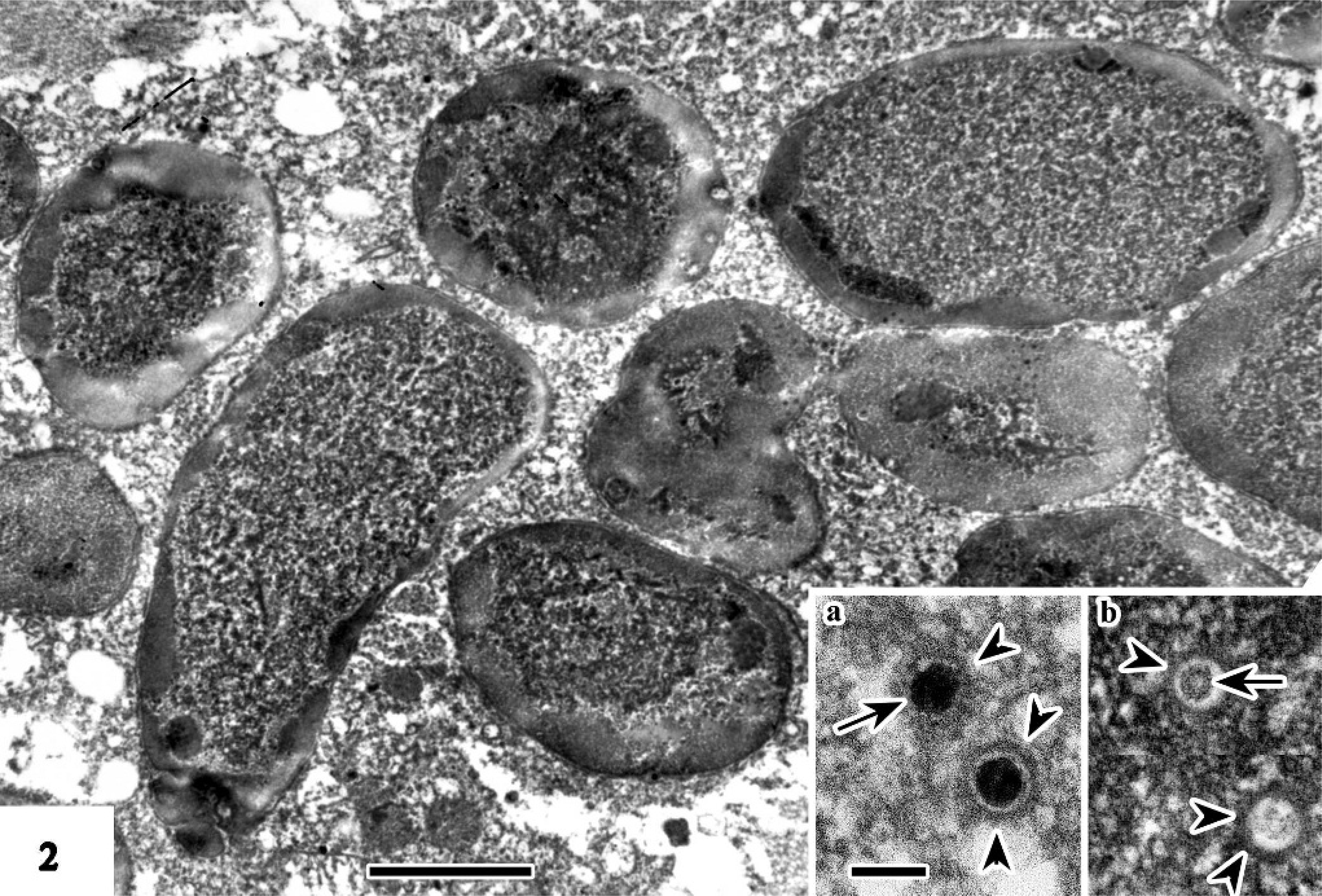
Skin; horse. Ultrastructural view of inclusion bodies within the nuclei of multinucleated giant cells present in a case of equine granulomatous dermatitis. Bar = 2 µm. Insets: cytoplasmic (a) and intranuclear (b) herpesvirus nucleocapsids. Each nucleocapsid is composed of a nucleoid (arrow) surrounded by an electron-lucent space and a blurred capsid (arrowheads). Bar = 100 nm.
The PCR revealed positive results for herpes virus (Fig. 3). This band was cut from the gel and submitted for forward and reverse sequencing. The forward and reverse sequences were assembled using the SeqMan program in the LaserGene Sequence Analysis Package (DNASTAR, Inc) and resulted in a 188-bp consensus sequence. A GenBank Blast search was performed (http://www.ncbi.nlm.nih.gov/Genbank.htmL) on the consensus sequence. A 100% identity nucleotide match was found for equine herpesvirus 2 (EHV-2) (GenBank accession number U20824). 16
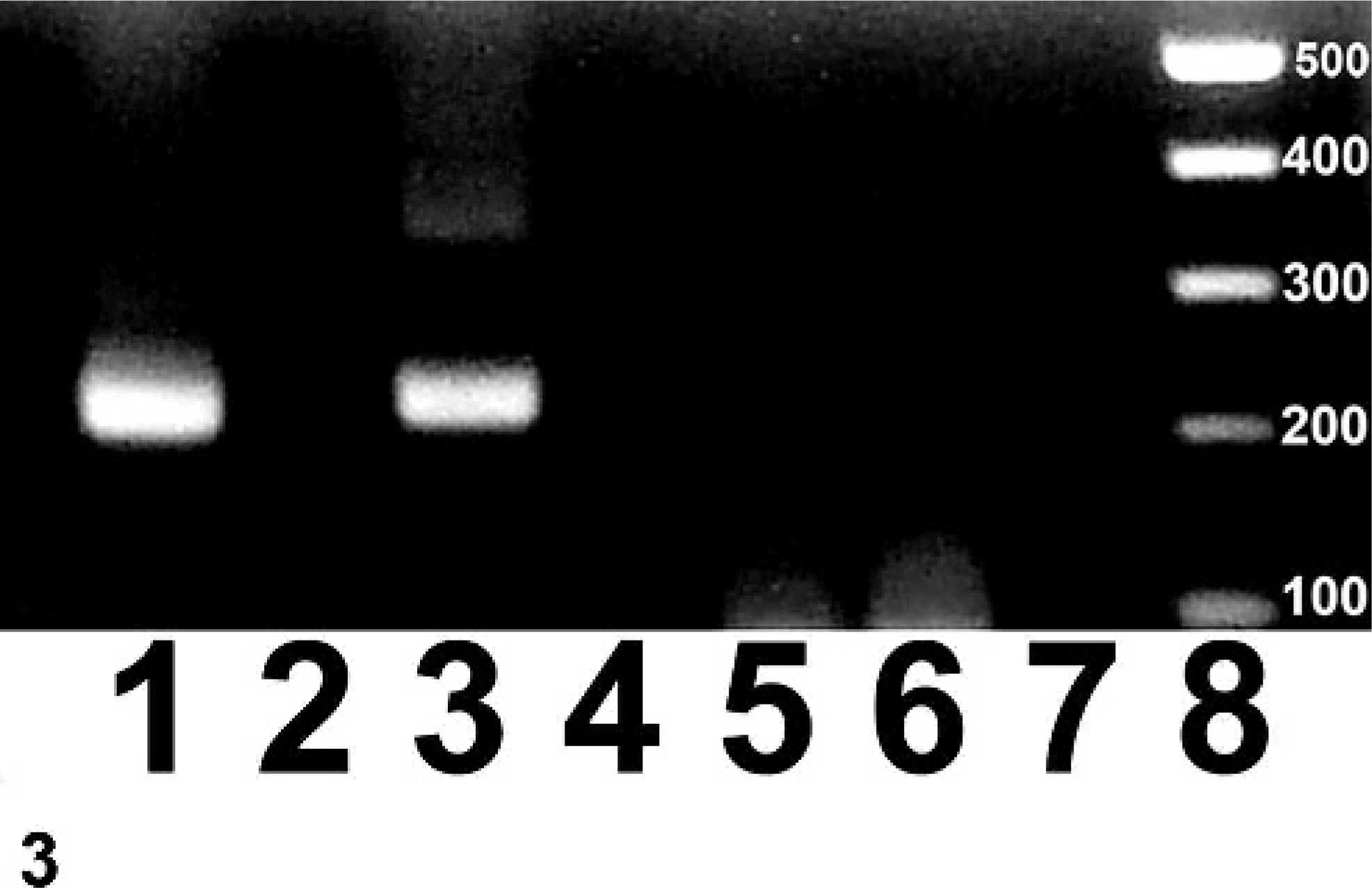
Skin; horse. Gel electrophoresis of the PCR product obtained by using degenerate primers in a consensus primer PCR method to target a highly conserved region within the herpesviral DNA, generally ranging from approximately 200 to 300 bp. The band representing the skin tissue from the horse is present in lane 1 (subsequently sequenced as EHV-2), lane 3 contains the positive equine herpesvirus (EHV-1) control, lane 5 contains the negative control that was subjected to both PCR reactions, lane 6 contains the negative control that was subjected to only the final (i.e., nested) PCR reaction, lanes 2, 4 and 7 are blank and lane 8 contains the molecular weight marker.
The pathogenesis behind these lesions is unknown at the current time. Given the distribution of histiocytic cells and giant cells with intranuclear inclusions within the lesion and surrounding areas of necrosis, the authors consider that EHV-2 is the causative agent rather than an incidental finding. EHV-2 is a gammaherpesvirus that occurs worldwide and may be isolated from healthy horses. 2, 9 It has been associated with pathologic changes in foals, including respiratory disease and pneumonia, and is thought to perhaps facilitate infection with Rhodococcus equi. 2, 9 The mechanism behind the tissue destruction in this case of EHV-2 associated dermatitis is unknown.
In past reports, herpesviruses have been shown to cause a variety of cutaneous lesions in many species. Equine herpes coital exanthema, caused by equine herpesvirus 3, is one such disease that can be recurrent and in which lesions are found associated mainly with progenital skin and mucosal membranes. 13 Coital exanthema is characterized by vesicular type lesions that can progress from papules to vesicles and often to pustules. Histologically, lesions of coital exanthema are located in the superficial layers of the skin and include ballooning degeneration and epidermal hyperplasia. These findings are inconsistent with the deep pyogranulomatous reaction seen in this case.
In cheetahs, feline herpesvirus 1 has been reported to cause cutaneous lesions that are most commonly associated with the eyes and mouth but also may be disseminated to areas of heavy grooming. 8 Gross descriptions of the cheetah cutaneous lesions are similar to those described in this case of equine herpesvirus dermatitis in that both are characterized as firm plaques that often ulcerate. In contrast, histologic examination reveals that the cheetah lesions are confined to the superficial layers of skin and inclusion bodies are seen in keratinocytes of the epidermis rather than in macrophages and giant cells of the deep dermis as seen in this case of EHV-2 associated dermatitis.
Herpesvirus diseases in cattle, including bovine herpes mammillitis and pseudo-lumpy skin disease, have been associated with cutaneous lesions. 13 Granulomatous folliculitis and mild epidermitis were reported in sika deer (Cervus nippon) infected with caprine herpesvirsus-2 (CpHV-2, goat-associated malignant catarrhal fever). 3 The granulomatous folliculitis was characterized by multinucleated cells, epithelioid macrophages, and few lymphocytes. Electron microscopic examination did not reveal viral inclusions within the dermal lesions of the sikas but the deer were positive by PCR for CpHV-2. 3 The cutaneous lesions in the sikas were not characteristic of malignant catarrhal fever, which commonly presents as a cutaneous vasculitis, and were in contrast to the deep dermal lesions noted in this case of EHV-2 associated dermatitis.
Finally, in humans, there are rare reports of granulomatous dermatitis occurring in the sites of previous cutaneous herpes zoster lesions. 4, 15 This dermatitis is characterized by a superficial to mid dermal infiltrate of histiocytes, multinucleated giant cells and lymphocytes. 4, 15 Only early (less than1 month) lesions tested positive for the herpesvirus suggesting that there may be a secondary component to the persistence and perhaps even the development of these lesions. 15 In contrast to this case of EHV-2 associated dermatitis, viral inclusions were not found nor were the lesions predominantly located in the deep dermis.
In summary, the primary differences noted in this case of EHV-2 associated dermatitis compared to previously described herpesvirus lesions of the skin are the affinity for the deep dermis and the cell type in which inclusion bodies are seen (i.e., macrophages and giant cells). This case supports the inclusion of herpesvirus (EHV-2) dermatitis on the etiologic differential diagnostic list in cases of equine granulomatous dermatitis. To our knowledge, this represents the first reported case of equine granulomatous dermatitis associated with a herpesvirus.
Footnotes
Acknowledgements
We wish to thank the Tifton VDIL, especially Dallas Ingram and Lisa Whittington, for assistance in processing tissues and investigating this case.