



Abstract

Nagata T, Nakayama H, Uchida K, Uetsuka K, Yasoshima A, Yasunaga S, Masuda K, Tsujimoto H, Kuwajima E, Nishimura R, Sasaki N, Doi K: Two Cases of Feline Malignant Craniopharyngioma. Vet Pathol
Figures 1–Figures 1–9 were inadvertently printed as one column when they should have been the width of two columns. Allen Press apologizes for this error.

Head; cat No. 1. MRI of an oval-shaped mass at the cranial base (arrows). The white opaque mass is located in the area of the hypothalamus to the optic chiasm.
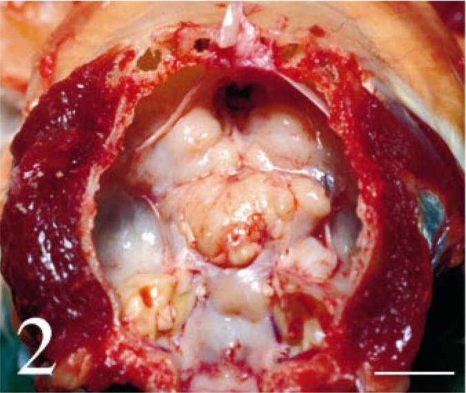
Intracranial tumor mass; cat No. 1. There is a white solid mass on the skull base. Brain was removed. Bar = 1.5 cm.
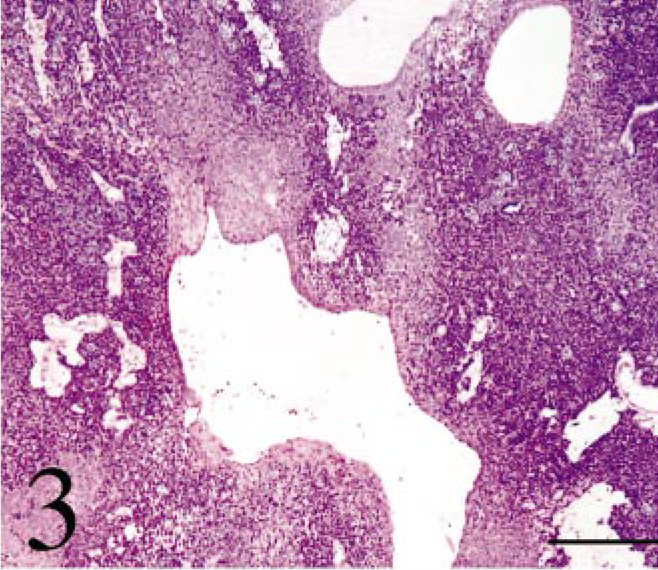
Intracranial tumor mass; cat No. 1. The tumor is composed of small multiangular- or spindle-shaped neoplastic cells proliferating in a solid mass. There are large cysts lined with flat cells. HE. Bar = 700 µm.

Intracranial tumor mass; cat No. 1. The small acinus part. Relatively large cells with pale cytoplasm compose small acini. HE. Bar = 12 µm.

Intracranial tumor mass; cat No. 1. The duct part. Small cuboidal cells compose ducts. HE. Bar = 25 µm.

Intracranial tumor mass; cat No. 1. The pavement part. Large multiangular-shaped cells proliferate in a pavement pattern. HE. Bar = 50 µm.

Intracranial tumor mass; cat No. 1. The tumor cells are positive for keratin. Immunostain counterstained with hematoxylin. Bar = 50 µm.

Intracranial tumor mass; cat No. 1. Electron microscopy of a tumor cell. Note the microvilli at the free margin (small arrows), tonofilaments in the cytoplasm (large arrow) and desmosome-like structures (large arrowheads). The cell binds to the basement membrane by hemidesmosome-like structures (small arrowheads). Bar = 1.2 µm.

Intracranial tumor mass; cat No. 2. The tumor tissue is composed of small cuboidal anaplastic epithelial cells, often making ducts and small or large cysts. HE. Bar = 100 µm.