
Abstract
A bilateral testicular neoplasm from an 11-year-old mixed-breed male dog was removed surgically and examined histologically. The neoplasm was nonencapsulated and composed of acinar and tubular structures lined by one or more layers of neoplastic polyhedral epithelial cells with an abundant mucinous secretion. On histochemistry, all neoplastic cells and associated secretions were periodic acid-Schiff positive. Some neoplastic cells and all associated secretions were positive on mucicarmine stain, and some neoplastic cells, all the stroma, and associated secretions were positive on alcian blue stain. On immunohistochemistry, the neoplastic cells had strong diffuse cytoplasmic immunoreactivity for cytokeratin and vimentin, weak scattered cytoplasmic immunoreactivity for carcinoembryonic antigen and neuron-specific enolase, and no immunoreactivity for S-100. On the basis of histopathology, histochemistry, and immunohistochemical findings, a diagnosis of mucinous adenocarcinoma of rete testis was made. Rete testis adenocarcinoma is a well known but very rare neoplasm in humans. To our knowledge, this is the first report of the mucinous variant of adenocarcinoma of the rete testis in a dog.
Adenocarcinoma of the rete testis is an extremely rare testicular tumor in humans. 9,19 Most patients with this tumor usually present with scrotal swelling and have an associated hydrocele. 17 The prognosis is usually poor, and metastasis to lymph nodes or lungs commonly occurs. 17 In laboratory animals, adenocarcinoma of the rete testis rarely occurs in Fischer 344 rats. 18 Exposure of rats to cadmium chloride have been associated with rete testis adenocarcinoma. 21 Spontaneous cases of this tumor have been reported in mice. 28 In addition, exposure of pregnant CD-1 mice to diethylstilbestrol has resulted in adenocarcinoma of the rete testis in male offsprings. 16 The exact etiology of the tumor is not known; however, the tumor has been associated with epididymitis, 17,19 hydrocele, 17 and maldescended testicles. 11 Diethylstilbesterol-induced adenocarcinoma of rete testis in mice had been proposed as a useful animal model to study the pathogenesis of the lesion. 15 In domestic animals, testicular adenocarcinoma has been reported only in a Merino ram. 24 In humans, the pathologic features of rete testis adenocarcinoma have been well documented by case reports and review articles. 9,17,19
In dogs, Sertoli cell tumors, seminomas, and interstitial cell tumors account for most of the testicular neoplasms. 5,25 These tumors usually have a benign behavior but occasionally metastasize. Other canine testicular tumors such as gonadoblastomas, schwannomas, and leiomyomas are rarely reported. 20,22,27 The detailed histopathologic, histochemical, and immunohistochemical features of adenocarcinoma of the rete testis in domestic animals are scarce. Although rete testis adenocarcinoma has been reported in dogs, 12 this is the first report of the mucinous variant of adenocarcinoma of the rete testis in a dog. This report describes the histopathologic, histochemical, and immunohistochemical findings of a rete testis mucinous adenocarcinoma in a 11-year-old mixed-breed dog.
A bilateral testicular mass was found in a 11-year-old mixed-breed dog. The testicles were surgically excised, fixed in buffered 10% formalin, and sent to the Tifton Veterinary Diagnostic and Investigational Laboratory of the University of Georgia for routine histopathologic examination. Paraffin sections, 4–5 µm thick, were prepared routinely and stained with hematoxylin and eosin.
Microscopic examination of the testicular tumor revealed an expansile and nonencapsulated neoplasm. The neoplasm consisted of variably sized and shaped tubules and acinar structures surrounded by a moderate fibrous stroma with transition from normal rete epithelium to tumor (Fig. 1A). Tubules and acini contained abundant mucinous secretions and were lined by moderately pleomorphic low cuboidal to columnar cells with well-defined cell borders (Fig. 1B). The cells had round to oval to elongate vesicular nuclei with mild amounts of eosinophilic cytoplasm. Mitotic figures were rare. Some tubules were ectatic and contained pale karyorrhectic cell debris, sloughed cells, and large amounts of pale basophilic mucinous secretions (Fig. 1C). There were mild multifocal areas of necrosis and hemorrhage with scattered infiltrates of neutrophils throughout the neoplasm.
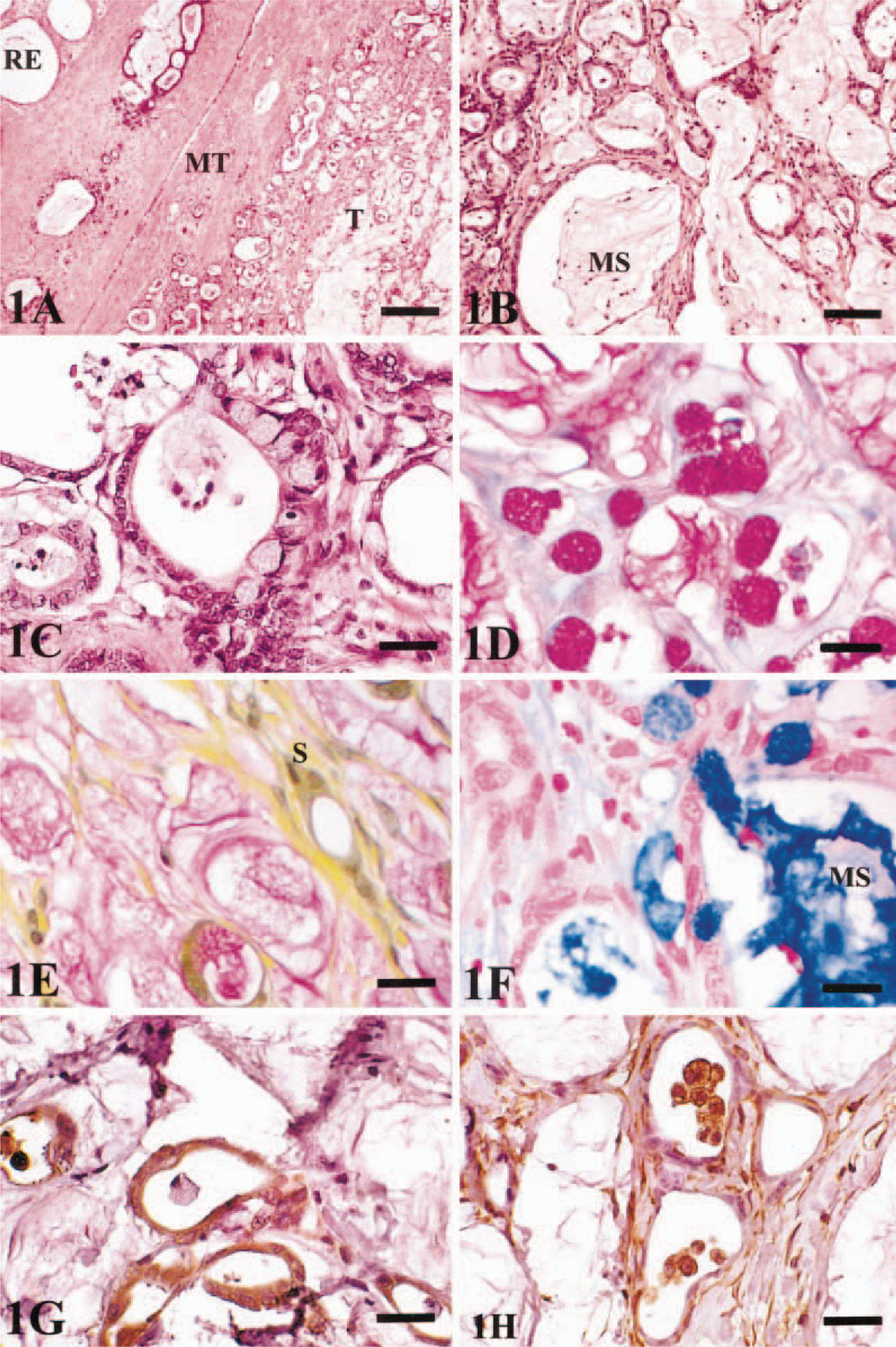
Testicular mass; dog.
To characterize the neoplasm, staining was performed with periodic acid–Schiff (PAS), alcian blue, and Mayer's mucicarmine, using standard methods. 4 In addition, immunohistochemical stains listed below were used as described previously. 8 The following primary antibodies were applied on testicular sections: anti-vimentin (mouse monoclonal, 1 : 40; Dako, Caprinteria, CA), anti-cytokeratin (rabbit polyclonal, 1 : 500; Dako), anti–neuron-specific enolase (NSE) (rabbit polyclonal, 1 : 200; Dako), anti-carcinoembryonic antigen (CEA) (monoclonal, 1 : 50; Zymed, So. San Francisco, CA), and anti–S-100 protein (rabbit polyclonal, 1 : 50; Dako). The streptavidin–biotin–peroxidase complex (Vector Laboratories, Burlingame, GA) and diaminobenzidine kits (Vector) were used to visualize the immune reactions.
Histochemistry demonstrated that all neoplastic cells and associated secretions were PAS positive (Fig. 1D). Some neoplastic cells and all associated secretions were positive on mucicarmine stain, but the stroma was negative (Fig. 1E). All associated secretions and the stroma and some of the neoplastic cells were positive on alcian blue stain (Fig. 1F).
Immunohistochemistry demonstrated that the neoplastic cells had a strong diffuse cytoplasmic immunoreactivity for cytokeratin (Fig. IG) and vimentin (Fig. 1H). Weak scattered cytoplasmic immunoreactivity for NSE and CEA was present. Neoplastic cells did not demonstrate immunoreactivity to S-100 protein.
The rete testis in dogs is part of the excretory ducts (ductus epididymis, ductus deferens, efferent duct, and vas deferens) of the testis that is surrounded by loose connective tissue (mediastinum testis). 7 The rete testis is composed of anastomosing channels that are lined by flat to simple cuboidal to columnar epithelial cells with large, deeply indented nuclei, numerous Golgi bodies, and spherical mitochondria. 7 The cells rest on a thick basal lamina beneath which collagen bundles, blood vessels, lymphatics, and nerve tissue are present. 7 Comparative rete testis ultrastructural studies indicate morphologic similarities in the rete epithelium of various animal species. 3,13 The following features have been reported: 1) collection of secretory granules, 2) prominent Golgi elements, 3) microvilli, 4) lipid droplets, 5) rod-shaped mitochondria, and 6) deep nuclear indentation. 13 We were unable to perform a detailed electron microscopic (EM) evaluation in this study because an initial examination by EM revealed poor cell preservation. However, prominent microvilli, some spherical and elongated mitochondria, indented nuclei, and cytoplasmic fine filaments were observed on EM examination of some sections of the neoplasm.
The following features have been reported as diagnostic criteria for human rete testis carcinoma: 1) primarily testicular mediastinum involvement, 2) no extension into the parietal tunica, 3) transition from normal rete testis to neoplastic rete testis epithelium, 4) lack of any other neoplasia, 5) lack of teratoma, and 6) transmission electron microscopic findings. 16,17 In this study, the neoplasm was primarily located within the testicular mediastinum, did not extend into the parietal tunica, exhibited some transition from normal to neoplastic rete testis epithelium, and no teratoma or other neoplasia was present.
Most of the rete testis carcinomas described in the literature had a predominant papillary growth pattern. 1,9,17 This papillary growth pattern was not seen in this study. A rete testis carcinoma in humans can have features similar to Sertoli cell tumors. 14 In this study, the neoplasm did not have features of a Sertoli cell tumor.
In this study, the tumor was positive for cytokeratin and vimentin and weakly positive for NSE. This is similar to what has been reported in rete testis adenocarcinoma in humans. 1 Adenomatous hyperplasia of the rete testis in humans is usually negative for vimentin. 10 In addition, weak vimentin positivity has been documented in human rete testis adenocarcinoma, whereas strong vimentin positivity was seen in this study. Alcian blue–, PAS-, and mucicarmine-positive staining indicate both acidic and neutral mucopolysaccharide nature of the associated mucinous secretions of the neoplasm.
In humans, mesothelioma and serous carcinoma testis of the tunica vaginalis are differentials for rete testis adenocarcinoma. 1 Mesothelioma is usually positive for cytokeratin and negative for CEA. 6 In this study, the tumor did not have the morphologic features of mesothelioma and was positive for CEA. Serous carcinomas usually have many psammoma bodies, 2 and these were lacking in this study.
The tumor in humans is mostly unilateral and rarely bilateral. 23 In this study, the tumor was bilateral. The tumor can metastasize to regional lymph nodes, bone, liver, and the lungs. 11,26 No evidence of metastasis has been reported in this dog thus far. Radical orchiectomy with retroperitoneal lymph node excision is usually the treatment of choice in humans. 1
On the basis of the histopathology, histochemistry, and immunohistochemical findings, a diagnosis of rete testis mucinous adenocarcinoma was made. To our knowledge, this is the first report of the mucinous variant of canine adenocarcinoma of the rete testis.
Footnotes
Acknowledgements
We would like to thank Lisa Whittington for her technical assistance and Dr. Eloise Styer for her help in electron microscopic examination.