
Abstract
An adult alpaca (Lama pacos) had a locally extensive area of hepatic atrophy involving the right lobe. Grossly, the atrophic lobe was light tan and firm and contained small, raised, white to yellow, partially mineralized circular nodules predominantly at the periphery of the atrophic tissue. Microscopically, viable hepatocytes were not present in the atrophic area, and the tissue consisted of diffuse biliary epithelial proliferation without any evidence of nuclear or cellular atypia or the presence of mitotic figures. The circular mineralized nodules consisted of granulomatous inflammation with intralesional parasitic ova surrounded by fibrous connective tissue. Morphologically, the ova were compatible with those of Fasciola hepatica. The severe biliary hyperplasia was unusual, and it was not clear whether it was caused by an aberrant host response to the parasitic infection or whether it was an unrelated event.
Pathology of the liver is not a commonly recognized problem in South American camelids, and only a limited number of etiologic agents are known to specifically affect the liver in these species.1 Here, we describe locally extensive hepatic atrophy with diffuse biliary hyperplasia and multifocal granulomatous hepatitis associated with ova of liver flukes in an alpaca (Lama pacos).
The affected animal was an adult castrated male alpaca of unknown age used as a control animal in an experimental vitamin D toxicity study. At the end of the experimental period, the alpaca was euthanatized, and a detailed necropsy was conducted.
The carcass was in fair nutritional state, and the most significant gross lesions were present in the liver. The right lobe of the organ was markedly atrophic (approximately 30% of normal size), light tan, and firm and contained numerous small circular foci (up to 4 mm in diameter) on both the diaphragmatic and visceral surfaces (Figs. 1, 2). These foci were predominantly present at the periphery of the atrophic lobe (Fig. 1) and were white to yellow and raised above the capsular surface, with firm to hard consistency. Cut sections of these foci revealed partially calcified material. Similar but fewer of these foci were also seen in other parts of the liver (Fig. 1). Multifocal irregular pale white foci were observed on the dorsal aspects of the diaphragmatic lung lobes. Histologically, these foci were areas of lipid pneumonia (alveolar histiocytosis), which are considered incidental and have previously been described in older llamas and alpacas.6
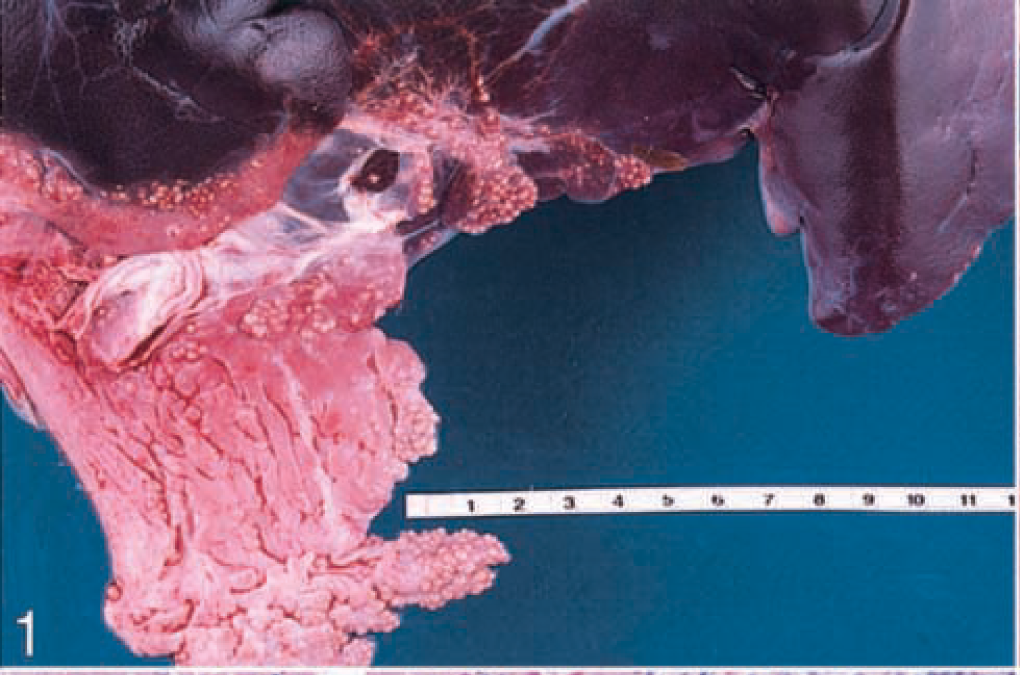
Liver; alpaca. Visceral surface, revealing marked atrophy of the right lobe and many small white to yellow raised circular nodules. Scale in inches.
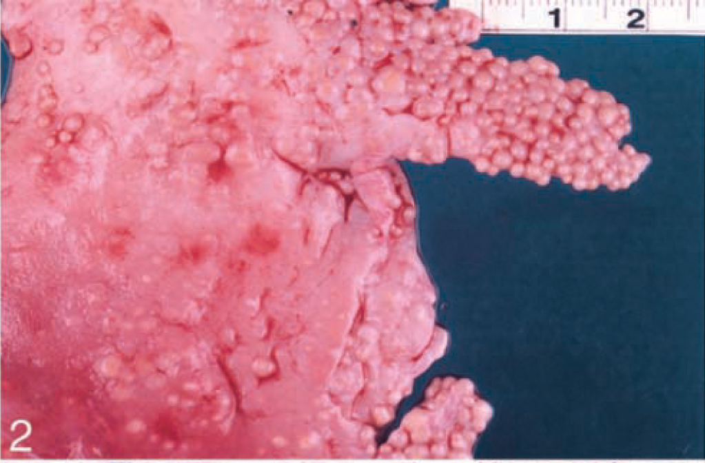
Right lobe, liver; alpaca. Diaphragmatic surface has many circular raised nodules. Scale in inches.
Microscopically, the atrophic lobe of the liver consisted of an accumulation of small bile ducts, some scattered inflammatory cells (predominantly lymphocytes), and a minimal amount of fibrosis (Fig. 3). No nuclear or cellular atypia or mitotic figures were seen in the biliary epithelium. Viable hepatocytes were not observed within this area.
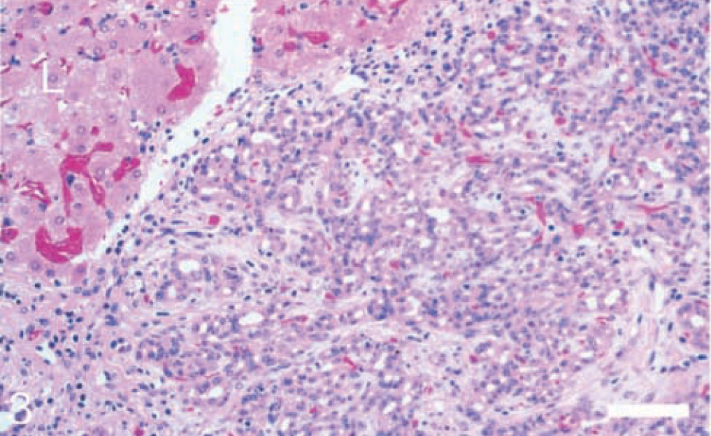
Liver; alpaca. An area of extensive bile duct proliferation is present near the periphery of normal liver tissue (L). HE. Bar = 100 μm.
The circular nodules seen on gross examination consisted of a peripheral area of dense fibrous connective tissue surrounding a central region of partially mineralized cellular debris. Variable numbers of partially degenerate parasitic ova were present in the core of some of the nodules (Fig. 4). The ova were approximately 60–90 × 130–150 μm and were morphologically compatible with ova of Fasciola hepatica.
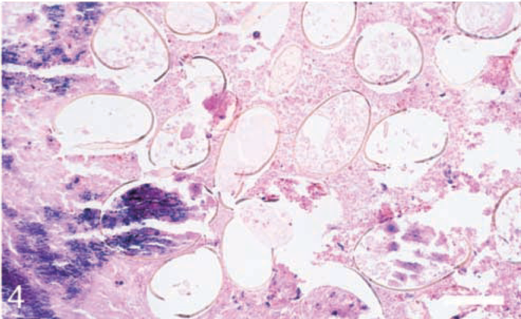
Liver; alpaca. A section of the central area of a partially mineralized granuloma with several parasitic ova of Fasciola hepatica. HE. Bar = 50 μm.
Although liver disease in South American camelids is not common, a number of examples of hepatic pathology have been reported, including metabolic, toxic, infectious (parasitic, bacterial, fungal, viral), and neoplastic conditions.1 Among the parasitic infections, liver flukes (F. hepatica, F. gigantica), immature cysts of tapeworms (Echinococcus granulosus, Taenia helicometra, T. hydatigena), and larval stages of nematodes (Lamanema chavezi) have been reported as etiologic agents.5
F. hepatica infections have been identified in a wide range of species, including the cow, sheep, horse, goat, dog, cat, rabbit, guinea pig, squirrel, deer, beaver, pig, and human.7 Fascioliasis has also been recognized in the llama and alpaca, both as a primary problem8 and as an incidental infection.3 Although there are no published reports of liver flukes in guanaco, F. hepatica has been identified as a significant problem in the phylogenetically related vicuña in a semicaptive research program in Argentina.2 Experimental infections in the llama have demonstrated that the prepatent period of 8–12 weeks is similar to that observed in other species10 and that uncontrolled infection can result in death.5
F. hepatica is endemic in the area where the alpaca had resided. The clinical presentation of F. hepatica infections in South American camelids is highly variable and quantitively related to the level of infection. Animals infected with a low number of parasites frequently show no clinical evidence of disease, whereas heavily parasitized animals most commonly present with symptoms of anorexia and lethargy.9
In South and North America, both acute and chronic forms of liver fluke infections have been reported in these animals,5 although the chronic form is most commonly observed. In this form, the flukes cause blockage of bile ducts and incite extensive fibrosis in the liver. In the affected alpaca, the findings were markedly different. The fibrotic response was minimal in the area of biliary hyperplasia but was extensive around the areas of granuloma formation. The most prominent lesion was an extensive area of biliary hyperplasia and multifocal granulomas containing parasitic ova.
The gross nodular lesions in the liver of this alpaca were also similar to the lesions caused by Lamanema chavezi, which is an important parasite in South American camelids that has not been reported outside of South America.4 Although grossly the hepatic lesions in animals infected with L. chavezi are multifocal and partially calcified (similar to the lesions in the affected alpaca), the microscopic findings are different (i.e., intralesional parasitic ova are not seen within the partially calcified nodules).
Although the host response to the parasitic eggs was unusual, the parasitic ova in this affected alpaca were determined to be those of F. hepatica, and the strange liver tissue reaction was attributed to individual biologic variation of the alpaca. Some may argue that the hyperplastic biliary change was the result of a blockage of a large bile duct, but no such lesion was identified at gross examination. Similarly, the hyperplasia could be attributed to an unidentified hepatotoxic agent(s). However, the biliary hyperplasia was confined to the right lobe, and the likelihood of toxins affecting a locally extensive area of liver is remote.
Footnotes
Acknowledgements
We thank Michael Schadt, Anita E. Sonn, and Mehmet Kupeli for providing technical assistance. This project was funded in part by the Dean's Office, College of Veterinary Medicine, Oregon State University, and the National Animal Disease Center, Agricultural Research Service, USDA.