
Abstract
Cryptosporidium spp. infection was associated with aural-pharyngeal polyps in three iguanas (Iguana iguana). All iguanas were presented for masses protruding from the ear canal, and the disease was characterized by a chronic clinical course. The masses consisted of nests of cystic glands surrounded by abundant fibrous connective tissue and lined by hyperplastic cuboidal to pseudostratified columnar epithelium that was moderately to heavily colonized by cryptosporidial organisms. Electron microscopy revealed that the majority of organisms were trophozoites.
Keywords
Cryptosporidium is a genus of protozoan parasites whose members infect a wide variety of mammalian, avian, piscine, and reptilian species. 1–7, 10 However, the Cryptosporidium species and the predominant site and lesion vary among mammals, birds, fish, and reptiles. C. parvum is an intestinal pathogen of mammals that is a significant cause of diarrhea in calves and humans, especially immunocompromised individuals. 3 In domestic chickens, C. baileyi infects both the gastrointestinal and respiratory tracts, but clinical signs and lesions are mostly associated with respiratory tract infections. 2 In contrast, C. meleagridis induces enteritis and diarrhea in turkeys. 2 In fish, Cryptosporidium infection in the stomach and intestine has been associated with severe wasting. 6 Most Cryptosporidium infections described in reptiles are also gastrointestinal and are often characterized by a chronic course of disease. Hypertrophic or proliferative gastritis is a common manifestation of Cryptosporidium infections in snakes, 1 and atropic gastritis has been described in lacertas. 5 Recently, a case of cryptosporidiosis associated with an aural polyp was described in an iguana (Iguana iguana). 4 Here, we describe three more cases of Cryptosporidium infection associated with aural-pharyngeal polyps in iguanas, implying the ear canal may be a site of infection in this species.
Clinically, all three affected iguanas presented for masses within the ear canal. The first animal presented was an adult male of unknown age with a 2–3-month history of decreased appetite and swelling of the head. Periaural abscesses were diagnosed, debrided, and cultured, but both anaerobic and aerobic cultures were negative. Two months later, the iguana presented with a 1-week history of anorexia. He was bright, alert, and responsive but was exophthalmic, and there was purulent debris near the eyes. A large mass extending from the base of the tongue and into the neck was observed on physical exam. Both tympanic membranes were closed, slightly swollen, and bulging. Because of the poor prognosis, the iguana was euthanatized and a full necropsy was performed. Gross necropsy findings consisted of bilateral firm white masses (diameter: right, 3 × 2 × 2 cm; left, 1.5 cm) located on either side of the intermandibular space and focally attached to the bottom of the skull (Fig. 1). The trachea and esophagus ran ventral to and between these masses. On cut surface, the masses were pedunculated, with cystic spaces and cavities of various sizes filled with granular yellow-brown material.
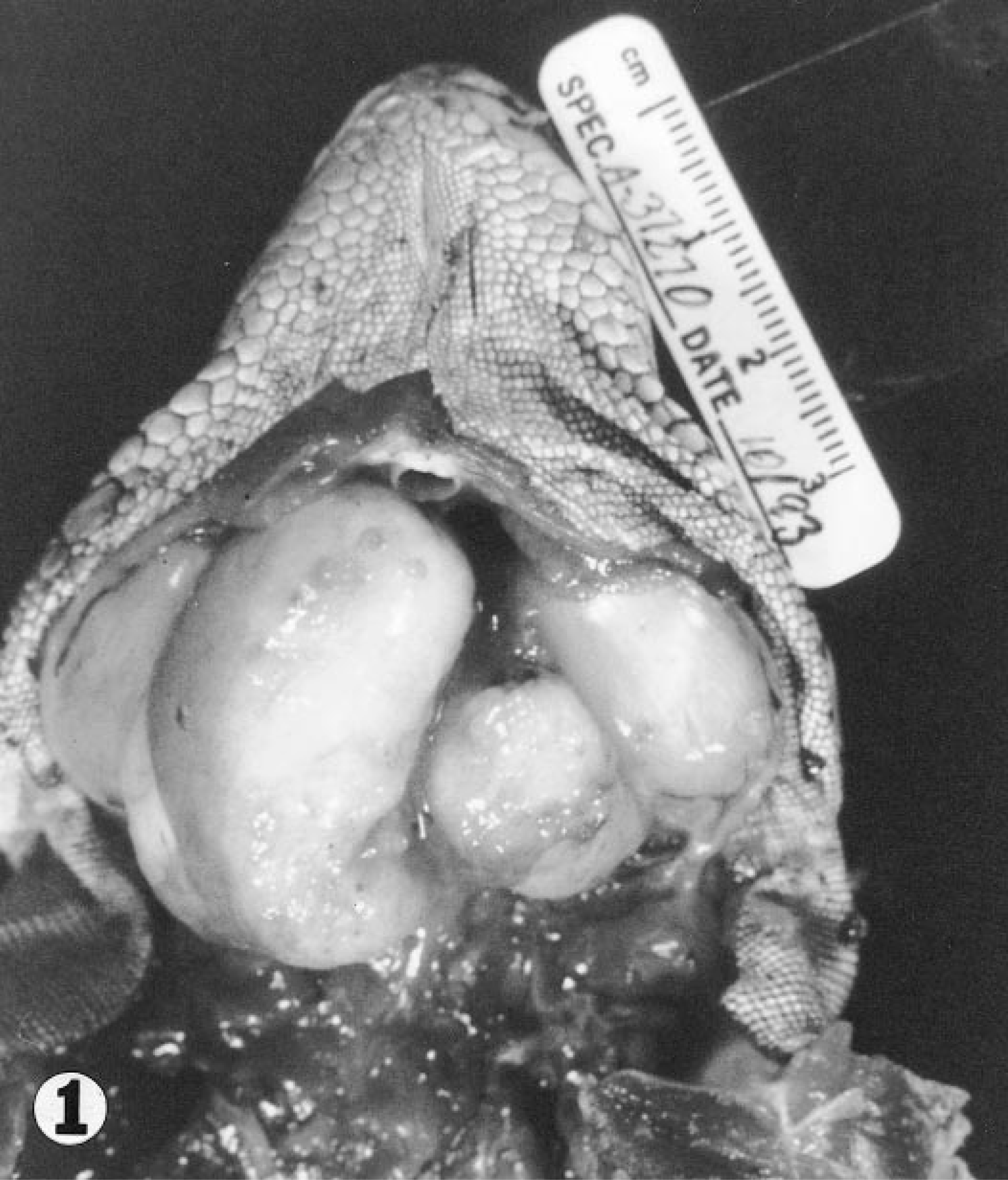
Bilateral aural-pharyngeal masses; iguana. The masses were pedunculated and on cut surface had cystic spaces of various sizes filled with granular yellow-brown material.
The second animal affected was a 5-year-old green iguana that initially presented for irregular growths protruding from the left tympanum. No diagnosis was made on initial presentation. The iguana presented 2.5 years later for inappetence and growths on the right and left tympanic membranes and a 1.5- × 1-cm oval, fluctuant growth protruding from the right auditory tube into the right oropharynx (Fig. 2). A fine-needle aspirate of the mass in the right auditory tube consisted of moderate amounts of necrotic debris, degenerative heterophils, and macrophages. Large numbers of extracellular spirochetes were also observed. Klebsiella oxytoca and Clostridium sp. were cultured from the aspirate. The masses were removed and submitted for histopathology. The third animal was a 2-year-old male iguana presented for surgical removal of a mass within the left ear canal.
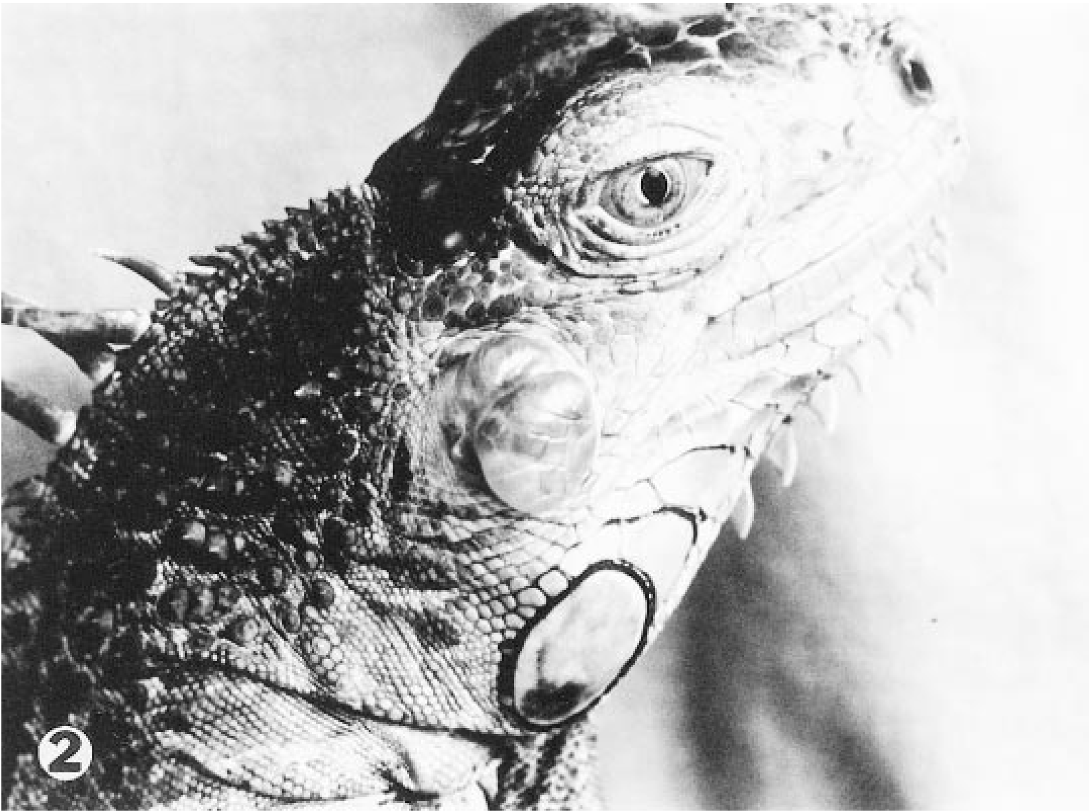
Aural-pharyngeal mass; iguana. Irregular growth extending from the left tympanum. A similar mass was present on the right side.
Histologically, the masses from all three iguanas were composed of fibrous tissue rimmed by oral mucosa containing widely separated ducts, glands, and cysts (Fig. 3). The mucosal epithelium varied from stratified squamous to cuboidal to pseudostratified columnar with numerous goblet cells. The cysts were lined by hyperplastic epithelium that varied from cuboidal to columnar. Some of the cysts, especially in the tissue from the second iguana, contained pools of erythrocytes and lakes of mucus. Other cysts were coalesced, leaving endophytic papillary remnants, or had ruptured, resulting in granulomas composed of aggregates of necrotic material surrounded by epithelioid macrophages. Intact cysts were commonly infiltrated by mononuclear inflammatory cells, predominantly lymphocytes and macrophages, and contained numerous round 3–8-µm protozoal organisms, consistent with Cryptosporidium sp., at the apexes of the epithelial cells. (Fig. 4).
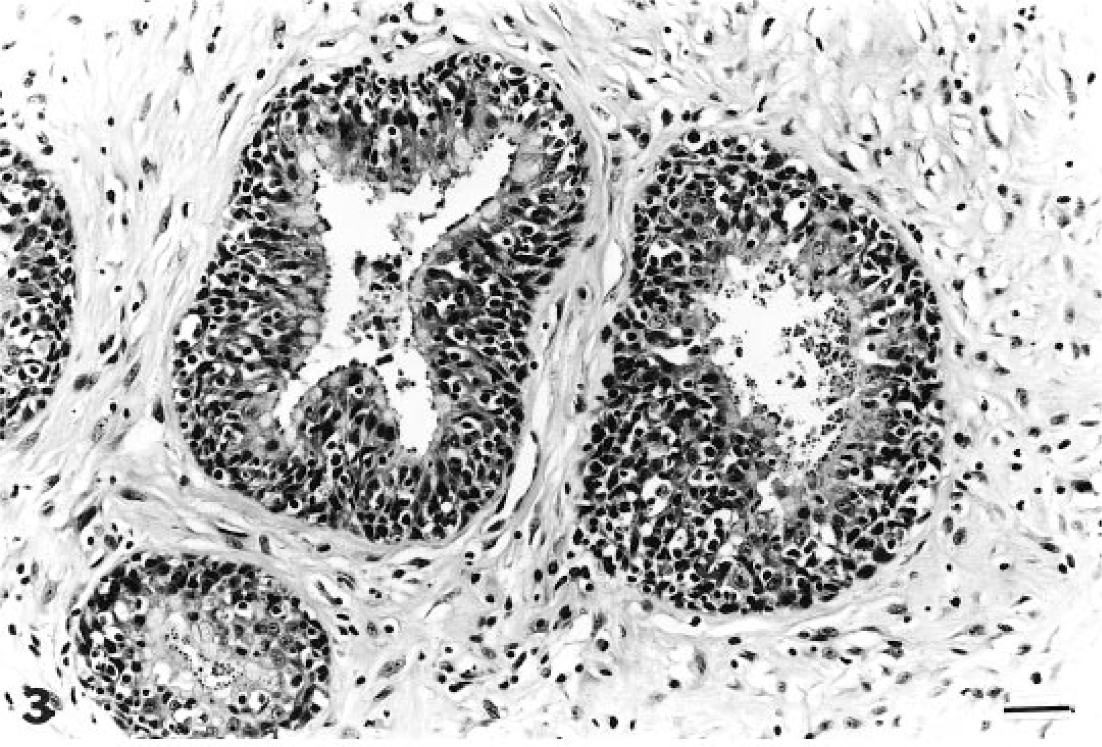
Aural-pharyngeal mass; iguana. The mass was composed of glandular cystic structures lined by hyperplastic cuboidal to columnar epithelium and surrounded by fibrous connective tissue. Numerous cryptosporidial organisms are present along the apical surfaces of the epithelial cells and within the cystic spaces. HE. Bar = 100 µm.
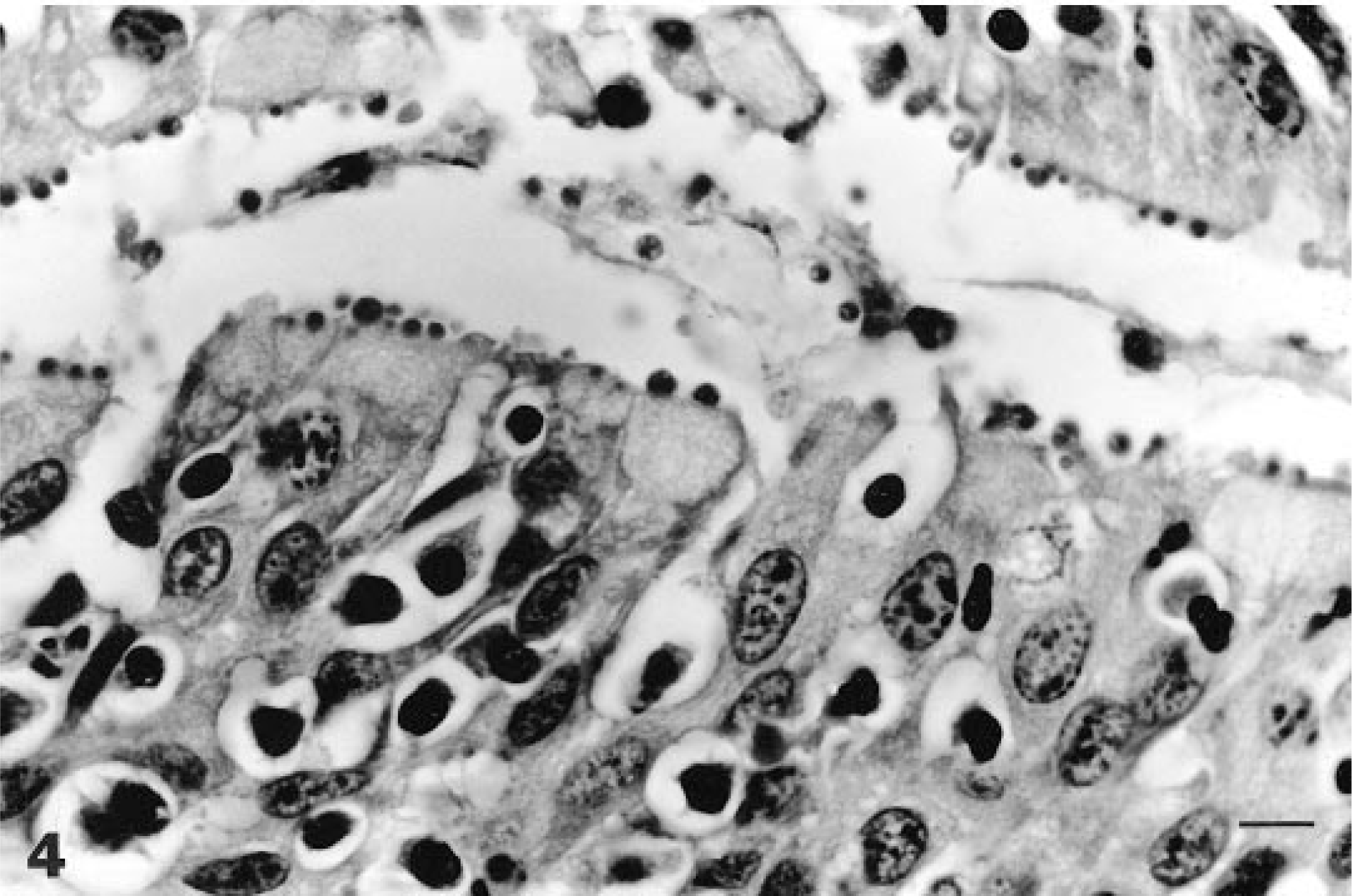
Aural-pharyngeal mass; iguana. Numerous cryptosporidial organisms are present along the apical surfaces of the epithelial cells. Mononuclear inflammatory cells, predominantly lymphocytes and macrophages, are infiltrating the epithelium. HE. Bar = 20 µm.
Sections from the first iguana were deparaffinized and fixed in 2% glutaraldehyde. Ultrathin sections were made and stained with 2% uranyl acetate and Reynold's lead citrate. These sections were examined using a Hitachi H-7000 transmission electron microscope. Many epithelial cells contained one or more cryptosporidial organisms, predominantly trophozoites, beneath the plasma membrane. Trophozoites were approximately 2 × 2.5 µm and were within a parasitophorous vacuole defined by a plasma membrane (Fig. 5). They were characterized by a single prominent nucleus and a distinct feeder organelle at the zone of attachment to the parasitized cell (Fig. 5).
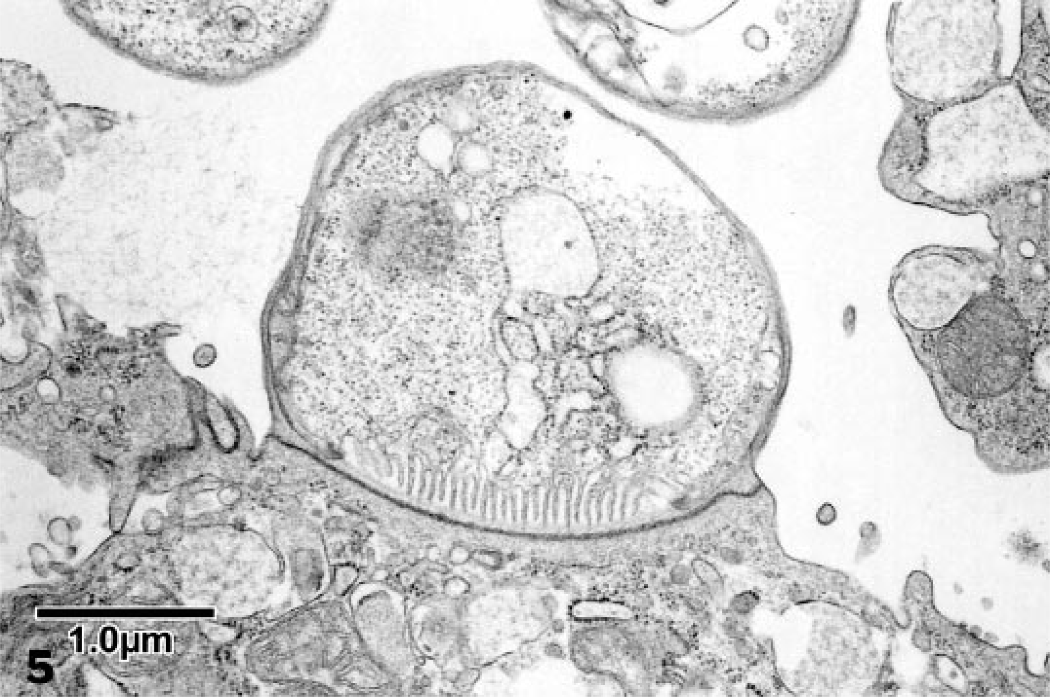
Electron micrograph. Aural-pharyngeal mass; iguana. Cryptosporidial trophozoite within a parasitophorous vacuole of an epithelial cell. The feeder organelle is present at the attachment site. Sections of two other trophoblasts are within the lumen. Bar = 1 µm.
Cryptosporidiosis in reptiles usually affects mature individuals, inducing chronic disease syndromes that are often associated with hyperplastic lesions. Hypertrophic gastritis is the most common presentation of C. serpentis infections in many species of snakes, 1 and one of us (E. W. Uhl) has seen several cases of proliferative enteritis associated with Cryptosporidium infection in leopard geckos. Both sites of infection are clinically associated with chronic wasting syndromes. The three iguanas described here also had chronic proliferative lesions, and the consistent location imply that the ear canal may be a preferred site of Cryptosporidium sp. infection in iguanas. A full necropsy was performed on only one of the three iguanas; however, no other sites of Cryptosporidium sp. infection were identified, and masses in the ear canal was the presenting complaint in all of the animals. In addition, there is one other report of cryptosporidiosis in an iguana, which also had a proliferative mass in the ear canal. 4
The proliferative lesions in these iguanas are similar to the nasopharyngeal polyps described in cats. 9 The exact etiology and origin of these masses in cats is not known, but they are believed to be secondary to chronic inflammation. 8 They usually consist of granulation tissue with an overlying respiratory epithelium that is infiltrated with neutrophils and lymphocytes. 8 The fibrous stroma of the aural-pharyngeal polyps in these iguanas was similar to that described in cats, but numerous glandular nests, which are not a common feature of polyps in cats, were also present. Recurrence of nasopharyngeal polyps after surgical resection in cats has been described 8 and was also observed in the second iguana.
Whether Cryptosporidium induced the formation of polyps, or was a secondary invader is not known. However, an association between this organism and polyps appears to be present; Cryptosporidium has been found in the aural polyps of four iguanas maintained by different owners in different parts of the country. The most likely route of infection is ingestion; however, definitive oocysts were not observed in these iguanas or in the iguana previously described. 4 Clinical and historic investigation, examination of secretions from the pharynx and ear canals of both affected and unaffected iguanas, and complete necropsies are needed to determine the sources, cycle of infection, and role of cryptosporidiosis in the pathogenesis of aural polyps in iguanas. Increased awareness of the association of cryptosporidiosis with these aural-pharyngeal polyps should help identify further cases for study.