
Abstract
Histopathologic and immunohistochemical examinations were conducted on a 5-year-old Holstein–Friesian cow with systemic κAL amyloidosis associated with bovine leukocyte adhesion deficiency. Amyloid deposits were present in the perivascular and intercellular spaces of the visceral organs, such as the liver, kidneys, pancreas, adrenal glands, and upper alimentary tract. Amyloid was stained positively with Congo red with or without 5% potassium permanganate pretreatment and had green birefringence observed under polarized light. Immunohistochemically, amyloid reacted strongly against anti-bovine IgG (H+L) and anti-bovine κ-light chain and reacted weakly against bovine λ-light chain antibodies but was negative for anti-human amyloid AA antibody. This is the first description of AL amyloidosis immunohistochemically related to immunoglobulin κ-light chains of precursor protein in cattle.
Amyloid is a pathologic proteinaceous substance deposited between cells in various tissues and organs of the body in a wide variety of clinical settings.5 Amyloidosis is divided into two types, systemic and localized forms, based on the localization of the amyloid. With reference to amyloid protein type, AL amyloid is associated with primary amyloidosis and immunocyte dyscrasias, and AA amyloid is associated with secondary amyloidosis and chronic inflammatory processes.6 AL amyloidosis is the most common form of human amyloidosis in the United States and is most often associated with multiple myeloma, a systemic neoplastic disease of plasma cells, but may be seen in other plasma cell abnormalities, such as Waldenstrom's macroglobulinemia, heavy chain disease, solitary plasmacytoma, and other types of B-cell lymphoma.5 AL amyloidosis is very uncommon in animals. A few cases have been reported in which AL amyloidosis was associated with myeloma and extramedullary plasmacytoma of cat and dog.2,3 In cattle, AA amyloidosis is the most common form and is usually associated with chronic inflammatory diseases of the mammary gland, joints, and respiratory system.5 However, AL amyloidosis has not been reported in cattle. Histopathologic and immunohistochemical examinations were conducted on systemic AL amyloidosis associated with bovine leukocyte adhesion deficiency (BLAD) in a cow.
A 5-year-old Holstein-Friesian cow had BLAD syndrome characterized as β2 integrin (CD11a,b,c/CD18) deficiency in leukocytes and clinically presented with emaciation, fever, chronic bronchopneumonia, severe neutrophilia (110 × 103 cells/μl), and persistent hypergammaglobulinemia (3.3–4.5 g/dl). Bone marrow transplanation was performed when the cow was 9 months old.7
Tissue samples collected from the liver, spleen, kidneys, heart, lungs, adrenal glands, pancreas, upper alimentary tract, urogenital system, bone marrow, and lymph nodes were fixed in 10% neutral buffered formalin and dehydrated in a series of serial alcohols. Sections for histopathologic examination were cut at 4 μm from paraffin-embedded blocks and stained with hematoxylin and eosin (HE), Congo red with and without pretreatment of 5% potassium permanganate for 5 minutes with examination under polarized light microscopy, and periodic acid-Schiff (PAS) with and without diastase digestion. Serial histologic sections of 10% neutral buffered formalin-fixed liver and kidneys were used for immunohistochemical analysis by the avidin-biotin-peroxidase complex (ABC) procedure (Vectastain Elite ABC kit, Vector Laboratories, Burlingame, CA). Specific antisera used in this study were anti-human amyloid AA monoclonal antibody (Kyowa Medix Corp., Tokyo), anti-bovine IgG (H+L) polyclonal antibody (The Binding Site, Ltd., Birmingham, UK), anti-bovine λ-light chain and bovine κ-light chain monoclonal antibodies (VMRD, Inc., Pullman, WA). The deparaffinized sections were treated in 0.1% trypsin solution for 10 minutes. After washing with distilled water, the sections were treated with 0.3% H2O2 in methanol for 10 minutes to suppress endogenous peroxidase activity and incubated with 10% normal goat serum for 30 minutes. The primary antibody was reacted at 4 C for 12 hours. The sections treated with the primary antibody were reacted with biotinylated rabbit anti-mouse IgG. The antibody reaction products were visualized with 3.3′-diaminobenzidine tetrahydrochloride and counterstained with Mayer's hematoxylin. Control incubations using excess antigen to immune or nonimmune sera as the primary antibody and omitting the primary antibody resulted in the absence of specific staining.
Macroscopically, multiple chronic pulmonary abscesses in anterior lobes of the lung and ulceration of the tongue and gingiva were observed. Histopathologically, an acellular, amorphous pale eosinophilic material (amyloid) was generally observed in the space of Disse of the liver and between and around the hepatocytes, separating them from the lumen of sinusoids (Fig. 1). In the kidneys, many glomeruli were affected and appeared as glomerular tufts enlarged by infiltrates of amyloid (Fig. 2). Amyloid was also deposited in the basement membrane of the collecting tubules and the loop of Henle in the medulla of the kidneys. Heavy deposition of amyloid was present in the wall of the sinusoids in the cortex of the adrenal glands. Multiple perivascular amyloid deposits were observed in the interstitial tissues of the pancreas. In the upper alimentary tract, perivascular amyloid deposits were observed in the lamina proprial mucosae of esophagus, rumen, reticulum, omasum, and abomasum. An acellular, amorphous pale eosinophilic material was stained positively with both PAS with and without diastase digestion and Congo red with and without 5% potassium permanganate pretreatment and had green birefringence observed under polarized light. There were no significant changes in the bone marrow and lymph nodes. Immunohistochemically, amorphous pale eosinophilic material in the liver and kidney reacted strongly to anti-bovine IgG (H+L) polyclonal antibody and anti-bovine κ-light chain (Figs. 3, 4) and faintly to bovine λ-light chain antibodies but was negative for anti-human amyloid AA antibody.
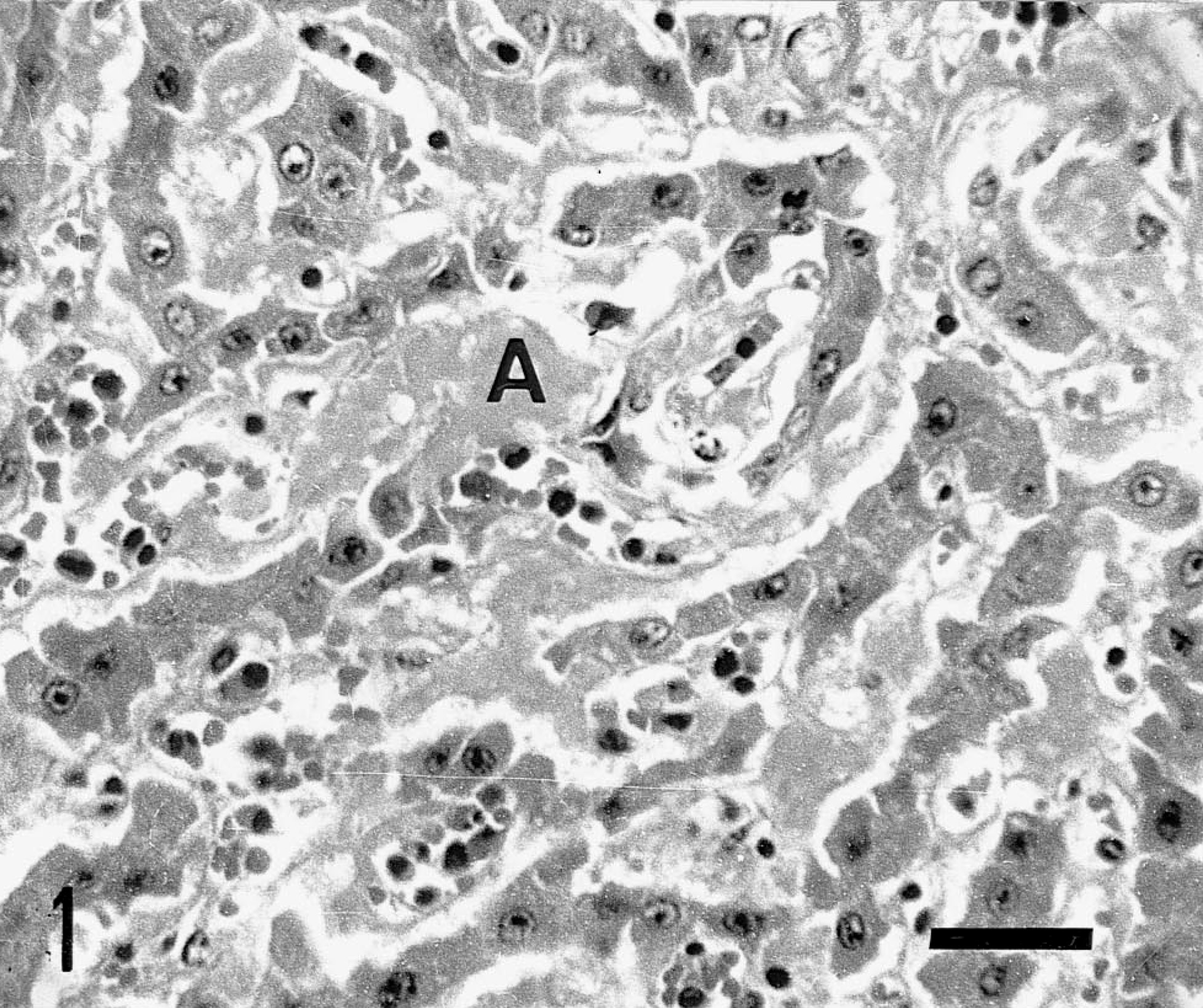
Liver; BLAD cattle. An acellular, amorphous pale eosinophilic material (amyloid; A) is present in the walls of the sinusoids of the liver and between and around the hepatocytes, separating them from the lumen of sinusoids. HE. Bar = 30 μm.
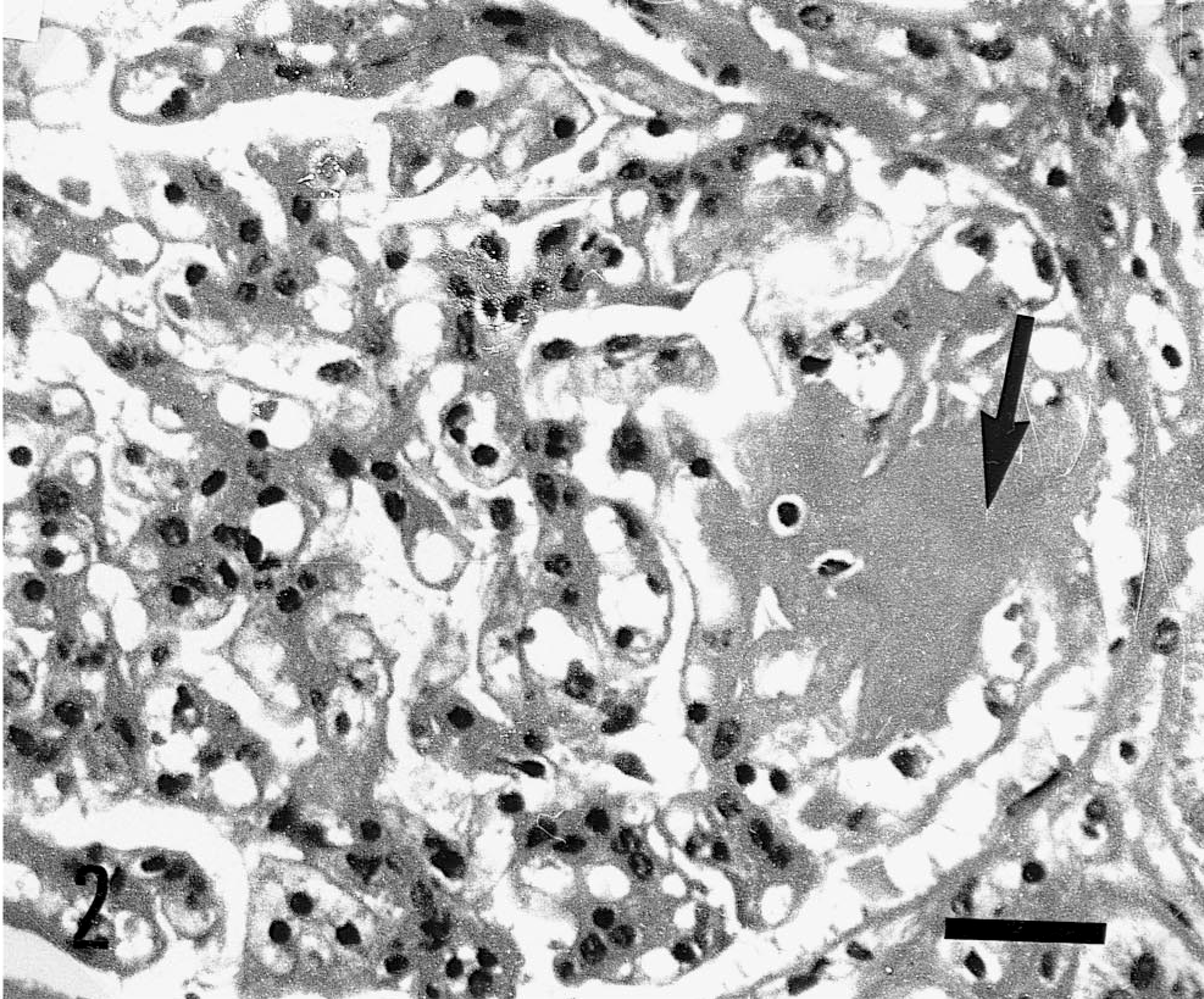
Glomerulus; BLAD cattle. An acellular, amorphous pale eosinophilic material (amyloid; arrow) is present in a glomerulus. Glomerular tuft is enlarged by infiltrates of amorphous material. HE. Bar = 30 μm.
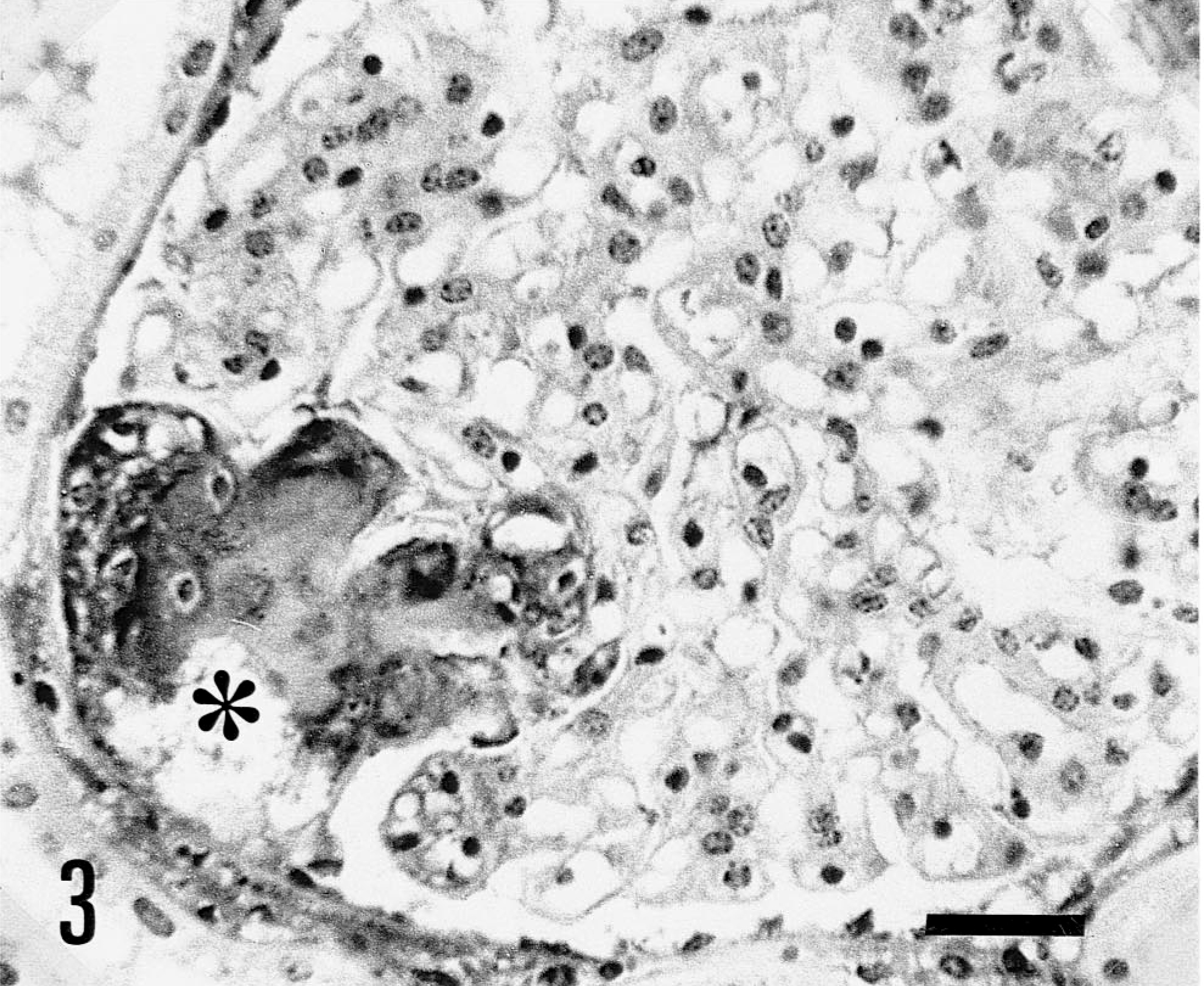
Glomerulus; BLAD cattle. An acellular, amorphous pale eosinophilic material infiltrating a glomerulus is stained with anti-bovine κ-light chain monoclonal antibody (∗). ABC method, Mayer's hematoxylin counterstain. Bar = 30 μm.
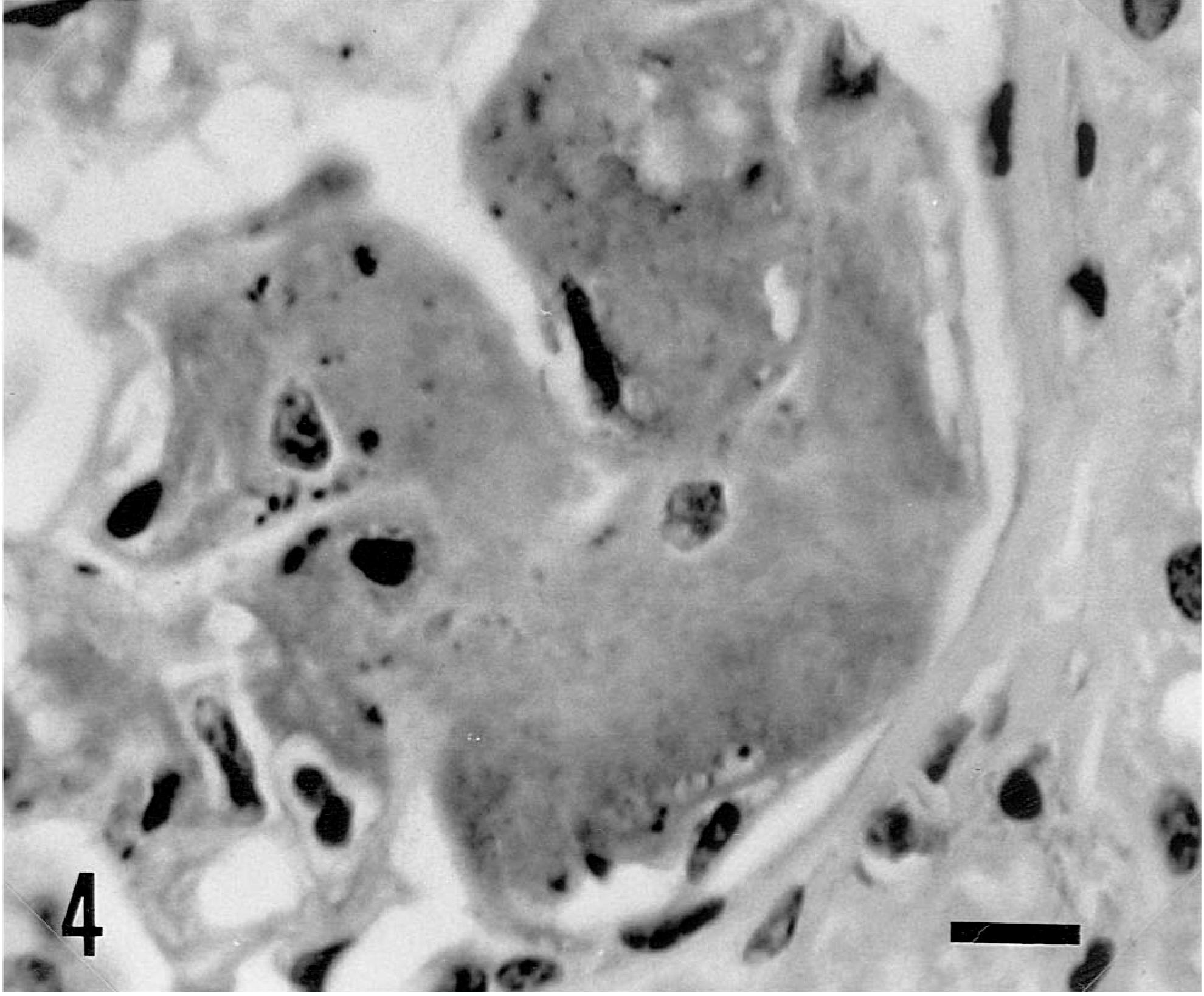
Glomerulus; BLAD cattle. Segmental deposition of bovine κ-light chain-positive amyloid in the wall of the glomerular capillaries. ABC method, Mayer's hematoxylin counterstain. Bar = 50 μm.
The histologic and immunohistochemical characteristics revealed that the amyloid protein was AL type derived from immunoglobulin κ-light chain. The weak reaction against λ-light chain antibody probably was due to absorption of some λ-light chains to the amyloid complex. In humans, primary amyloidosis is occasionally associated with multiple myeloma of B-cell lymphomas. From 10% to 15% of patients with multiple myeloma develop AL amyloid. In most human patients, AL amyloidosis is not associated with multiple myeloma, systemic neoplastic conditions, and other types of basal diseases.6 Chemically, AL amyloid usually consists of the variable region of immunoglobulin light chains, and it can be derived froom either the κ or λ moieties. In most human patients, AL amyloid consists of amyloid protein derived from λ-light chain, but in a few cases, it is derived from κ-light chain.6
Systemic AL amyloidosis has not been reported in cattle. A calf with plasma cell tumor was described previously, but no amyloid deposit was observed.8 There have been no descriptions of BLAD cattle with amyloidosis, although many precise histologic studies of the disease have been reported.1,4,8,10 This is the first description of AL amyloidosis immunohistochemically related to immunoglobulin κ-light chains of precursor protein in cattle.
BLAD in Holstein cattle, formerly termed bovine granulocytopathy syndrome, is characterized as a β2 integrin (CD11a,b,c/CD18) deficiency in leukocytes.9 Persistent hypergammaglobulinemia due to unknown causes was often observed in the chronic stage of BLAD, although multiple myeloma, solitary plasmacytoma, and B-cell lymphoma were not described in the previous reports of BLAD cattle.1,4,8,10 No systemic neoplastic disease of plasma cells or plasma cell abnormality was found in the cattle in this report. BLAD cattle often had bronchopneumonia consisting of heavy infiltrates of neutrophils in bronchi, bronchioles, and alveoli, with granulation tissues in the chronic stage. However, AA amyloid associated with a chronic inflammatory process was not observed in either the present or previous studies.1,4,8,10 The present findings suggest that the deposition of AL amyloid derived from immunoglobulin κ-light chains of precursor protein may have been the result of hypergammaglobulinemia of unknown causes.