
Abstract

Written informed consent to publication has been obtained from the patient or next of kin
SM wrote the case report; LRJ was the operating surgeon and edited the manuscript and accompanying images; both authors read and approved the final manuscript
Duodenal diverticula (DD) should be considered in the differential diagnosis of a cystic lesion seen adjacent to the head of pancreas.
Introduction
The duodenum is the second most common site for gastrointestinal diverticulum. 1 Patients with duodenal diverticula are often asymptomatic.1–3 Diagnosis of this condition is often made incidentally in 14.5% of barium examinations of the upper gastrointestinal tract. Prevalence of this condition is reported to be 22% of cadavers on autopsy. 1
The clinical presentation is typically characterized by non-specific abdominal symptoms. 3 However, patients can present with symptoms of duodenal diverticulitis, perforation, bleeding, cholelithiasis, ascending cholangitis, ulceration or intestinal obstruction.1,2 Computed tomography (CT) or magnetic resonance imaging (MRI) is usually diagnostic for DD when diverticulum is filled completely with gas or a combination of fluid and gas. 3 Lack of either gas or oral contrast within these lesions makes distinction from cystic pancreatic masses challenging, and further evaluation with upper gastrointestinal barium examination may prove diagnostic. 1
A case of symptomatic DD is reported here misinterpreted as a cystic tumour of pancreas on pre-operative images.
Case report
A 51-year-old woman presented with recurrent right upper quadrant pain without jaundice or weight loss.
Ultrasound and magnetic resonance cholangio-pancreatography (MRCP) revealed calculi in her gallbladder and a prominent common duct without intrahepatic duct dilatation or stones. A 15 mm focal small cystic pancreatic lesion with fluid collection lying adjacent to the posterior uncinate process was also reported on MRCP. An endoscopic ultrasound (EUS) showed this cyst to be a multiseptate cystic lesion in the head of pancreas raising possibility of intraductal papillary mucinous tumour (IPMT). She had deranged liver function tests (LFTs). Tumour markers including CA19-9, CEA and CA 125 were within the normal range.
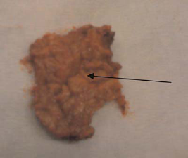
Following discussion at our multidisciplinary meeting, the patient underwent a laparotomy with a view to perform a duodenum preserving pancreatic head resection to remove this cystic lesion and cholecystectomy. After Kocherisation to mobilize the duodenum, the resection of the head of pancreas was commenced during which there suddenly appeared to be a diverticulum arising from the second part of the duodenum close to the ampulla of Vater (Figure 1). This was embedded within the substance of the head of the pancreas cavitating the uncinate process (Figure 2). The diverticulum was then invaginated with 3-0 prolene sutures. After intravenous injection of Secretin, a tiny pancreatic duct was revealed at the excision bed. A Roux en Y loop was brought up to form a pancreaticojejunostomy at the head of pancreas with insertion of a pancreatic stent.
Intraoperative picture. Photograph showing the duodenal diverticulum-pointed by the surgical forceps Photograph showing the cavitated uncinate process of pancreas-black arrow
The patient had a protracted postoperative period which was complicated by postoperative sepsis, bilateral pleural effusions and a contained anastomotic leak which was managed conservatively. Patient was discharged home two months following surgery. The patient has been doing well 36 months post surgery.
Discussion
Cystic lesions of the pancreas are being detected with increasing frequency with advancements in abdominal imaging technology often for investigating non-pancreatic-related problems. 4
Cystic lesions of the pancreas are broadly classified into neoplastic and non-neoplastic cysts. Non-neoplastic cysts are benign and include pseudocysts, retention cysts, benign epithelial cysts, abscesses, duodenal wall cysts (duodenal diverticula), lymphoepithelial cysts, and mucinous non-neoplastic cyst. The predominant cystic neoplasms are intraductal papillary mucinous tumors (IPMT), mucinous cystic neoplasms, serous cystic adenomas, and solid pseudopapil-lary neoplasms. Except for serous cystic adenomas, which are almost always benign, cystic pancreatic neoplasms are considered to be either malignant or premalignant. 5
DD are common anomaly which poses clinicians a diagnostic dilemma. DD are smooth round outpouchings of the duodenum, usually located in the medial periampullary region. 1 Fluid-filled DD on imaging, owing to their proximity to the head of pancreas can be mistaken for cystic pancreatic masses. 2
DD can be classified as congenital or acquired. While congenital DD are rare, the more common acquired ‘pseudodiverticula' are known to result from increased intraluminal pressure leading to herniation of the mucosa and submucosal coat through the intestinal wall usually at a point weakened by the entry of or the point of bifurcation of blood vessels.2,3
On CT/MRI, DD often appear as a thin-walled, round fluid collection in the periampullary region along the medial border – containing gas and/or oral contrast/fluid on CT/MRI. Lack of either gas or oral contrast within these lesions makes distinction from cystic pancreatic masses challenging, and further evaluation with upper gastrointestinal barium examination may prove diagnostic. 1
Traditionally, CT has been used as a first line of diagnosis for cystic lesions of the pancreas. However, the limited resolution of CT scanning prevents reliable differentiation of the pancreatic cysts. 6
Endoscopic ultrasound (EUS) has been used to image the pancreas with improved resolution and is well suited for assessment of cystic lesions because it can provide images of the cystic wall and septations. EUS permits sampling of cyst fluid, mass lesions and lymph nodes. However, EUS has not been able to accurately differentiate between benign and malignant cystic neoplasms. 7
Fine needle aspiration (FNA) cytology can supplement EUS for diagnostic yield enhancement. However, the results of cyst fluid analysis has not been a promising one. 8
A pilot ex-vivo study conducted by Iftimia et al. has suggested that optical coherence tomography (OCT), a high resolution structural imaging technology based on low coherence interferometry, could be used by clinicians in the future to more reliably differentiate between benign and potentially malignant pancreatic cysts. 9
Cystic pancreatic neoplasms occur in 0.7% of people. 4
The decision with respect to observation or surgery in patients with cystic pancreatic lesions can be quite challenging owing to the low sensitivity and specificity of imaging studies and endoscopic cyst aspiration which fail to accurately differentiate between benign, pre-malignant and malignant pathologies. 4
A survey conducted by Abraham et al. at a high volume medical centre elicited that 9.2% of pan-creaticoduodenectomies are performed for benign disorders where there is preoperative suspicion of malignancy. Whipple resection is widely considered justifiable in patients who do not have a preoperative tissue diagnosis. 10
In the case presented, the preoperative investigations (i.e. MRCP and EUS) were strongly indicative that the cystic lesion encountered was potentially malignant and hence the patient was scheduled to have duodenum preserving pancreatic head resection. Intraoperative diagnosis of a DD embedded in the head of the pancreas was made following resection of the uncinate process of the pancreas.
Conclusion
Our case highlights the importance of considering the various differential diagnosis in case of a cystic lesion seen adjacent to the head of the pancreas.
Our case also highlights the fact that despite a combination of (repeated) imaging and EUS aspiration, the diagnosis of DD remained elusive indicating that upper gastrointestinal contrast studies should be pursued prior to surgery in patients with unchanged cystic pancreatic lesions on repeated imaging with/without suspicious results on EUS aspiration.
Footnotes
Acknowledgements
The authors thank the patient for permitting them to publish the case