
Abstract

DECLARATIONS
None declared
None
Not applicable
KNS
All authors contributed equally
Edward Absoud
An aortoduodenal fistula may be overlooked as a cause of major upper gastrointestinal bleeding in patients with an abdominal aortic aneurysm.
Introduction
Abdominal aortic aneurysm (AAA) is a very serious and rare communication between the lumen of the aorta and that of the duodenum. When such a fistula occurs in the absence of previous aortic aneurysm surgery, it is called a primary aortoduodenal fistula (PADF), a condition much less frequent than a secondary aortoduodenal fistula which occurs as a result of previous aortic aneurysm grafting. The diagnosis can be delayed due to the rare occurrence of the condition, nonspecificity of abdominal signs, and frequent initial absence of records alerting the clinician to a known AAA, thereby resulting in increased morbidity and mortality.
Salmon described the first case report of a PADF in 1843. 1 Since then about 250 cases have been reported in literature. 2 7 The incidence at autopsy is between 0.04% and 0.07%.4,7 The incidence of secondary aortoduodenal fistula is 0.5-2.3%. 6
In this article we present a further case of PADF and discuss the pathophysiology, diagnosis and management of the condition, highlighting the fallibility of diagnostic investigations and the importance of having a high index of clinical suspicion.
Case report
A 69-year-old man was brought to our emergency department after collapsing at home following a bout of acute abdominal pain and vomiting approximately 500 mLs of fresh blood. He had a blood pressure of 75/45 mmHg and heart rate of 110 bpm. Abdominal examination evinced mild epigastric tenderness and was otherwise unremarkable.
He had a history of an AAA which was being monitored and measured 4 cm on ultrasound scan seven months previously. Other relevant past medical history included carcinoma of the bladder which was treated with chemotherapy two years previously and diverticular disease. He was a 30-pack-a-year smoker till two years previously before giving up completely.
After intravenous fluid resuscitation, an urgent oesophagogastroduodenoscopy was performed which showed a large haematoma and some fresh blood in the stomach which the endoscopist was unable to clear. A repeat oesophagogastroduodenoscopy the following day revealed infraampullary duodenal ulceration and an adjacent blood clot but no active bleeding. The patient was started on proton pump inhibitors. After returning to the medical ward, he continued to pass dark red blood per rectum and had a haemoglobin level of 10 G/dL which had dropped from 13 G/dL at admission 24 hours earlier.
An abdominal computed tomographic (CT) scan (Figure 1) was performed the same day and showed a posterior duodenal ulcer as well as a 4 cm infrarenal AAA with suspicion of a small false lumen but no convincing evidence of a leak.
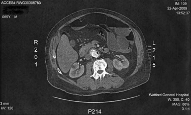
CT with intravenous contrast showing an abdominal aortic aneurysm (arrow) but no signs to suggest a leak
The patient was transfused blood and appeared to be settling over the next two days. However, on day five he had a sudden large haematemesis (about 1.5 L) and complained of renewed epigastric pain. Following surgical referral at this point, he was taken urgently to the operating theatre and an exploratory laparotomy was performed which revealed a PADF involving the third part of his duodenum. The aorta and duodenum were meticulously separated, the aneurysm repaired with a Dacron tube graft and the duodenal defect closed in two layers with 3/0 PDS. Omentum was interposed between aortic and duodenal repairs. Tissue from the aortic and duodenal edges of the PADF was sent off for histology, and atheroma from the AAA for culture and sensitivity. Laboratory results did not show any evidence of infection. The patient was transferred to ITU where, after steady progress, he succumbed to a chest infection on his 14th postoperative day.
Discussion
PADFs are thought to result mostly from direct wear and inflammatory destruction of an aortic aneurysm. Such fistulae, arising from an atherosclerotic AAA, comprise 73% of all PADFs, while 26% are caused by traumatic or mycotic aneurysms. 8 11 The most common infectious agents responsible for mycotic aneurysms are Klebsiella and Salmonella, 11 although Staphylococcus and Streptococcus have also been implicated. 15 Much rarer causes such as radiation, tumours, ulcers and ingestion of foreign bodies account for the remaining 1%. 12 Due to anatomical proximity, the third part of the duodenum is most frequently involved. About two-thirds of PADFs occur at this site, whereas the fourth part of the duodenum is affected in one-third of cases (Figure 2). 7

Arrangement of retroperitoneal organs in the upper abdomen. The stomach, liver and peritoneum have been removed for demonstration. Note the proximity between the abdominal aorta and the third and fourth parts of the duodenum
The importance of PADFs lies mainly in their being a rare and easily-missed cause of gastrointestinal bleeding and their association with a very high mortality rate if left untreated. In the absence of treatment, the mortality rate is almost 100%. With surgical intervention, survival ranges from 18-93%.6,7,13 - 18 As many as 40% of operated cases develop complications, and the overall postoperative mortality rate is greater than 30%.3,6 PADFs associated with infected aneurysms have a worse prognosis, with a postoperative mortality rate exceeding 50%.11,15
The most common clinical features of PADF are upper gastrointestinal bleeding (64%), abdominal pain (32%) and a pulsatile abdominal mass (25%).3,19 However, these symptoms and signs are concomitantly present in only 10% of cases.5,7 Other symptoms that may be present include back pain, melaena, fever, sepsis and shock. Just as in our case, an intermittent herald bleed often precedes major haemorrhage. An intermittent bleed is followed by transient closure of the fistula by a thrombus and contraction of bowel around it. Early recognition of this symptom and its clinical importance is parmount. Occurrence of a herald bleed should critically arouse suspicion of a PADF and immediate action.
The three most useful diagnostic modalities for detecting PADF are abdominal CT scan with intravenous contrast, endoscopy (oesophagogastroduodenoscopy) and arteriography. Of these, CT is by far superior as it is less invasive, more convenient and more expedient than either oesophagogastroduodenoscopy or arteriography. CT also has another advantage in that it poses no risk of dislodging the aortic thrombus. These qualities make CT the investigation of choice in diagnosing PADF.19,20 The presence of a PADF may be evinced by detection of air in the retroperitoneum and in the thrombus, as well as by a loss of the fat plane between aorta and duodenum. Appearance of contrast within the duodenum is a confirmatory sign. However, despite its advantages, CT can miss the presence of a PADF, 20 as happened in our patient. A high index of clinical suspicion is, therefore, critical in this situation.
Oesophagogastroduodenoscopy is an excellent investigation to rule out other causes of upper gastrointestinal bleeding such as ulcers and varices. It should be performed only on a haemodynamically stable patient. A negative oesophagogastroduodenoscopy does not rule out the possibility of a PADF, although the presence of an ulcer or erosion adjacent to a thrombus in the duodenum, with or without an extrinsic pulsatile mass, is highly suggestive.21,22 The acute angle between the third and fourth parts of the duodenum can make visualization of a fistula distal to the third part very difficult. 19
Arteriography has a role in planning aortic reconstruction but, with the great improvements that have taken place in CT imaging, has a very limited place in the acute setting nowadays. 23 A clinical suspicion of PADF together with unremitting gastrointestinal haemorrhage exigently requires an exploratory laparotomy.
Surgical treatment of PADF consists of repairing the duodenal defect, preferably in two layers, and performing a prosthetic repair of the aorta with a Dacron or polytetrafluoroethylene graft. In cases where contamination is minimal, placement of an in situ graft is feasible. Interposition of omentum between duodenal and aortic repairs is desirable. 24 Where contamination is present, or in case of a primary mycotic aneurysm, an extraanatomical aortic graft is preferred and extensive debridement required. 11 Intraoperative cultures should be taken and empirical antibiotic therapy initiated. Specific antibiotics should be administered once the culture and sensitivities result is obtained.
In recent years endovascular repair of PADFs has been reported as a successful alternative to open surgery. 25 27 This approach is very useful where open repair is not feasible due to anatomical reasons, for instance in patients who have had previous radiation therapy resulting in extensive retroperitoneal fibrosis, or in unstable patients who are poor candidates for major surgery. In the latter group of patients, endovascular repair can serve as a valuable initial therapeutic option, allowing control of bleeding, prior to a later definitive repair.
Conclusion
PADF is a rare complication of aortic aneurysms and a rare cause of gastrointestinal bleeding. As delay in diagnosis and treatment carries a very high mortality rate, a high index of clinical suspicion is advised and any patient in whom a readily identifiable source of gastrointestinal haemorrhage has not been found should be considered to have a PADF unless proven otherwise. CT scan is the investigation of choice to diagnose PADF, although oesophagogastroduodenoscopy is useful in ruling out other causes of gastrointestinal bleeding. However, whereas these diagnostic tools can certainly help to corroborate or refute the diagnosis of a PADF, they are no substitute for astute clinical judgement. Early surgical intervention improves the chances of successfully managing this rare, lethal, clinical conundrum.
Footnotes
Acknowledgements
None