
Abstract
This case report describes the results of the gross pathological, histological and immunohistological examinations of a fibrosarcoma that spontaneously developed in the left neck region of an aged male Lewis rat that participated in a long-term facial nerve regeneration study. A 10 mm long polylactic acid (PLA) tube was implanted into a 7 mm critical defect gap of the buccal branch of the left facial nerve of a male eight-week-old Lewis rat. Forty-two weeks after implantation, an approximately 8 cm × 9 cm × 2.5 cm rapidly growing mass with ulceration of the overlying skin was found in the left neck region. Gross examination of the tumour and the surrounding tissues before tumour excision revealed that the tumour had clear boundaries, and had not invaded any facial tissues, the facial nerve and the PLA nerve guide. Gross examination of the tumour's cut surface revealed that the tumour comprised numerous smooth nodules with a homogeneous white tan colour. Examination of the haematoxylin and eosin-stained sections of the tumour revealed that the tumour was predominantly composed of bundles of spindle-shaped atypical proliferating cells that were mixed with bundles of collagen fibres and arranged in a storiform pattern. Tumour emboli and skin invasion were also observed. Immunohistological examination revealed that the tumour cells were weakly positive for vimentin, but negative for keratin, α-smooth muscle actin, S-100 protein and CD34. From the results of these analyses, the final pathological diagnosis of this tumour was a fibrosarcoma.
Aged Wistar and Sprague-Dawley rats are frequently used as the control comparator in carcinogenicity studies because they develop spontaneous neoplasms of various types. 1–4 By contrast, aged Lewis rats are not used in such studies because little is known about their background and the incidence of spontaneous neoplasms. 5 Consequently, no reports that describe the results of immunohistological examinations of spontaneously arising neoplasms in aged Lewis rats have been published in the medical and/or veterinary scientific literature. During a study whose aim was to develop an artificial biocompatible nerve conduit using Lewis rats, one of the rats spontaneously developed a fibrosarcoma in the neck region. This case report describes the results of the gross pathological, histological and immunohistological examinations of this tumour.
Case report
A male eight-week-old Lewis rat was purchased from Charles River Laboratories Japan (Tokyo, Japan) for use in a long-term facial nerve regeneration study in which the degeneration status of the conduit after its implantation was assessed. For this purpose, a 10 mm long polylactic acid (PLA) tube was implanted into a 7 mm critical defect gap of the buccal branch of the left facial nerve. Forty-two weeks after implantation, an approximately 8 cm × 9 cm × 2.5 cm rapidly growing mass with ulceration of the overlying skin was found in the left neck region. The tumour and the ulcerated overlying skin were surgically excised from the rat by circular resection under 4% isoflurane anaesthesia. Examination of the tumour and surrounding tissues before tumour excision revealed that the tumour had clear boundaries (Figure 1a), and had not invaded any facial tissues, the facial nerve and the PLA nerve guide. After excision, the circular skin defect of the rat was closed by a reading man local flap procedure.
6
(a) The spontaneous tumour that was surgically removed from the left neck of an aged male Lewis rat. (b) The tumour's cut surface revealed that the tumour comprised numerous smooth nodules with a homogeneous white tan colour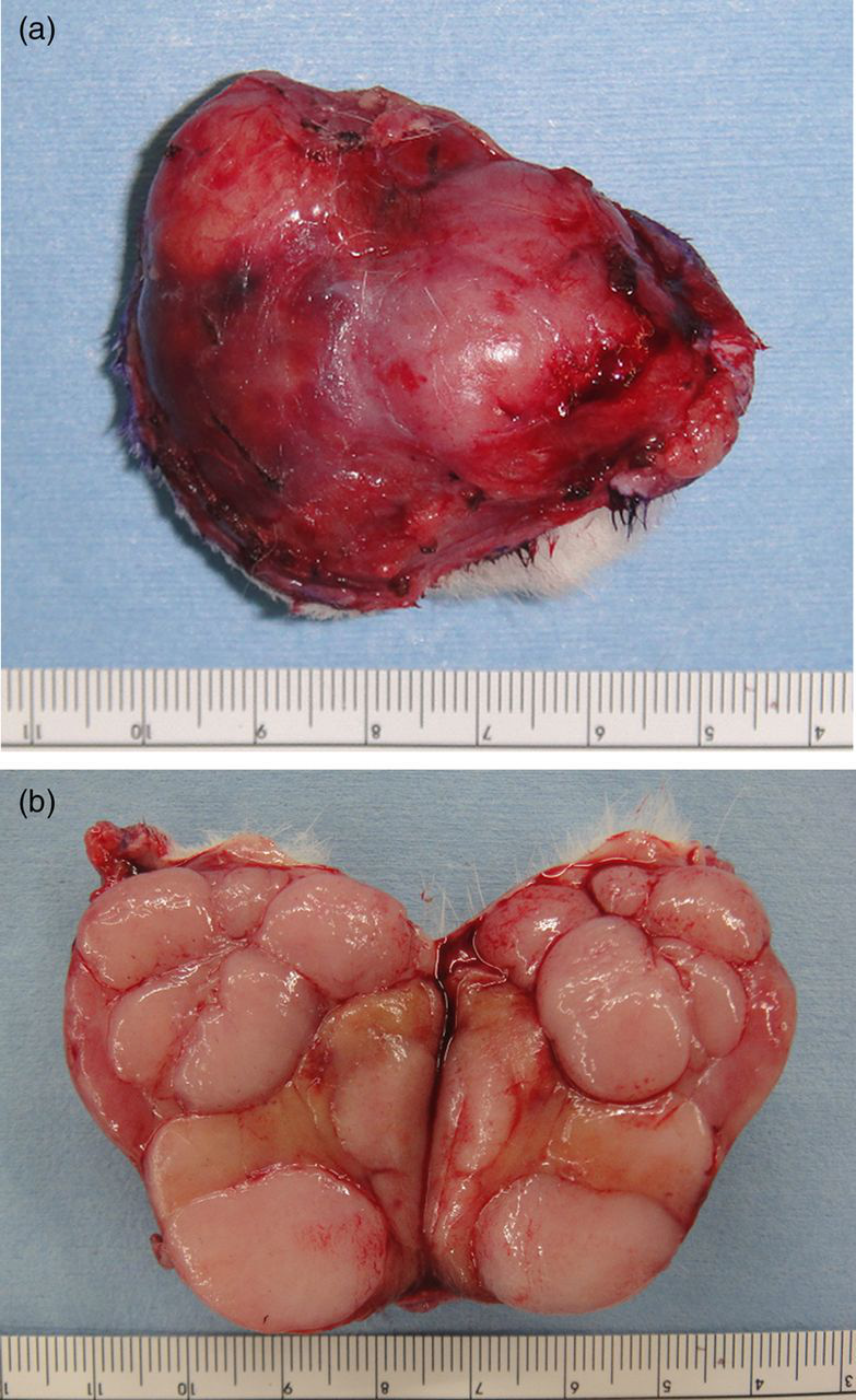
Gross examination of the tumour's cut surface revealed that the tumour comprised numerous smooth nodules with a homogeneous white tan colour (Figure 1b). The tumour was tentatively diagnosed at gross examination as being a lymphoma or a lymph node metastasis of a malignant tumour. The surgically resected tumour was then fixed in 10% neutral-buffered formalin and then prepared for histological and immunohistochemical analyses.
For the histological analysis, histological slides were prepared from the formalin-maintained specimen after its embedding in paraffin blocks for haematoxylin and eosin (H&E) staining using standard procedures. For immunohistochemical analysis, the paraffin-embedded sections were first deparaffinized with xylene, and then subjected to a heat-induced antigen retrieval process by autoclaving the deparaffinized slides at 121°C at a pressure of two atmospheres for 15 min with 0.01 mol/L citrate buffer, pH 6.0. After being treated with a protein blocking reagent (DakoCytomation, Glostrup, Denmark), the sections were incubated overnight at 4°C with various primary antibodies at the following dilutions: a polyclonal keratin antibody (1:700) (DakoCytomation), which was used as an epithelial cell marker; a monoclonal vimentin (clone V9) antibody (1:50) (DakoCytomation), which was used as a mesenchymal cell marker; a monoclonal α-smooth muscle actin (α-SMA) (clone 1A4) antibody (1:50) (DakoCytomation), which was used as a smooth muscle and myofibroblastic differentiation marker; a polyclonal S-100 protein antibody (1:800) (DakoCytomation), which was used as a nerve cell marker; and a monoclonal CD34 (clone QBEnd 10) antibody (1:50) (DakoCytomation), which was used as an endothelial cell and histiocyte marker. Endogenous peroxidase activity was quenched by immersing the sections in 3% hydrogen peroxide for 5 min. The sections were then incubated with a detection agent (Histofine Simplestain Rat MAX-PO® [M] and [R]) (Nichirei Biosciences, Tokyo, Japan), and the antibody–antigen complexes were visualized by staining the sections with 3,3′-diaminobenzidine. Positive control tissues of the epidermis, vascular pericytes, peripheral nerves and vascular endothelial cells were prepared and stained with the keratin, vimentin, α-SMA, S-100 protein and CD34 antibodies in the identical manner to confirm that the staining technique and antigen retrieval procedure were correctly done in the tumour sections. Isotope controls using mouse and rabbit IgG antibodies (Abcam, Cambridge, MA, USA) showed no background staining. The sections were counterstained with Mayer's haematoxylin solution and examined (×40) under a Leica DFC-6000B microscope to which a charge-couple device camera (DFC500; Leica Microsystems, Heerbrugg, Switzerland) was attached.
Examination of the H&E-stained sections of the tumour revealed that the tumour was predominantly composed of bundles of spindle-shaped atypical cells that were arranged in a storiform pattern. The cells appeared to be proliferating and were mixed with bundles of collagen fibres (Figure 2a). There were also dense clusters of small round cells that also appeared to be proliferating (Figure 2b). Emboli were also found in the tumour (Figure 2c), and the tumour had invaded the overlying skin. Examination of the immunostained sections of the tumour revealed that the tumour cells were weakly positive for vimentin (Figure 2d), but were negative for keratin, α-SMA, S-100 protein and CD34 (data not shown). According to these findings, a final pathological diagnosis of a fibrosarcoma with skin invasion was made.
Histopathological features of the spontaneous tumor in the neck of an aged male Lewis rat. (a) A haematoxylin and eosin (H&E)-stained section of the tumour which shows that the tumour is predominantly composed of bundles of spindle-shaped atypical cells that were arranged in a storiform pattern (magnification × 40). (b) Dense clusters of small round cells that also appeared to be proliferating in an H&E-stained section of the tumour (magnification × 40). (c) Emboli in the tumour (magnification × 40). (d) An immunostained section of the tumour which shows that the tumour cells are weakly positive for vimentin (magnification ×40)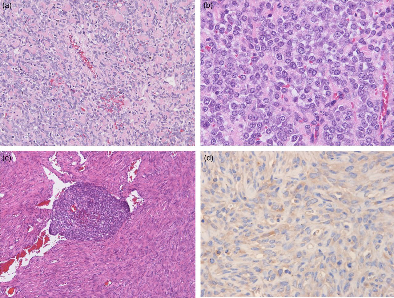
Two months after the surgery, the rat was humanely killed as scheduled for assessing the extent of facial nerve regeneration and the degeneration status of the conduit. On necropsy, we found an approximately 2 cm × 1 cm × 0.5 cm soft elastic mass in the left axilla which was removed and then prepared for H&E staining using standard procedures. Examination of the H&E-stained sections of this mass revealed that it was a lipoma, and no evidence of recurrence of the fibrosarcoma was found.
Discussion
According to Walsh and Poteracki, 3 the most common neoplasms in aged Wistar rats are pituitary adenomas, mammary fibroadenomas and adenocarcinomas, adrenal cortical adenomas and endometrial stromal polyps. Prejean et al. 1 reported that the incidence of spontaneous tumours in untreated Sprague-Dawley rats in an 18-month series of carcinogenesis experiments is 45%. In this study, Prejean et al. 1 also reported that (a) the incidence of spontaneous tumours in the female rats was almost double that found in males, and this difference was mainly due to the high incidence of mammary gland tumours in the females, and (b) most tumours occurred in the endocrine system, and mainly in the pituitary and adrenal glands. Prejean et al. 1 also reported that they found spontaneous fibrosarcomas in the integument and soft tissue in only one of the 360 Sprague-Dawley rats that they monitored for 540 days. In their study of spontaneous tumours in 2669 aged Sprague-Dawley rats (1340 males), Chandra et al. 2 reported that the incidence of spontaneous fibrosarcomas in the skin and subcutis was 0.37% in males and 0.6% in females. In their comparative review of carcinogenicity bioassays that comprised 930 control Wistar rats, Poteracki and Walsh 4 reported that spontaneous fibrosarcomas in the integument and soft tissue were present in 3.2% of the male rats and none were present in the female rats. In their study on the life expectancy and spectrum and incidence of spontaneous tumours in 629 inbred Lewis rats (325 males), Baum et al. 5 reported that the mean lifespans of male and female Lewis rats are 32.5 ± 6.6 (standard deviation) months and 27.7 ± 5.1 months, respectively. They also reported that (a) the high incidences of tumours of the haemopoietic system in males (27.7%) and endometrial carcinomas in female (45.2%) are characteristic features of aged Lewis rats, and (b) spontaneous fibrosarcomas in the skin are uncommon in males (0.9%) and do not develop in the females (0%).
Kirkpatrick et al. 7 reported that malignant mesenchymal tumours can frequently be found around a subcutaneously implanted biomaterial in Fischer rats. Specifically, they found macroscopic tumours at 25.8% of the implantation sites, 26–110 weeks after the implantation, and that malignant fibrous histiocytomas (MFH) and pleomorphic sarcomas were the most frequently found tumours. In this report, we describe a spontaneous tumour in an aged Lewis rat, 42 weeks after its engrafting with a PLA nerve guide. Although this tumour involved its overlying skin, the tumour had not invaded any facial tissues, the facial nerve and the PLA nerve guide. Nakamura et al. 8 reported that tumours, which were predominantly fibrosarcomas and MFHs, were found in 22 of 50 male Wistar rats, two years after the implantation of a subcutaneous PLA plate. Interestingly, they found that 20 tumours were at the implant sites, and two tumours were at sites that were distant from the implant. The role of PLA in the molecular mechanism of spontaneous tumour occurrence in rats is still unknown. Nevertheless, the existence of a relationship between PLA and spontaneous tumour development in this aged Lewis rat at a site approximately 20 mm from the site of the implanted PLA nerve guide, 42 weeks after its implantation, cannot be excluded.
From our histological analysis of the H&E-stained sections of the tumour, we diagnosed the tumour in this aged male Lewis rat to be a fibrosarcoma. Other possible diagnoses of this tumour that were considered were an MFH, a leiomyosarcoma, an angiosarcoma, a malignant peripheral nerve sheath tumour, a poorly differentiated carcinoma and the rare dermatofibrosarcoma protuberans (DFSP). Since the immunohistological analysis showed that the tumour cells were weakly positive for vimentin, but negative for keratin, α-SMA, S-100 protein and CD34, we were able to exclude leiomyosarcoma, angiosarcoma, a malignant peripheral nerve sheath tumour, a poorly differentiated carcinoma and DFSP from the differential diagnosis. We were also able to exclude MFH from the differential diagnosis because no giant cells with multiple nuclei were observed in the H&E-stained sections of the tumour. Accordingly, our final pathological diagnosis of this tumour was a fibrosarcoma.
Footnotes
ACKNOWLEDGEMENTS
This study was partially supported by the Global COE program, Multidisciplinary Education and Research Centre for Regenerative Medicine (MERCREM) from the Ministry of Education, Culture, Sports, Science, and Technology (MEXT), Japan. The authors would like to acknowledge Dr Arieh Bomzon, ConsulWrite (