
Abstract
Unilateral (left eye) optic nerve hypoplasia was detected in a six-month-old male Beagle dog. Vision testing indicated that the left eye had poor vision and testing the pupillary light reflex showed the left eye to have an absence of the afferent pathway of the reflex but it had a normal efferent pathway. Ophthalmoscopy revealed a small-sized optic disc, winding retinal artery and dilated retinal vasculature in the left globe. Electroretinography showed no abnormal findings even in the left globe. Histopathologically, the left optic nerve was markedly hypoplastic and was composed of sparse neural elements and a moderate amount of connective and glial tissues. In the retina of the left globe, the nerve fibre layer and the ganglion cell layer were reduced in thickness, although a small number of ganglion cells were still present. There were no abnormal findings detected in the right globe and the right optic nerve. The brain appeared normal macroscopically.
Optic nerve hypoplasia (ONH) is an uncommon congenital abnormality in animals, although ONH has been reported in several canine breeds (Saunders 1952, Gellat & Leipold 1971, Vestre & Brightman 1980, Kern & Riis 1981, Spiess et al. 1991, Turnquist et al. 1991, Termote 1998). The incidence of ONH in dogs has been estimated to be 0.1% based on an ophthalmological examination of 1400 Beagle dogs (Rubin 1974). Ophthalmological examination done on Beagle dogs in our laboratory from 2000 to 2006 revealed that the incidence of ONH was 0.47% (6/1284) for males and 0.16% (2/1251) for females. This paper describes the details of unilateral ONH observed in a male Beagle dog in our laboratory in 2006.
Materials and methods
Animal
The dog was a male chosen from a group of 20 male and 20 female six-month-old Beagle dogs purchased from Covance Research Products Inc, Virginia, USA in 2006. The dog was housed individually in a stainless-steel cage in an animal room under controlled conditions (temperature: 22 ± 4°C; relative humidity: 55 ± 25%; air ventilation: 13–15 times/h; lighting: 12 h/day). The dog was given 300 g of pelleted diet everyday (DS-A, Oriental Yeast Co Ltd, Tokyo, Japan) and allowed free access to tap water via an automatic water supply system.
The dog was handled according to the ‘Law Concerning the Protection and Control of Animals’ and ‘Guideline for Animal Experimentation’ in Japan. In addition, this study was approved by the Regulation and Animal Welfare Committee of Bozo Research Center Inc, Tokyo, Japan.
Examinations
A behavioural vision test was carried out in a maze and obstacle course after covering each eye in turn with CobanTM Self-Adherent Wrap (3M Health Care, Minnesota, USA). A pupillary light reflex and an ophthalmic examination were also performed. Following instillation of a mydriatic agent (tropicamide and 0.5% phenylephrine hydrochloride/parasympathetic blocker, Mydrin® P, Santen Pharmaceutical Co Ltd, Osaka, Japan) to both eyes, the cornea, lens and anterior vitreous were examined using a hand-held slit-lump biomicroscope (SL-14, Kowa Co Ltd, Tokyo, Japan), and the fundus was examined using an indirect ophthalmoscope (Omega 200, Heine Optotechnik, Herrshing, Germany). After the dog was placed for at least 30 min in a dark room, both eyes were instilled with a mydriatic agent to dilate the pupils. Then both eyes were anaesthetized using a surface anaesthetic for ophthalmology (0.4% oxybuprocaine hydrochloride, Benoxil® 0.4% solution, Santen Pharmaceutical Co Ltd). The xenon lamp was set at 30 cm from one eye of the dog, and after instillation of one drop of the corneal protector (Scopisol® 15, Senju Pharmaceutical Co Ltd, Osaka, Japan) on the concave side of a contact lens-type electrode (Kyoto Contact Lens Co Ltd, Kyoto, Japan, diameter, about 20 mm), it was gently attached to the cornea of the targeted eye, and the ground electrode was attached on one earlap. Standard electroretinography (ERG) (luminous energy: 20 J, low-filter: 1 Hz; high-filter: 1 kHz; the evoked potential detection device [MEB-9102, Nihon Kohden Co, Tokyo, Japan] and the photic flash stimulator [SLS-3100, Nihon Kohden Co]) was recorded by single flash. The dog was again placed for at least 30 min in the dark room, and oscillatory potential (OP) (luminous energy: 20 J, low-filter: 50 Hz; high-filter: 1 kHz) was recorded by single flash. After examination, the surface of the corneas was sufficiently washed with saline.
After these examinations, the dog was euthanized by an overdose of sodium pentobarbital. The left and right globes with the optic nerve were fixed in phosphate-buffered 0.5 v/v% glutaraldehyde/1.5 w/v% paraformaldehyde solution. Paraffin sections were stained with haematoxylin & eosin (H&E) for microscopic examination. Some sections were also stained with Masson's trichrome, Klüver-Barrera's and Bodian's staining and by immunostaining for glial fibrillary acidic protein.
Results
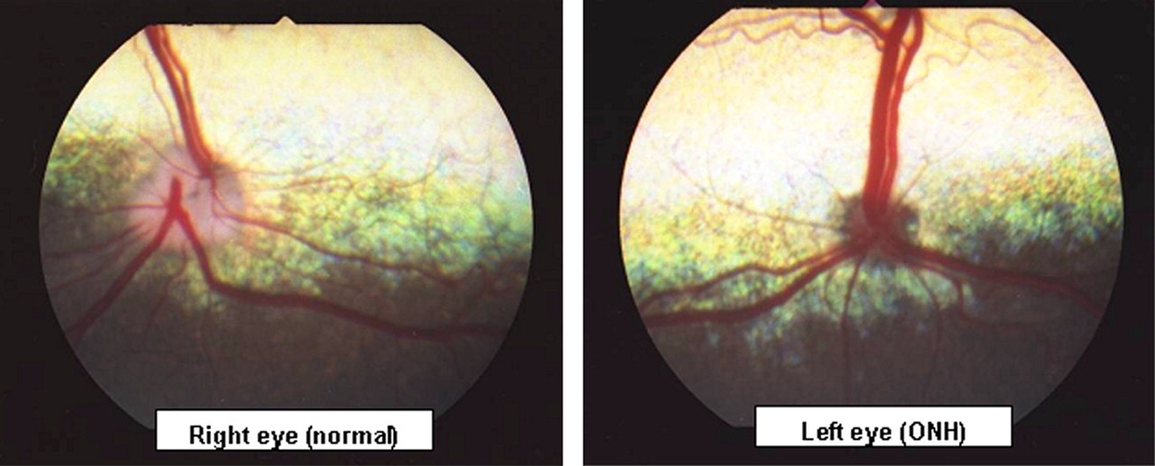
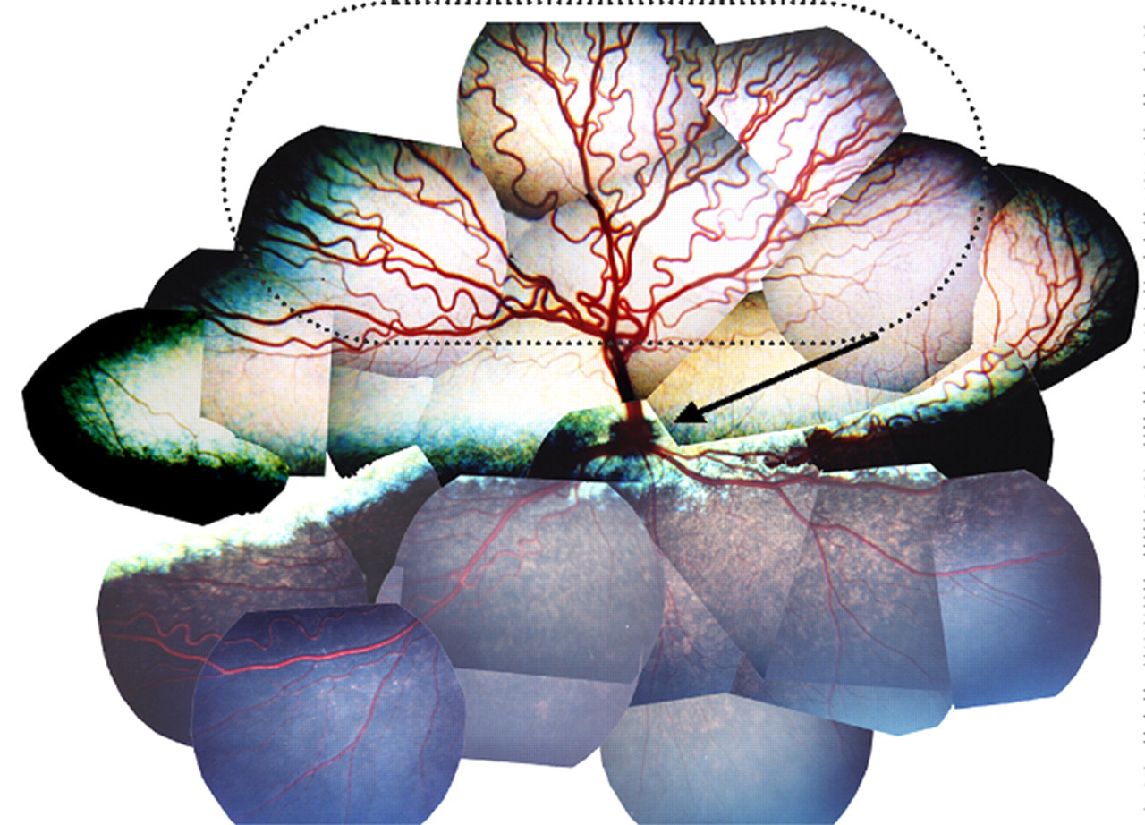
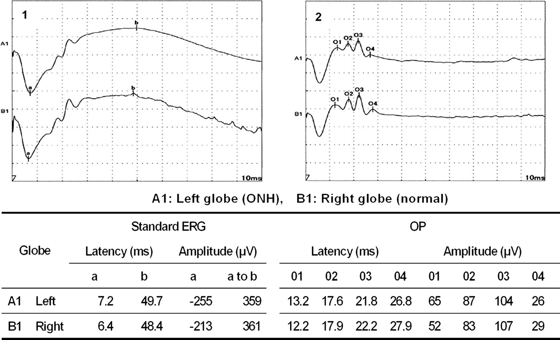
The behavioural vision test suggested that functional vision of the left eye was poor. Pupillary light reflex to the left eye did not induce pupillary response in either eye, while that to the right eye brought about contraction of the pupil in both eyes. Ophthalmoscopy revealed a small-sized optic disc, tortuous retinal arterioles and dilated retinal vasculature in the right globe (Figures 1 and 2) but not in the right one. Standard ERG and OP were normal in both eyes (Figure 3).
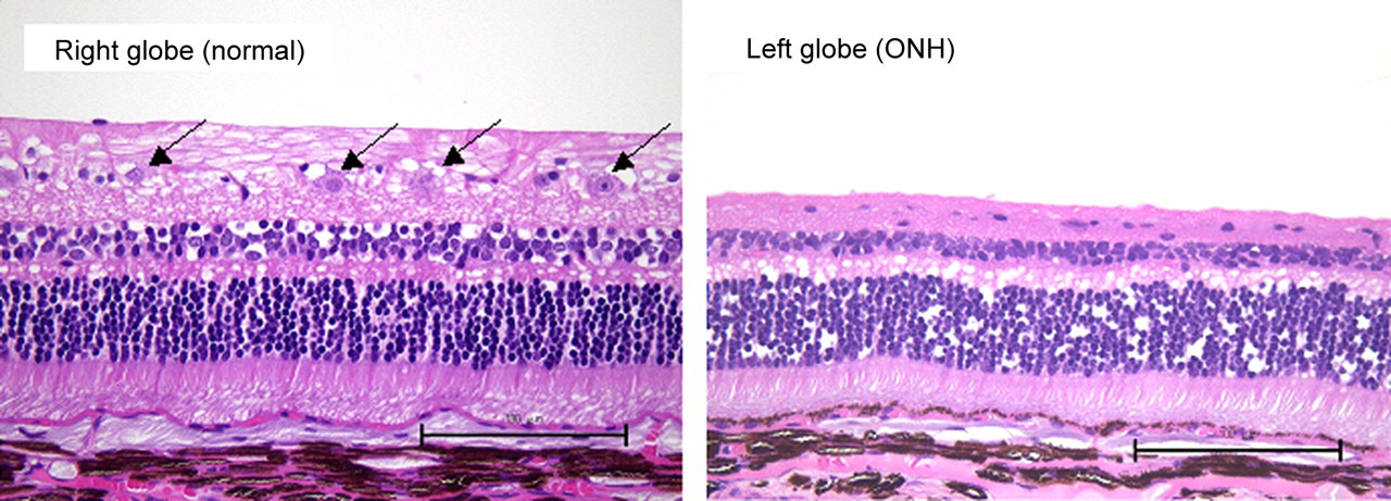
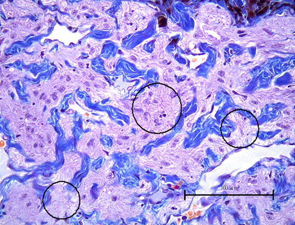
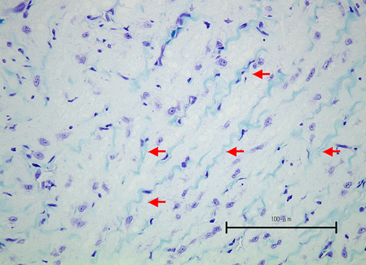
At autopsy, the size of both globes was normal. The right optic nerve appeared normal while the left one was markedly hypoplastic. The optic nerve remnant near the optic chiasm was barely discernible (Figure 4), but there was a normal-sized optic nerve-like tissue near the left globe (Figure 5). The brain appeared normal. Histologically, the right globe and the right optic nerve were normal. In the retina of the left globe, however, the nerve fibre layer and the ganglion cell layer were reduced in thickness, although a small number of ganglion cells remained (Figures 6 and 7). The outer layers of the retina were normal. The optic nerve-like tissue near the left globe was composed of sparse neuronal elements, such as axons (Figure 8) and myelin sheath (Figure 9) and a moderate amount of glial and connective tissues.
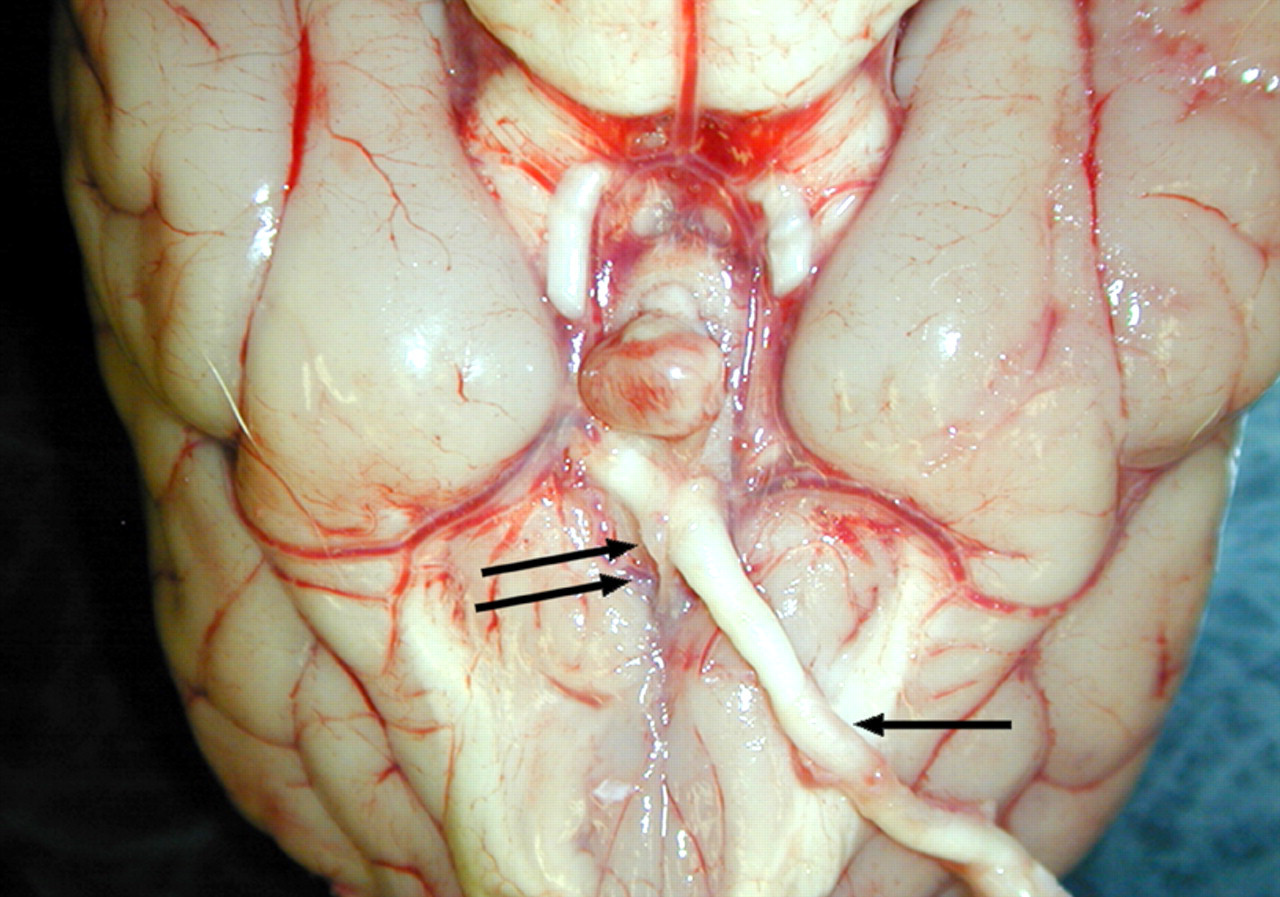
The right optic nerve (arrow) is normal while the left one is barely discernible near the optic chiasm (double arrows)
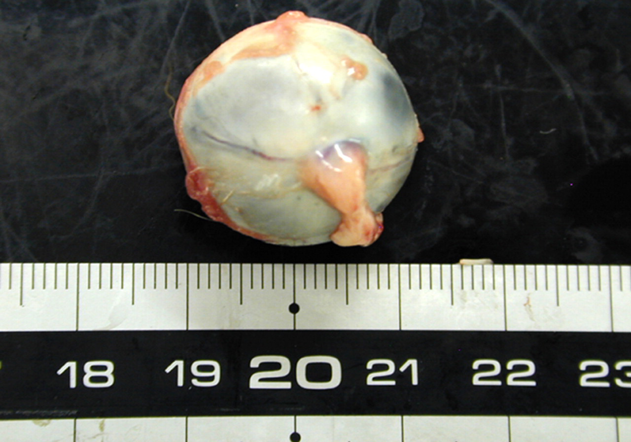
Optic nerve-like tissue is present near the left globe
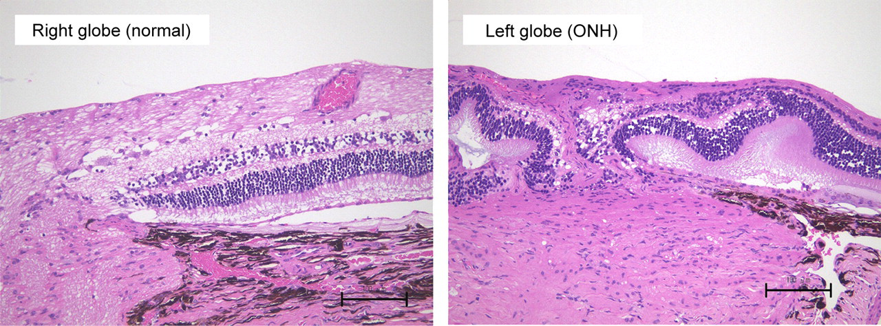
The retina surrounding the optic papillary in both globes, the nerve fibre layer and ganglion cell layer are markedly reduced in thickness. Haematoxylin and eosin stain, × 200
Discussion
A case of unilateral ONH in a six-month-old male Beagle dog observed in our laboratory was examined from multiple viewpoints.
In this case, the behavioural vision test showed that functional vision of the left eye was poor, and the results of the pupillary light reflex suggested that the efferent pathway controlling the iris constrictor muscle functioned normally, while the afferent pathway transmitting the stimulus of light from the retina to the brain was absent in the left eye. Moreover, ophthalmoscopy revealed a small-sized optic disc, a tortuous retinal arteriole and dilated vasculature in the left eye. Previously, Ernest (1976) and Kern and Riis (1981) reported that, except for a small-sized optic disc, there was no abnormality in the retinal vasculature in canine cases of ONH. Standard ERG and OP of the affected eye were normal as previously reported by Kern and Riis (1981), suggesting that some functioning neuronal cells remained. The right eye appeared normal in these examinations. These findings indicated that only the left eye was affected in the present case.
The left optic nerve was markedly hypoplastic and composed of sparse neuronal elements and a moderate amount of connective and glial tissues. In the retina of the left globe, the thickness of the nerve fibre layer and the ganglion cell layer was reduced as previously reported (Ernest 1976, Kern & Riis 1981), although there were a small number of ganglion cells observed. There were no abnormalities detected in the right globe and the right optic nerve.
Most reported cases of ONH in animals have been bilateral (Gellat & Leipold 1971, Ernest 1976) although unilateral ONH with hydrocephalus was reported in a three-year-old Pekingese dog (Turnquist et al. 1991). The present case was also unilateral but was not accompanied with macroscopic abnormality of the brain. A genetic basis for ONH has been postulated but not proven in animals, although it was reported that the defect was familial in rare cases in man (Hackenbruch et al. 1975).
ONH is also an uncommon congenital abnormality in humans. An increased risk of ONH in humans has been associated with several environmental and teratogenic factors (Hittner et al. 1976, Petersen & Walton 1977, Hoyt & Billson 1978, Tornqvist et al. 2002). Investigation of ONH in Beagle dogs, which have been routinely checked for functional abnormalities of the eyes before being used in toxicity studies, can be valuable in elucidating more details of ONH in humans because there are many similarities between canine ONH and human ONH.