
Abstract
Hepatosplenic involvement is a rare manifestation of abdominal tuberculosis in children. We describe the case of a 7-year-old girl with persistent fever, cough, and hepatosplenomegaly. Typical lesions were shown in the liver and spleen by ultrasound and computed tomography. Colonoscopy showed a nodular, ulcerated mass that partially obstructed the cecum. Microbiological and histopathological findings of intestinal and liver biopsy confirmed the clinical suspicion of tuberculosis.
Keywords
Tuberculosis (TB) is an important public health problem, which is increasing in frequency in developed countries owing to high immigration (1). In the last two decades, there has been a progressive change in the clinical presentation of TB. Indeed, classical manifestations of pulmonary TB are gradually being replaced by extrapulmonary forms, either in the HIV-positive or in the HIV-negative population (2, 3). This modified disease presentation of TB should be taken in mind by pediatricians and general physicians in the diagnostic approach to a child presenting with enlarged liver and spleen, especially when risk factors for TB are present.
Case report
A 7-year-old Chinese girl presented to our department with a 3-week history of low-grade fever and cough. Her previous history was unremarkable. Physical examination revealed a soft abdomen with a tender liver edge 3 cm below the right costal margin and a palpable spleen tip; the chest was found to be normal. Routine laboratory tests were normal except for hemoglobin 9.9 gr/dL (nv 11.5-14.5) and erythrocyte sedimentation rate (ESR) of 79 mm/h (nv < 38). Tuberculin skin test was positive with 20 mm induration. Quantiferon-TB Gold test was 59 IU/mL (nv < 0.34). HIV test was negative. Chest X-ray showed mild thickening along the right major fissure and bilateral hilar enlargement. Abdominal ultrasound (US) performed using a 3.5 mHz convex transducer (Acuson Sequoia 512, Siemens, Mountain View, CA, USA) revealed mild hepatomegaly, multiple hypoechoic nodules up to 13 mm in diameter scattered throughout the liver and spleen (Fig. 1); slight wall thickening of the last ileal loop and ascending colon, and multiple enlarged mesenteric lymph nodes were also shown. Multidetector computed tomography (CT) of the abdomen was performed using 16-slice CT scan (LightSpeed, GE Healthcare, Milwaukee, WI, USA) with 5-mm slice thickness (reconstructed 2.5 mm) and low dose technique (80 kv, 50-90 mAs) after i.v. contrast medium administration. CT revealed multiple rounded, low-density lesions in the liver and spleen, ranging in size from 2 to 18 mm (Fig. 2); peripancreatic, periportal, para-aortic, and paracaval lymph nodes up to 15 mm in diameter with no calcification, and diffuse small bowel wall thickening were also shown. CT of the chest demonstrated small opacities in the right lower lobe and multiple enlarged lymph nodes in the hilar, paraesophageal, and paratracheal regions (Fig. 3). Direct smear microscopy for acid-fast bacilli in the sputum and gastric lavage was negative, but the culture for Mycobacterium tuberculosis was positive. Colonoscopy showed a nodular, ulcerated mass that partially obstructed the cecum. Intestinal biopsy and image-guided needle biopsy of the liver revealed granulomatous tissue with caseous necrosis. Both direct smear microscopy for acid-fast bacilli and culture for Mycobacterium tuberculosis of intestinal and liver biopsy specimens were negative.

Abdominal US (para-sagittal view) at tine diagnosis: multiple hepatic hypoechoic nodules (calipers). GB, gall bladder; IVC, inferior vena cava
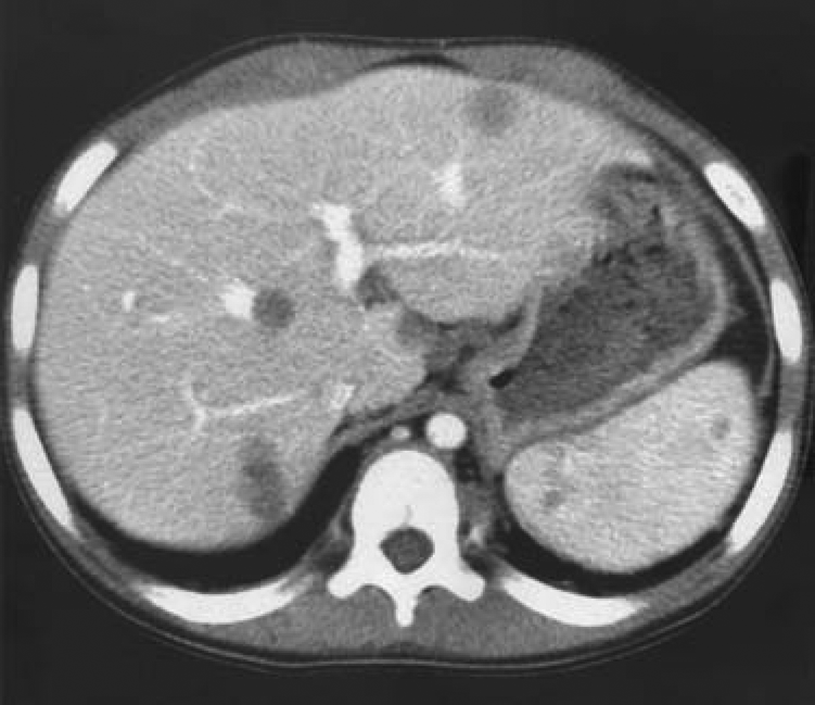
CT of the abdomen: multiple rounded, low-density lesions in liver and spleen

CT of the chest: small opacities in the right lower lobe along the major fissure; enlarged lymph nodes in the hilar region (white arrow) and azygos-oesophageal lymph nodes (black arrow)
Diagnosis of macronodular hepatosplenic TB with concomitant intestinal and lung involvement was made, and a three-drug anti-TB therapy consisting of isoniazid, rifampicin, and pyrazinamide was planned. After 9-month therapy, there was a marked improvement in the general condition, and the liver and spleen were no longer palpable. Abdominal US revealed reduction in size and peripheral calcification of the hepatosplenic lesions (Fig. 4); abdominal lymph nodal groups were normal.

Abdominal US (axial view) after 9-month therapy: peripheral calcification of the liver lesions with posterior echo-shadows (arrow)
Discussion
Hepatosplenic involvement of abdominal TB may occur by hematogenous dissemination either through the hepatic artery in miliary TB or through the portal vein from gastrointestinal lesions (4). Isolated or simultaneous involvement of the liver and spleen usually presents in a fine miliary pattern with nodules ranging from 0.5 to 2 mm in diameter (micronodular form) which cannot be detected on CT scan (5, 6), but may result in a “bright liver and spleen” at US imaging (6). Macronodular involvement is less frequent and is manifested by single or multiple focal, low-density lesions ranging in size from 1 to 3 cm, with or without peripheral rim enhancement on CT (5-7). Enlarged abdominal lymph nodes and calcified lesions in the liver and spleen are frequently reported either on CT or US (4-8). Abdominal lymphadenopathy in TB may occur either due to abdominal origin of TB, or to regional drainage of lung infection in patients with pulmonary TB (9). Despite advances in imaging techniques, radiological aspects of hepatosplenic TB are not specific. They do not allow differentiating TB from lymphoma, abscesses, fungal infection, or metastasis, thus often requiring the need for liver biopsy (4-8).
Our case is worth mentioning for several reasons. First, the macronodular form of hepatosplenic TB is extremely rare in children (3). Second, concurrent involvement of the lung and gastrointestinal tract has been infrequently reported in hepatosplenic TB (5). Third, the diagnosis of TB was confirmed by both histopathological and microbiological findings, that is more the exception than the rule in such setting (3, 8). Indeed, several studies in children with abdominal TB have faced difficulties in microbiological confirmation, thus necessarily relying on histopathological diagnosis alone (10), as in our patient.
Both US and CT play a role in the evaluation of abdominal TB. As it does not involve ionizing radiation, US may be useful as initial diagnostic tool and is excellent for radiological follow-up. CT is considered the imaging modality of choice for diagnosis, because of its ability to better identify solid organs involvement, caseating lymph nodes, and bowel-wall thickening (11).
In conclusion, the diagnosis of abdominal TB represents a major challenge for physicians. Unfortunately, despite the recent resurgence in industrialized countries, this condition is still poorly considered and the diagnosis is often delayed (12). A high clinical index of suspicion, laboratory tests including microbiology, and careful interpretation of radiological imaging may mean the difference between prompt diagnosis and unacceptable delay in recognizing the great mimicker.