
Abstract
The incidence of gastrointestinal carcinoids appears to be increasing, and the rectum is the third most common location. Transcatheter arterial embolization (TAE) with trisacryl gelatin microspheres (Embosphere®) has been reported as an effective method for hepatic metastases of rectal carcinoids. Complications are uncommon and usually of minor consequence. We report an unusual case of a 34-year-old man with tumor lysis syndrome following TAE with Embosphere® in a patient with multiple hepatic metastases of a rectal carcinoid. Early detection and effective treatment are essential for this rare but potentially catastrophic complication.
Keywords
For treatment of hepatic metastases of rectal carcinoid tumors, many different modalities are reportedly effective, including cytoreductive surgery, radiofrequency ablation, hepatic artery embolization, chemotherapy, somatostatin analogues, and liver transplantation (1–4). Liver embolization with trisacryl gelatin microspheres (Embosphere®) has been reported as a feasible and relatively safe treatment for patients with liver tumors (4,5). In hepatic metastases of rectal carcinoid tumors, transcatheter arterial embolization (TAE) with Embosphere® particles can lead to partial response and symptomatic improvement of the disabling endocrine symptoms (4). We report a case of a 34-year-old man who developed tumor lysis syndrome following TAE with Embosphere® for hepatic metastases from a rectal carcinoid tumor.
Case report
A 34-year-old man was admitted to our hospital for treatment of multiple hepatic metastases from a rectal carcinoid tumor. He had been treated with local extirpation of a rectal tumor 4 years previously. Abdominal computed tomography (CT) 25 months later revealed multiple liver metastases. The diagnosis was confirmed by liver biopsy, and the tumor cells were immunoreactive to synaptophysin. The patient was then treated with Yttrium-90 selective internal radiation therapy about 1 year and 7 months before this admission. Yttrium-90 resin microspheres (SIR-spheres; Sirtex Medical, Sydney, Australia) mounted with a radioactive load of approximately 50 Bq per sphere were used for radioembolization. One year and 2 months after the Yttrium-90 therapy, a follow-up abdominal CT showed liver metastases in progression. Therefore, further embolization with Embosphere® (BioSphere Medical, Inc., Rockland, MA, USA) was performed.
The patient had no past medical history of serious illness such as hypertension, diabetes mellitus, or chronic kidney disease, nor any hospitalization other than treatment for the carcinoid tumor.
On physical examination, a huge mass was palpable in the upper abdomen. There was no ascites or signs of encephalopathy. On the day of admission, laboratory results showed hemoglobin 11.3 g/dL, platelet count 207000/mm3, alanine aminotransferase (ALT) 37 LU/L, aspartate aminotransferase (AST) 35 LU/L, albumin 3.9 g/dL, total bilirubin 0.57 mg/dL, lactate dehydrogenase (LDH) 402 IU/L, creatinine 0.8 mg/dL, potassium 4 mmol/L, and prothrombin time 13.5 s. The Child-Pugh class was A.
On unenhanced and contrast-enhanced CT, there were more than 20 hepatic tumors in both lobes, with the largest being > 18 cm in diameter (Fig. 1a and b). Hepatic angiography was performed via a right femoral artery approach (Fig. 2a and b). A 4-French catheter and a 2.7-French microcatheter were used for celiac trunk and superselective hepatic (segmental and subsegmental) angiography, respectively, revealing multiple tumor stains in both hepatic lobes. TAE was performed by injection of 2 mL 100-300μm Embosphere® particles in the left hepatic artery's supplying branches and 2 mL 100-300 μm, 2 mL 300-500 μm Embosphere® particles injection in the right hepatic artery's supplying branches until there was sluggish hepatic arterial flow. The coil shown in the picture (Fig. 2) was placed during the procedure of Yttrium-90 therapy. Coil embolization of the right gastric artery and gastroduodenal artery could avert reflux and thereby decrease the risk of gastric ulcer during Yttrium-90 therapy.
CT scan demonstrated multiple metastases in both hepatic lobes before embolization. (a) Unenhanced CT scan; (b) contrast-enhanced CT scan
Angiography showed multiple tumor stains at bilateral hepatic lobes. (a) Common hepatic artery angiography; (b) left hepatic angiography
Three days after TAE, the patient complained of general fatigue and poor appetite. He also became oliguric. Laboratory tests showed ALT 430 IU/L, AST 1910 IU/L, total bilirubin 7.63 mg/dL, LDH 16300 IU/L, creatinine 6.54 mg/dL, potassium 7 mmol/L, phosphorous 10.7 mg/dL, calcium 6.2 mg/dL, and uric acid 13.4 mg/dL. As the patient developed hyperkalemia, hyperuricemia, hyperphosphatemia, hypocalcemia, and acute renal failure, the diagnosis of acute tumor lysis syndrome was made. He was first treated with 0.9% saline hydration, then alkalinization of urine with sodium bicarbonate, allo-purinol for hyperuricemia, kalimate, and calcium gluconate for hyperkalemia were applied 3 days after TAE. However, anuria developed and he received emergency hemodialysis. Rasburicase was applied for hyperuricemia on the fifth day after TAE.
After intensive care, the patient gained satisfactory recovery of both renal and hepatic functions on day 68 after TAE. Laboratory tests showed ALT 16 IU/L, AST 16 IU/L, total bilirubin 0.58 mg/dL, creatinine 0.79mg/dL, BUN 12 mg/dL, potassium 3.9 mmol/L, and prothrombin time 12.6 s. Follow-up unenhanced CT scan on day 34 after TAE showed large areas of necrosis (Fig. 3).
Unenhanced CT scan demonstrated large areas of necrosis 1 month after embolization with Embosphere® particies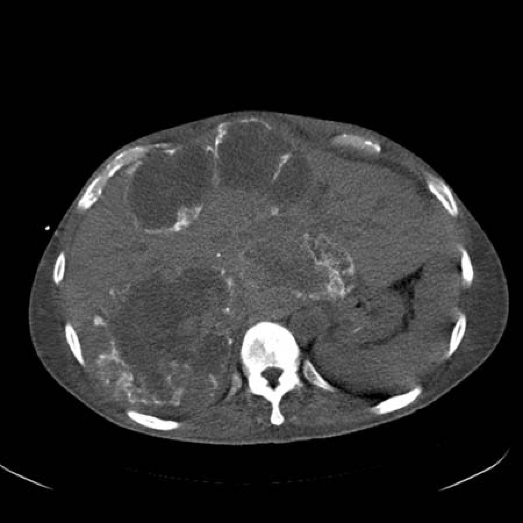
Because of disease progression 1 year later, TAE was performed once more with Embosphere® after prevention for tumor lysis syndrome (adequate hydration and the usage of rasburicase). Neither tumor lysis syndrome nor acute renal failure occurred after this procedure. Finally, the patient died of progressive disease 4 months after the second TAE.
Discussion
Carcinoid tumors are morphologically and biologically heterogeneous neuroendocrine tumors that have malignant potential. They are most commonly found in the gastrointestinal tract, with the rectum as the third most common location (6). In the United States, the age-adjusted incidence of colorectal carcinoid tumors is about 1 in 100,000, and the incidence of rectal carcinoid tumors has increased about 10-fold over the last 35 years (7). Rectal carcinoid tumors also present with metastasis in 4-18% of cases (8).
Many different kinds of treatments have been reported to be effective in hepatic metastases of carcinoid tumors, including somatostatin analogues, alpha-interferon, chemotherapy, radiofrequency ablation, liver embolization alone or with chemotherapy (chemoembolization), cytoreductive surgery, and liver transplantation (1–4). Liver embolization, as part of a multimodality treatment protocol, may lead to partial radiological response as well as symptomatic improvement of disabling endocrine symptoms (2–4, 9).
Several types of particles have been used in TAE, including trisacryl gelatin microspheres (Embosphere®). Embosphere® particles are not degradable and are more homogenous of size than particles previously used (e.g. polyvinyl alcohol or gel-foam). Granberg et al. have reported that TAE with Embosphere® particles is a safe and effective treatment for patients with metastatic carcinoid tumors (4). Our patient with metastatic carcinoid tumors underwent superselective TAE with Embosphere® particles, however, tumor lysis syndrome developed after the treatment. To our knowledge, this is the first reported case of tumor lysis syndrome after TAE with Embosphere® for metastatic carcinoid tumors.
Most cases of tumor lysis syndrome occur after treatment of hematopoietic malignancies (10). However, it may occur, albeit rarely, after TAE of solid tumors. Hsieh et al. reported a few cases that developed tumor lysis syndrome after TAE of hepatocellular carcinomas. Larger tumor size is one of the risk factors (11). In our patient, there were more than 20 metastatic carcinoid tumors in both lobes of the liver, with the largest one being > 18 cm in diameter. Moreover, vessel occlusion by Embosphere® particles may be more permanent because they are non-degradable.
The frequency of tumor lysis syndrome is increasing among patients who have tumors that used to be only rarely associated with this complication. It occurs when tumor cells release their contents into the blood, leading to hyperuricemia, hyperkalemia, hyperphosphatemia, and hypocalcemia (10). Deposition of uric acid and calcium phosphate crystals in the renal tubules can cause acute renal failure. Standard treatment includes the correction of electrolyte imbalance and acidosis. Adequate intravenous hydration is essential to prevent acute renal failure. Allopurinol can be used for hyperuricemia but may cause serious allergic reactions. Recombinant urate oxidase (rasburicase) is an effective alternative for lowering uric acid levels. If patients fail to respond to medical treatment, urgent hemodialysis is indicated (12).
The cause of a high attenuation rim around the necrotic masses is not clear (Fig. 3). Lipiodol was not mixed with Embosphere®. Furthermore, contrast medium retention was also less likely as CT was performed on day 34 after TAE. One possible explanation is dystrophic calcification after tissue necrosis (13).
In conclusion, TAE for hepatic metastases of carcinoid tumors is a feasible treatment that may give relief of symptoms. Tumor lysis syndrome is an uncommon but potentially catastrophic complication that should always be considered and treated immediately when it occurs.
Acknowledgements
This work was supported by grants from the Taipei Veterans General Hospital (VGH 100C-178) and from the National Science Council (NSC 98-2314-B-075-029). The authors also thank the Division of Experimental Surgery of the Department of Surgery, Taipei Veterans General Hospital for their assistance.