
Abstract
In April 2010, a Medicines and Healthcare Products Regulatory Agency safety alert concerning all metal-on-metal (MOM) hip replacements recommended measuring chromium and cobalt concentrations when managing patients with painful prostheses. The need for this review is illustrated by the recent surge in requests for these blood tests from orthopaedic surgeons following this alert. The aim is to provide guidance to laboratories in assessing these requests and advising clinicians on interpretation. First, we summarize the basic terminology regarding the types of hip replacements, with emphasis on the MOM type. Second, we describe the clinical concerns over implant-derived wear debris in the local tissues and distant sites. Analytical aspects of the measurement of the relevant metal ions and what factors affect the levels measured are discussed. The application of inductively coupled plasma mass spectrometry techniques to the measurement of these metals is considered in detail. The biological effects of metal wear products are summarized with local toxicity and systemic biological effects considered, including carcinogenicity, genotoxicity and systemic toxicity. Clinical cases are used to illustrate pertinent points.
Introduction
Clinical chemistry laboratories in the UK are receiving increased numbers of requests for blood cobalt (Co) and chromium (Cr) concentrations because orthopaedic surgeons have shown a relationship of concentrations of these metals to clinical events affecting hip replacements. This trend has been accelerated by the recent issue by the Medicines and Healthcare Products Regulatory Agency (MHRA) in the UK of a Medical Device Alert advising that all patients with a metal-on-metal (MOM) implant be followed annually for at least five years, with blood Cr and Co measurements when indicated. 1
Hip replacements were first used in the 1930s by Dr Phillip Wiles from the Middlesex Hospital in the UK, but it was not until Sir John Charnley's cemented metal-on-plastic (MOP) hip replacements in the 1960s that a design suitable for widespread use was widely available. The first implants used a stainless-steel ball with a Teflon cup. The Teflon was later replaced with high-density polyethylene due to the high incidence of osteolysis due to wear debris from the Teflon. Variants of this design have proliferated, and a variety of materials have been used, including titanium stems and titanium cup shells, different plastics and ceramics.
Every year one million hip replacements are implanted worldwide and 70,000 in the UK (UK data from the UK National Joint Registry,
MOM replacement
The use of MOM hip implants was led by Europe (initially Germany with small 28-mm diameter femoral head components and latterly the UK with large diameter [>36 mm] femoral head components). The latest data from the USA show that 35% of all hip implants are now MOM. We have estimated that the total number of MOM implants that have been implanted is approximately one million, with the majority over the last 10 y. Many studies have shown that these have good early (<5 y) and possibly medium-term (5–10 y) results.
MOM re-surfacings have the advantage that the metal components can be made thin enough, yet maintain strength and the ability to bond to bone (often with a coating or roughened surface) so that large diameter femoral head components are feasible. Large diameter femoral head components are desirable because they offer increased range of motion (if used with a narrow femoral neck component), reduced risk of dislocation and the ability to re-surface or cap the femoral head.
All materials used for hip implants will wear in use. Particles released from MOP implants, primarily polythene particles, are typically tens of micrometres in diameter and can clearly be seen in the surrounding tissue by light microscopy. They are responsible for osteolysis and implant loosening in approximately 90% of patients at 10 y postoperatively. MOM implants generate nanometre-sized metal particles and metal ions at approximately one trillion per year (one million particles generated per step times one million steps walked per year). 2,3 These 40-nm particles are not visible using light microscopy, although it is possible that aggregates may be visible.
The MOM implants have two bearing surfaces of a Co–Cr–molybdenum (Mo) alloy (most commonly ASTM F75 4 in the ratio of Co:Cr:Mo of 60:30:7, although other alloys such as F90, F799, F562 and F1537 may also be used) that are separated by a thin film of synovial fluid when tested in a hip simulator. However, when used in patients it is likely that this lubricant film is not constant and thick enough to prevent the surfaces from contacting and generating wear debris, as evidenced by the reports of a wide range in blood metal ion concentrations. Typical blood levels of Co and Cr in patients with unilateral, well functioning hip implants are 30 and 45 nmol/L (1.7 and 2.3 μg/L), respectively. However, blood Co concentrations can rise as high as 6550 nmol/L (387 μg/L) and Cr 3400 nmol/L (179 μg/L). 5 We have recently recorded concentrations of these metals in fluid surrounding these hips of 400 mg/L of Cr and 22 mg/L of Co.
There is an important distinction between MOM hip replacement and hip re-surfacing. In the former, a stemmed (femoral) component is used, whereas in the latter the femur is capped or re-surfaced. Hip re-surfacing therefore conserves femoral bone by capping the femoral head rather than removing it. If a later redo/revision operation is required, then a conventional ‘stemmed’ hip replacement is straightforward on the femoral side and so hip re-surfacing is favoured for younger patients who are likely to need more than one hip replacement during their lives. For the purposes of this review, we have used the term MOM hip replacement rather than refer to the subset of hip re-surfacing because it is likely that the most important and pertinent clinical problems relate to the bearing surface.
Since 1996, approximately one million patients have had MOM hips, with revision due to poor biocompatibility ranging from 1% to 20% at five years postoperatively, depending on the type of prosthesis. The full economic cost of a revision has been estimated at up to £30,000.
There are many designs of these implants, but all use the same Co–Cr–Mo alloy, although there are manufacturer differences in the treatment of the alloy in the manufacturing process. The implants are based on alloys predominantly manufactured from Co, Cr and Mo, but other metals such as tungsten, nickel and titanium may also be present. It is unclear whether these have any effect on the wear of the implant or on metal release. The lubrication of the space between the head and the cup by a thin film of synovial fluid 6 will remove any released metal from the joint.
The mechanism of metal release is primarily by wear of the bearing surfaces, but there may also be an element of corrosion especially in the shaft. The linear wear rate of these implants is approximately 5 μm/y, 7 but in some failed implants we have measured wear rates up to 257 μm/y. 8 Metals may also be released from implants by corrosion. This may be caused by the action of body fluids on the surfaces or by an electrochemical couple being formed between different metal components.
Local effects of MOM hips
Soft tissue inflammatory reactions surrounding MOM implants have only recently been reported, partly because imaging techniques such as magnetic resonance imaging (MRI) previously suffered from artefact due to the prosthesis. These reactions have been described as pseudotumours. 9,10 Their true prevalence will be unknown until large-scale MRI screening studies are carried out. However, studies of small numbers of failures have found that females are more affected than males, with one study reporting 14% of young females with MOM implants affected. Even if the prevalence for the whole population is much lower, the problem is important because it can cause destruction of bone and muscle, which may be either unreconstructable or leave the patient with long-term poor function. One type of MOM implant (the Ultima; Depuy, Warsaw, IN, USA) was withdrawn by the manufacturers because of this problem. 11,12 The Depuy ASR implant has recently been withdrawn due to a higher than expected failure rate as shown in statistics from the National Joint Registry (NJR, a register of all orthopaedic joint replacements performed in England and Wales).
The British Hip Society in collaboration with the NJR and the MHRA has recently conducted an audit of this problem and we await their findings. Meanwhile, surgeons are requesting blood Co and Cr concentrations on patients with MOM implants as a means of understanding how well the hip is performing in vivo. Research in this area has increased dramatically. This review summarizes the evidence that has stimulated surgeons to request these tests and highlights the gaps in understanding that may be useful to clinical biochemists when understanding the clinical interpretation of blood concentrations of Co and Cr in patients with MOM hip replacements.
Systemic effects of MOM hip replacements
Cr(VI) is a well-known environmental and occupational carcinogen and mutagen, so many reports have focused on the carcinogenic and mutagenic potential of these implants. Some reports have shown increased occurrence of some cancers, but the same reports also showed decreased incidence of other cancers in patients with MOM hip replacements. 13–15 A recent report has suggested that there may be an increased incidence of some cancers in rheumatoid and osteoarthritis patients who have had knee replacements, 16 although this increased cancer risk in such patients may possibly be related to the inflammatory disease. 17
Concern over the effects of metal ions has prompted various authorities to examine the potential carcinogenic effects of MOM hips. Firstly, the UK Government's Committee on Mutagenicity of Chemicals in Food, Consumer Products and the Environment published a statement saying that there was evidence of genetic damage in patients with all types of MOM and some MOP implants, but not from stainless steel on polythene implants. 18 The committee concluded that ‘However, it was not possible to make any definite conclusions as to which metal ions, or interactions between metal ions or particulate metals might be responsible for the observed genotoxicity…There was limited evidence available to suggest a possible interaction between chromium and cobalt ions and possible mutagenicity/DNA damage in vitro but not in vivo. There was no convincing evidence for metal-specific effects of wear debris with regard to potential for clastogenicity or aneugenicity’. A series of papers from Learmonth and Case in Bristol 19–21 was considered by the committee. They showed that DNA damage, including aneuploidy and chromosomal translocations, could be linked to Cr–Co containing implants, but not to stainless steel-containing implants. However, the report also concludes that evidence for an association of these effects with implants is not in itself evidence of a causal relationship.
Secondly, a review by the National Institute for Health and Clinical Excellence (NICE) recommended further research into many of the unknown questions surrounding metal ions from MOM implants. 22 Thirdly, in 2006, the British Orthopaedic Association organized a national two-day meeting entitled, ‘Biological Implications of Metal on Metal Hip Replacements’ which concluded with the need for more research into this area and has recently conducted a UK-wide audit of all failed MOM implants with adverse soft tissue changes. Lastly, the US Food and Drug Administration (FDA) have recommended that the first 350 patients of the initial cohort receiving MOM hip re-surfacing devices should have blood concentrations of metal ions measured for 10 y postoperatively. 23,24 More recent advice from the FDA 25 is more cautious than the MHRA, ‘At the current time, there is no evidence to support the need for checking metal ion levels in the blood or special imaging if patients with MOM hip implants have none of the signs or symptoms described above and the orthopaedic surgeon feels the hip is functioning’. No advice on action limits for metal concentrations is given, and the advice is to interpret concentrations in the context of the overall specific clinical scenario including symptoms, physical findings and other diagnostic results.
The British Hip Society has issued revised guidance, 26 specifically aimed at larger diameter implants. The advice is that the MHRA advice still applies, but that patients may need to be followed up for the life of the implant.
NICE guidance on the use of MOM hip re-surfacings 22 has highlighted the need for further studies on the effects of metal wear particles from these products, but emphasized the safety of these products and general lack of adverse effects. However, there is a relatively high rate of revision of these operations, and unexplained pain in many patients has been related to elevated blood metal concentrations and increased blood metal concentrations have been correlated to the position of the implant. 5,27,28 There have been little published data on appropriate reference ranges for these metals or the clinical utility of the data.
Measurement of Cr, Co and other metals
Cr and Co are nowadays usually measured in biological samples by inductively coupled plasma mass spectrometry (ICPMS), although electrothermal atomization atomic absorption spectrometry (ETAAS) has also been used, and was commonly used before the widespread introduction of ICPMS. The sensitivity of ETAAS for these elements is limited; for example, in a method for measuring Co in serum, multiple injections into the graphite tube were needed to attain the desired sensitivity. 29 The large amount of sample required to obtain the needed sensitivity will cause a large background absorbance which may have stretched the capabilities of older instruments. Nevertheless, the concentrations found in these subjects are well within the range of ETAAS, and this technique has been used in several studies. 30–32
ICPMS has lower detection limits than ETAAS and is also capable of simultaneous measurement of Cr and Co, and of many other elements 33,34 and is used in the majority of studies considered here. Accurate measurements can be made using either collision/reaction cell quadrupole ICPMS (Q-ICPMS) or high-resolution ICPMS (HR-ICPMS). The use of either of these is essential to cope with the interference on the measurement of 52Cr from 40Ar12C. All of the available collision/reaction cell instruments are capable of reducing this interference to acceptable levels while retaining good sensitivity. Although there is in principle interference in the measurement of 59Co from ions such as 44Ca14N, there does not seem to be any significant effect of these, and measurements can be made in standard mode.
High resolution ICPMS, using a double focusing magnetic sector mass spectrometer, is available in a few laboratories and overcomes the uncertainties of interference reduction with collision/reaction cells. 35–37 The interferences can usually be completely resolved, but the expense of the hardware will preclude the use of this technique in all but a few specialist and research laboratories. Krachler et al. 38 looked in detail at method validation, especially the use of currently available quality control (QC) samples. The accuracy of values assigned to their product by one manufacturer of QC materials was questioned.
The electrochemical technique anode stripping voltametry has been used occasionally, 39 but is not common. This is a much more labour-intensive technique and requires a total acid digestion of the sample before analysis.
Other metals may also be measured. Mo is a component of the alloy commonly used, but measurement will add little diagnostic value. Some other implants may contain other metals such as iron, nickel, vanadium and titanium in the alloy used or in other components such as the shell surrounding the cup and in the shaft. Measurement of Ni or Ti may be of value in some circumstances where metal release may be due to corrosion rather than wear at the bearing surface. Mo and vanadium may also be measured by Q-ICPMS. The sensitivity for Mo measurement is limited by the number of isotopes of the element, but it is readily measured without the need for the use of collision cell technology. Vanadium suffers from interference from 35Cl16O, but this is readily removed by collision cells. Nickel may also be measured using collision cell Q-ICPMS. Titanium measurements by Q-ICPMS are difficult in clinical samples at normal concentrations, although it is possible to use quadrupole instruments to measure higher concentrations of titanium. Some workers (Andrew Taylor, personal communication) have claimed to be able to measure titanium using optical emission spectroscopy. Measurement of the major isotope, 48Ti, is interfered with by 48Ca, which although only 0.19% of total calcium, would still be in considerable excess over titanium. Other titanium isotopes, 47Ti and 49Ti, although the abundance of each is less than 10%, can be measured by HR-ICPMS using medium resolution settings. Ti measurements are not practicable using Q-ICPMS.
There are no Certified Reference Material quality standard reference materials available, and there is a limited number of commercial products including both elements at suitable concentrations. Until now, most quality assessment materials and External Quality Assurance programmes have focused on the industrial exposure requirements for samples. Cr and Co, in blood, serum and urine, are now included in the monthly Trace Element Quality Assessment Scheme (TEQAS) quality assessment scheme in the UK (part of NEQAS, National External Quality Assessment Service) organized from the University of Surrey in Guildford and are also included in the Quebec Multielement Eternal Quality Assessment Scheme organized by the Institut National de Santé Publique in Quebec, Canada.
Metal contamination from blood collection system
Equine blood was analysed by high-resolution inductively coupled plasma mass spectrometry before and after standing in trace element-free blood collection tubes (BD K2EDTA tubes, BD, Oxford, UK) and after being aspirated into the tube via the Vacutainer needle. All results expressed as nmol/L (μg/L), mean and standard deviation of six measurements. The Ni assay in the ‘blood’ samples showed high contamination
There have been some reports of urine metal concentrations in these patients, 36 but urinary metal measurements are rarely used. Although urine is a non-invasive specimen, there is potentially a higher variation in urinary excretion attributable to factors including renal function and urinary concentration. A random collection is suggested, with the metal concentration normalized to creatinine concentration.
Synovial fluid metal concentrations can also be measured, 41 and may be of value in some situations. The fluid surrounding the implant can have a very high concentration of metal. Samples of fluid may be taken by needle biopsy prior to operation for microbiological testing and to reduce the fluid pressure in the cavity. A high metal content in this fluid will be an obvious indicator of wear in the implant, and has been shown to correlate with blood concentrations, but does not always add any diagnostic value. Metal analysis of the tissue removed during the revision operation is also possible, and can complement the histological findings, but is largely of research interest.
Blood, serum and urine can be analysed by direct analysis after simple dilution but synovial fluid and tissue samples need prior digestion with acid. This is often performed with a microwave digestion system, but digestion can also be effectively performed with a heated digestion block. The need for digestion of synovial fluids is in part due to the variable nature of the matrix: many of the samples presented to the author's (BS) laboratory have a large amount of solid material, presumably protein aggregates, which may contain bound metal and thus should not be excluded from analysis. Fluid samples may also contain intact wear particles which may not be totally vaporised in the analytical system.
Reference concentrations
Unexposed population
Reference intervals for metals from non-surgical studies
HR-ICPMS, high-resolution inductively coupled plasma mass spectrometry; ETAAS, electrothermal atomization atomic absorption spectrometry
There are less data available on some of the other metals mentioned, although there is monitoring of all in occupationally exposed subjects, usually in urine.
Exposed population
Studies of metals in blood, serum and urine in patients with hip replacements and other surgical implants
AAS, atomic absorption spectroscopy, ASV, anode stripping voltametry, Q-ICPMS, quadrupole inductively coupled plasma mass spectrometry, HR-ICPMS, high resolution ICPMS, THR, total hip replacement (type not specified), BHR, Birmingham hip replacement (Smith & Nephew, London, UK); MTHR, Metasul bearing hip replacement (Zimmer, Warsaw, IN, USA); MOM, metal-on-metal
Data have been re-calculated to show molar concentrations in addition to mass units where appropriate. Papers have been listed in ascending order of publication
Reported studies on other joint replacements
There have been few studies of metals in tissues, largely because such studies, looking at tissues other than local to the implant, have to be at postmortem. Case et al. 53 looked at patients primarily with stainless steel implants, but also some Cr–Co implants. Metal was found in lymph nodes, liver and spleen. 54 Similar results from patients with hip or knee replacements were reported by Urban et al. 55 More widespread dissemination of metals was seen in patients with a failed implant. In at least one case, chromium phosphate particles were detected. Polyethylene particles from MOP implants were also detected. In both of these studies, the cause of death was unrelated to the implant. More recently, Hart et al. 56 have studied tissue immediately surrounding the implant using X-ray synchrotron spectroscopy. Widespread deposition of Cr was found, with few Co particles remaining in the tissues. The Cr was found to be almost entirely chromium phosphate. Co and Mo were found as isolated metallic particles.
Causes of increased metal concentrations
Implant-derived metal debris is a consequence of wear, corrosion or both. Modular components have potential for increased metal release – when compared with one-piece (monobloc) components such as a hip re-surfacing – because of the additional wearing surfaces. Clinical studies relating the mechanism of wear in these implants to blood metal concentrations and in vitro studies using hip simulators have been important in defining some of the factors affecting metal release. The factors shown to be important include: orientation of the components (particularly the cup); implant design; head size and patient activity. Khan et al. 57,58 measured the rise in serum metal concentrations after exercise. Significant increases in serum Cr and Co concentrations were found after just one hour of exercise, with differences between implant types. The BHR (Birmingham Hip Replacement; Smith & Nephew, Memphis, TN, USA) and Cormet (Corin, Cirencester, UK) implants, with a 46.8- or 48-mm diameter head, gave 8.5 and 6.5 times larger increases than Metasul (Zimmer, Warsaw, IL, USA) implants, with a 28-mm diameter head. A much lower effect of activity on metal concentrations was reported by Heisel et al. 59 Marginal increases in blood metal concentrations were found even after treadmill exercise, and no change in normal exercise. In a more recent study, no correlation was found between activity and metal concentration. 60
There are reports of a strong positive correlation between cup inclination angle and either wear rate of removed MOM hips 61 or blood metal ion levels. 5,62,63 However, there are confounding variables that may also influence wear rate; these can be divided into the following groups: patient factors (small [<50 mm] femoral head size, 61,62 gender, activity); surgical factors (horizontal femoral offset, 64 cup version angle, 62 incorrect sizing); and manufacturing factors (metallurgy and design). Interestingly, among the five most used types of MOM hip (BHR, Cormet, ASR, Metasul, Adept [Finsbury Orthopaedics, Surrey, UK]), there are no proven statistically significant differences in metal ion release and wear rates despite differences in metallurgy (e.g. heat-treated versus non-heat-treated) and design (e.g. amount of hemisphere, clearance between two bearing surfaces). The angles of inclination and version or abduction, which define the orientation of the cup within the pelvis, are both important. Blood metal concentrations in patients with an angle of inclination of greater than 55° have been shown to be significantly raised. This seems to be a more important parameter than the duration of the implant or the manufacturer.
Biological effects of wear products and metal toxicity: systemic effects
Cr and Co are both essential trace elements. The role of Co in vitamin B12 is well known. Cr is thought to be essential for glucose metabolism, and probably helps the binding of insulin to cell receptors. 65 Mo is also essential, but only as Mo co-factor for sulphite oxidase and xanthine oxidase. 66 There is some evidence that nickel 67 and vanadium 68 may have some biological function, but this is controversial. There is no evidence that titanium has any biological role. However, the concern in this context is the possible toxic hazard from the metal ions and particles. Toxicity has most been studied in the metal working industries, but other toxic effects are also well known.
The effects of the metal wear debris on the local tissues have dominated the recent debate regarding the use of MOM hips, but for many years concerns about the potential biological effects of metal particles have been raised. The areas of concern are primarily the chemical form of the metal released from the implant and the metabolic fate of the metal. The toxicity of Cr and Co has been recognized as a concern in industrial uses of these metals for many years. 69,70 Fears of carcinogenicity of particles were raised as early as 1971, 71 and a report by the International Agency for Research on Cancer in 2000 72 concluded that alloys containing nickel and Co are possibly carcinogenic and that there are insufficient data for other more complex alloys. The current state of knowledge of hazards from wear metals has recently been reviewed. 73
The main concern has been the possibility that Cr is present as Cr(VI), which is a well-known carcinogen. There are reports that the Cr released from implants may be as hexavalent Cr, 74 but this is not supported by other reports. Clinical studies have linked MOM hip replacements to white blood cell DNA and chromosomal damage and immunological function disturbances, 19–21,75–81 although as noted previously, epidemiological studies have not found any evidence for an overall increase in cancer risk.
Co poisoning is known to be associated with cardiomyopathy 82,83 and in vitro studies have shown neurotoxic 84 effects. Although not so well known, there have also been reports of neurotoxicity following treatment of anaemia with cobalt chloride 85 and occupational exposure to Co. 86 There have been a few case reports 87–89 linking high Co concentrations in these patients to neurotoxicity. It seems likely that this may be a more significant risk in orthopaedic patients.
One other aspect of concern, especially now that these operations are being offered to younger patients, is the potential for reproductive effects. There are little data published on the effects of metals on semen, but there are suggestions that Mo may have adverse effects on semen quality. 90 Cr and Co have not been reported as having any adverse effects. Brodner et al. 91 found that metal concentrations in cord blood were undetectable, in contrast to maternal blood concentrations. However, Novak et al. 92 found that metal concentrations in blood from neonates whose mothers had implants were higher than those in controls.
Biological effects of wear products and metal toxicity: local effects
There is also concern that the metal wear debris causes exaggerated periprosthetic tissue inflammation surrounding MOM hips and will cause loosening of the joint, 93 bone loss 94 or tissue damage. 5 This may be the cause of the high rate of unexplained pain in failed MOM hips when compared with MOP hips, 43% and 12% respectively. 95 In addition, these severe inflammatory changes have been seen surrounding the Ultima MOM hip 96 and were responsible for its withdrawal from the UK health-care market by the MHRA. The large mass that can develop surrounding the tissue, sometimes referred to as a ‘pseudotumour’, can be readily visualized by MRI scans. 9,10 There is also a typical inflammatory response in the tissue which has been termed ALVAL: aseptic lymphocytic vasculitis-associated lesion. 97 There are no published data suggesting that measurement of circulating concentrations of inflammatory markers, such as serum C-reactive protein, has any diagnostic value.
Hur et al. 98 published a short study of five patients with a MOM implant who had renal failure. The patients with renal failure had higher Co concentrations than patients with normal renal function, but the Cr concentrations were similar. It was not stated whether the patients were dialysis-dependant, although one patient underwent renal transplantation, and concentrations in comparative patients with renal failure but without a hip replacement or other surgical implant were not given.
Clinical value of metal measurements
MOP, metal-on-plastic, COC, ceramic-on-ceramic, MOM, metal-on-metal
A potential use of blood metal ion concentrations is to identify those patients who may go on to develop further problems. High concentrations in an asymptomatic patient may be an indication of increased wear which may indicate need for an early revision before tissue necrosis becomes a problem. Lower concentrations can be used to reassure non-symptomatic patients and minimize the need for long-term follow-up.
Clinical case examples
Case 1 – Silent osteolysis in a 29-year-old woman
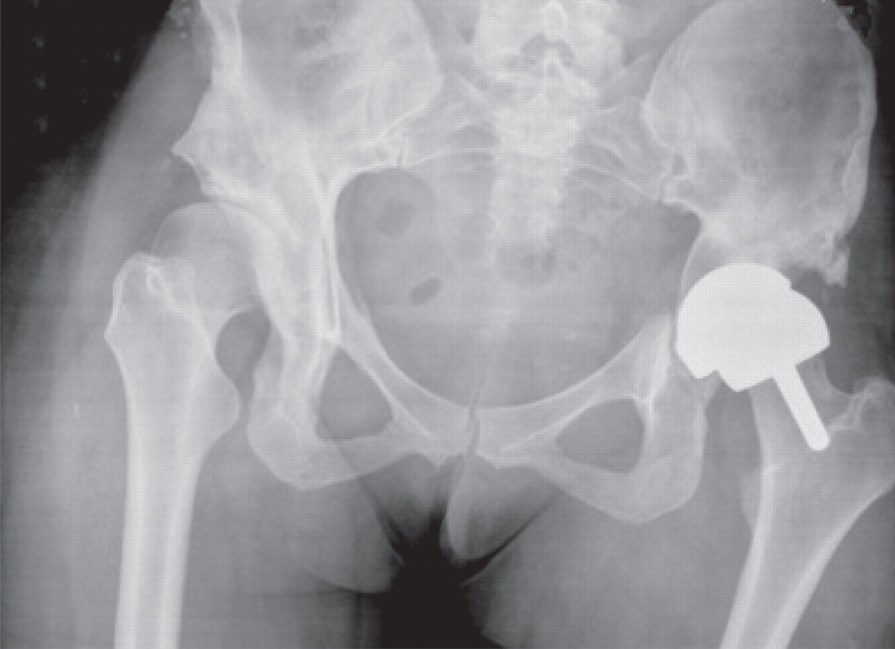
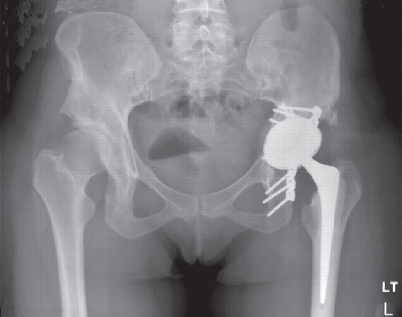
A 29-year-old woman presented with sudden onset of left hip pain and inability to bear weight following a low-energy wakeboarding incident. She had had a pelvic osteotomy in 2000 and subsequently a Birmingham Hip Resurfacing (Smith and Nephew) in 2001. In 2006, a plain antero-posterior (AP) radiograph of the pelvis showed evidence of neck narrowing (Figure 1). There appears to be rarification and reduced bone density with thinning of the iliopectineal cortical outline medially. Blood metal ion levels were Co 621 nmol/L (36.6 μg/L) and Cr 730 nmol/L (38 μg/L). Figure 2 shows an AP radiograph from 2007 immediately after her wakeboarding accident. There is a fracture of her pelvis adjacent to the left hip re-surfacing. Cup inclination measured 55° with uncovering of head and edge loading. Component head size and outer shell diameter were 42 and 48 mm, respectively. Note the right hip dysplasia with evidence of previous periacetabular osteotomy and a gracile femur. Metal concentrations were also measured in synovial fluid: Cr 228,000 nmol/L (11,850 μg/L) and Co 12,600 nmol/L (745 μg/L). Figure 3 shows an AP radiograph postfixation and total hip replacement. Case 1. Plain antero-posterior radiograph of the pelvis showed evidence of neck narrowing. There appears to be rarification and reduced bone density with thinning of the iliopectineal cortical outline medially Case 1. Antero-posterior radiograph from 2007 immediately after her wakeboarding accident. There is a fracture of her pelvis adjacent to the left hip re-surfacing. There appears to be rarification and reduced bone density with thinning of the iliopectineal cortical outline medially Case 1. Antero-posterior radiograph postfixation and total hip replacement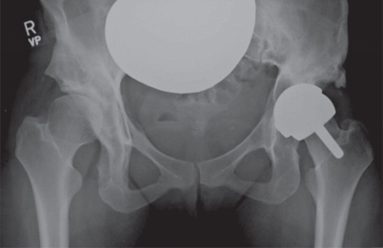
The clinical ‘metal ion’ questions raised by this case are: did blood metal ion levels return to normal following revision? This was very important to the psychological wellbeing of the patient who was of reproductive age, planning a family and had read about carcinogenicity following MOM hips. Subsequent metal concentrations were Cr 199 nmol/L (10.3 μg/L) and Co 28 nmol/L (1.7 μg/L) in October 2008 and Cr 91 nmol/L (4.7 μg/L) and Co 13 nmol/L (0.8 μg/L) in October 2009. As in other patients with metallosis, Cr concentrations can remain elevated after revision, while Co concentrations can fall relatively quickly.
Case 2 – Silent osteolysis and imminent acetabular fracture and high metal ion levels in a 65-year-old man
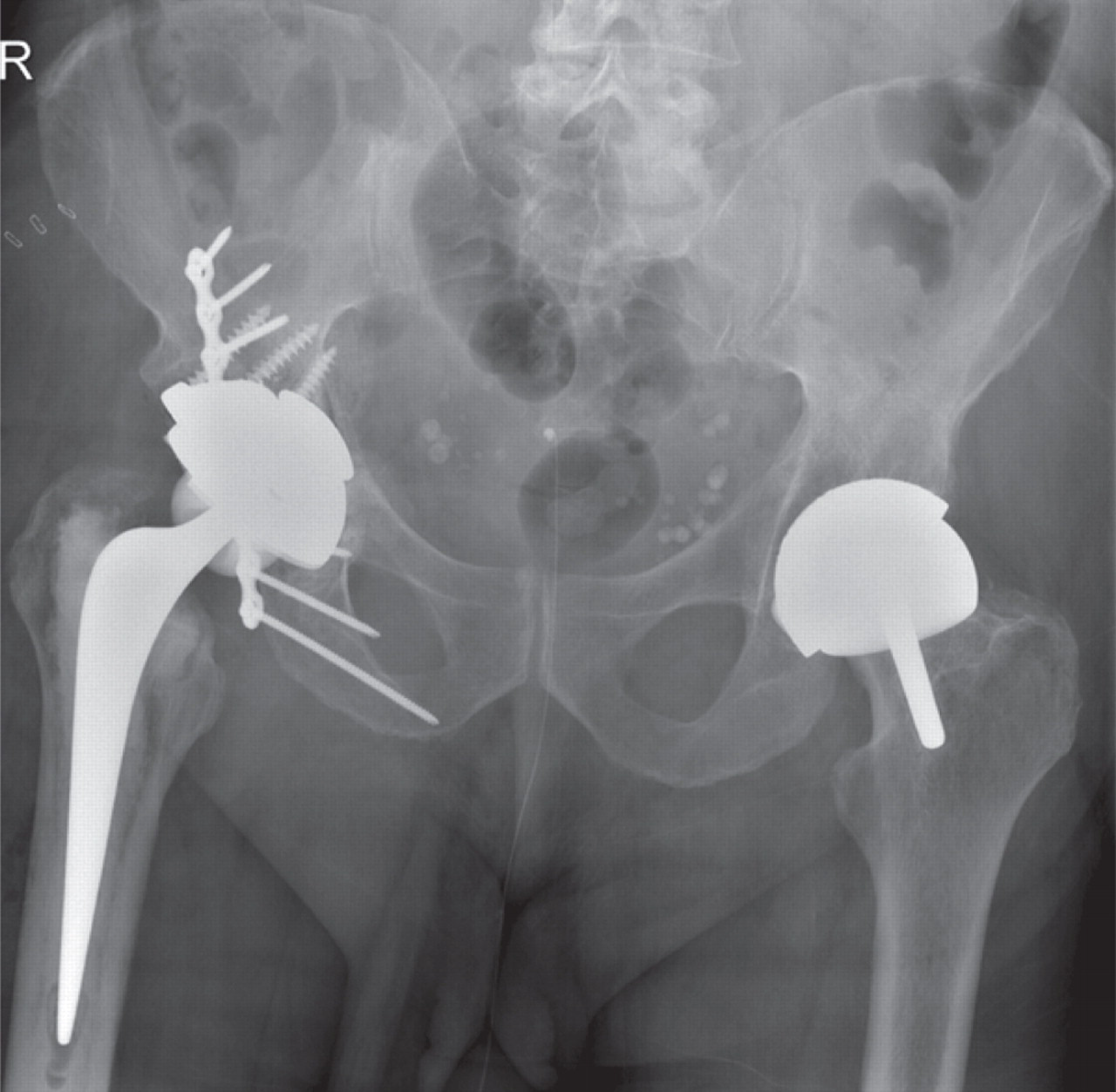
Figure 4 shows an AP radiograph from a 65-year-old man who presented with a painful right MOM hip re-surfacing (Birmingham Hip Resurfacing, Smith and Nephew) five years postoperatively. The femoral component was loose and the acetabular bone was so thin that fracture was imminent. High blood metal ions were recorded (Cr 698 nmol/L [36.3 μg/L] and Co 1212 nmol/L [71.4 μg/L]), and may have predicted such severe osteolysis if they had been measured prior to this. A postrevision AP radiograph (Figure 5) shows the extent of the reconstruction needed to stabilize the bony defects found at operation. By six months postrevision, he was able to return to sailing the Atlantic Ocean. A series of later pre- and postoperative measurements of blood Co and Cr revealed a dramatic rate of decay for Co levels and a slower rate for Cr (Figure 6). The blood metal concentrations had increased in the period following the initial outpatient appointment. Case 2. Antero-posterior radiograph from a 65-year-old man who presented with a painful right metal-on-metal hip re-surfacing five years postoperatively. The femoral component is loose and the acetabular fracture was so thin that fracture was imminent Case 2. Antero-posteriorradiograph showing the extent of the reconstruction needed to stabilize the bony defects found at operation Case 2. Pre- and postoperative changes in blood chromium and cobalt concentrations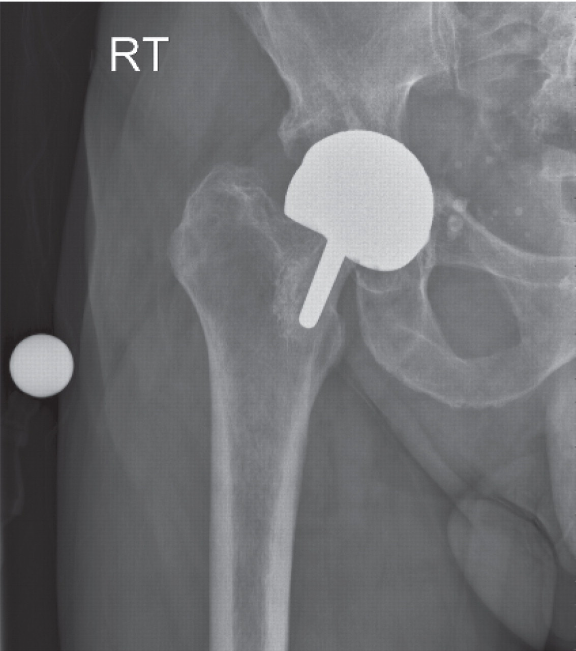
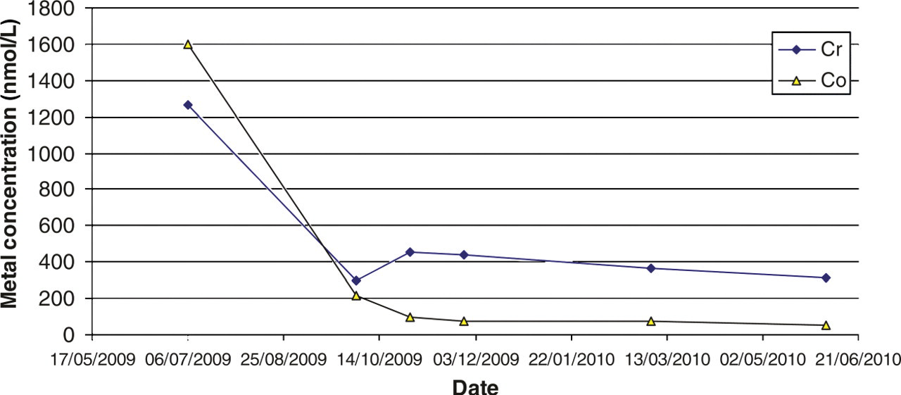
The clinical ‘metal ion’ questions posed by this case are: is the other hip at risk? Did the levels of metal ions return to that of a typical unilateral MOM hip? Was the loss of bone caused by local metal ion poisoning? We once again show that the Co concentration falls to normal concentrations much faster than does Cr concentration. There can be no direct proof in an individual case that the loss of bone density is directly caused by metallosis, but we suggest that it is a reasonable assumption that this is the case.
Case 3 – Silent muscle necrosis with very high metal ions and rapid deterioration requiring revision and inability to reconstruct muscle
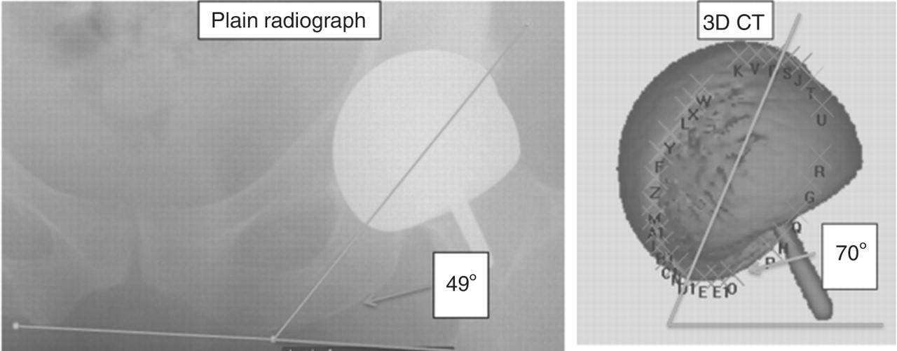
A 54-year-old lady attended a research clinic with a good hip function score and very high blood metal ion concentrations with an ASR hip re-surfacing (DePuy, Leeds, UK). Blood Cr concentration was 2700 nmol/L (140 μg/L) and Co concentration was 6551 nmol/L (386.5 μg/L). An AP radiograph is shown in Figure 7. Two years later her hip function deteriorated dramatically. This might have been better predicted from the high metal ion concentrations. Three-dimensional computed tomography measurement (Figure 8) revealed cup angles of 70° inclination (the optimum is 45° because angles higher than this cause edge loading and high rates of wear of the hip) and 49° anteversion (the optimum is between 5 and 25°, depending on the femoral version). Metal artefact reduction sequence MRI 99 revealed a large trochanteric bursa (Figure 9) with evidence of nodularity within the bursal sac and an irregular wall. A large amount of black fluid surrounded the hip at revision operation. Synovial fluid analysis showed Cr concentration 5063 μmol/L (263.3 mg/L) and Co concentration 207.1 μmol/L (12.2 mg/L). Analysis of the explanted hip revealed a wear rate that was 100 times greater than predicted by hip simulator studies. Case 3. Antero-posterior radiograph from a 54-year-old lady who attended a research clinic. She had a good hip function score and very high blood metal ion concentrations Case 3. Three-dimensional computed tomography (3D CT) measurement showing cup angles of 70° and 49° anteversion Case 3. Metal artefact reduction sequence magnetic resonance imaging revealed a large trochanteric bursa with evidence of nodularity within the bursal sac and an irregular wall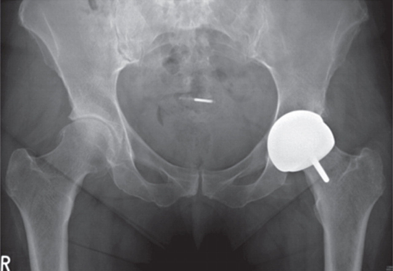
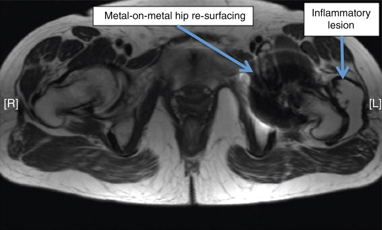
The clinical ‘metal ion’ questions raised by this case are: the patient wanted to know why this had happened? Did the metal ion levels return to normal? Could other patients be spared the muscle destruction if earlier intervention is taken? The change in blood metal concentrations following the operation is shown in Figure 10. The blood Co concentration showed a significant fall in the weeks before the revision surgery, probably due to her reduced activity as a result of the pain from the joint. There is an immediate and ongoing fall in metal concentrations postsurgery, although concentrations remained very high for a considerable time after the operation, and Cr continues to remain high more than 12 months later. We are continuing to monitor metal concentrations to see if there is an eventual fall to ‘acceptable’ levels. The conclusion is that there remains a slow release pool of Cr, possibly with the capsular tissues. There appears to be no toxic effect of this very high metal concentration. The very high metal concentrations seen preoperatively are a clear indication of high wear in the joint. The patient was reviewed for a year before the revision operation. It may be that the final tissue damage may have been less severe had the operation been performed sooner during this period. This highlights the need to take clinical action when raised metal concentrations are seen in these patients, even if there is as yet no pain. Case 3. Pre- and postoperative changes in blood chromium and cobalt concentrations. Preop, preoperative; Postop, postoperative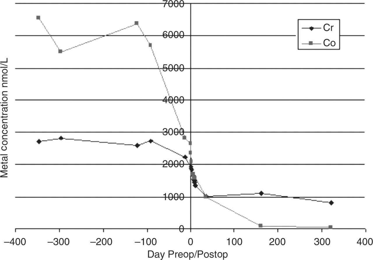
Conclusions
Increased circulating concentrations of Cr and Co can be used to monitor wear in MOM hip replacements. These operations are very successful but a minority of patients suffer wear in the joint, which will be reflected in increased metal concentrations. These may be used in conjunction with radiological and clinical assessments to inform the decision about revision of the original operation. The recent MHRA safety alert will serve to bring this potential problem to the attention of all concerned and expedite the clinical decision before major clinical problems arise.
DECLARATIONS